



Abstract
Background: To investigate the effect of Ashwagandha (ASH) Withania somnifera (WS) in preventing the damage caused by Fluoxetine (FLX) on testis.
Methods: Adult thirty-two male Wistar rats were randomly divided into four groups (eight rats in each): a control group, an Ashwagandha-treated group (200 mg orally) according to Bhargavan et al (2015), a FLX -treated group (10 mg/kg orally), and a combined FLX and ASH-treated group. Hormonal profiles (testosterone, FSH, and LH) and sperm parameters (count, motility, abnormality, and semen fructose levels) were evaluated. Testicular tissues were analyzed for biochemical markers, including glutathione peroxidase (GSH), superoxide dismutase (SOD), glutathione S-transferase (GST), malondialdehyde (MDA), and nitric oxide (NO), as well as for histopathological changes.
Results: Biochemical parameters revealed tissue oxidative stress, in FLX group which was evidenced as increase in MDA, NO level and reduction in GSH, GST, SOD activities in testes which were reduced in co-treatment group. in addition, spermatogenesis was inhibited as indicated by the decrease (testosterone, LH and FSH), (sperm count, motility). Moreover, ASH treatment leads to an increase in testosterone (TS), LH, FSH levels, sperm count and motility.
Conclusion: These observations suggest that the antioxidant properties of Ashwagandha may have played a role in ameliorating the testicular toxicity induced by Fluoxetine.
Keywords: Ashwagandha, adaptogen, infertility, selective serotonin reuptake inhibitors (SSRIs), fluoxetine, lipid peroxidation, oxidative stress, spermatogenesis
Introduction
Natural products are the source of over half of modern pharmaceuticals [1]. Ashwagandha (Withania somnifera), known as Indian ginseng or winter cherry, is a widely used adaptogenic herb found in Asia, Africa, and the Middle East [2]. It has been traditionally used for stress reduction, immune modulation, and enhancing male fertility [3,4]. ASH root extract has demonstrated antioxidant, anti-inflammatory, and neuroprotective effects, as well as the ability to improve hormonal balance and semen quality [5]. It acts through direct antioxidant mechanisms and by restoring reproductive hormone levels and sperm parameters [6].
Oxidative stress, caused by excessive reactive oxygen species (ROS), impairs sperm function and DNA integrity, leading to infertility and pregnancy loss [7]. Hormonal regulation of spermatogenesis involves GnRH, LH, FSH and LH stimulates Leydig cells, while FSH supports Sertoli cell function. Disruption in these hormones can result in poor sperm quality and infertility [8].
Medications, especially antidepressants like SSRIs, have been linked to male infertility through hormonal imbalance and oxidative damage [9] SSRIs such as fluoxetine (FLX) are widely prescribed but can cause side effects including sexual dysfunction, reduced testosterone, and impaired spermatogenesis [10]. Chronic FLX exposure has been shown to decrease sperm quality, TS and FSH levels, and increase oxidative stress in rat models [11] Herbal remedies like ASH may offer a protective effect against such drug-induced reproductive toxicity [12]. Therefore, this study aims to investigate the efficacy of ASH in ameliorating FLX-induced testicular dysfunction in rats by assessing biochemical, hormonal, histological, and semen-related parameters.
Materials and Methods
Materials
Fluoxetine
Fluoxetine (Philozac®) was obtained as capsules from Amoun Pharmaceutical Company, Cairo, Egypt. Each capsule contained 10 mg fluoxetine hydrochloride. FLX was dissolved in distilled water and was given orally to animals by gavage at a dose level of 10 mg/kg body weight (equivalent to the therapeutic dose for human according to Laurence & Bacharach [13] daily for eight weeks.
Plant extraction
The ethanolic extract of ASH dry powdered roots was carried out according to the modified method of Filipiak-Szok et al., [14]. In brief, 2 gm of powder were soaked in 20 ml 70% ethanol at room temperature for 24 hrs. under continuous stirring; then the mixture was filtered through sterile filter paper (Whatman number 42, England). The solvent was evaporated using a rotary evaporator, and the extract was stored at –20 °C until further use.
Animals
The study was performed on thirty-two male albino rats (Sprague-dawaley) weighing 150-200 gm. The rats were obtained from an animal house at Assiut University, Faculty of Medicine. They were housed under controlled laboratory conditions (12-h light/ dark cycle at 25 ±1 °C), provided with the standard diet of rodent pellets and water. All the experimental procedures were performed by the guidelines of the Institutional Animal Care and the norms of Ethical Committee of the Faculty of Science Al-Azhar University, Assiut.
Ethics approval
Experiment was approved by the Ethics Committee of Faculty of Science Al-Azhar University, Assiut; Certificate Reference Number: (AZHAR 2/2021). Effort was taken to minimize the number of animals used, as well as their pain and suffering during experiments.
Animal grouping
The animals were randomly divided into four groups with eight animals in each. First group served as control. In the second group rats were orally administrated with FLX at a dose of 10 mg/kg body weight equivalent to the therapeutic dose for human according to Laurence & Bacharach [13]. Third group; animals were treated with ASH at a dose of 200 mg/kg body weight, according to Anwer et al. (2012) daily for 8 weeks. Fourth group; animals were given FLX followed by ASH daily for 8 weeks. All rats were handled blindly, with randomization applied during group allocation [15].
Blood and tissue sampling
At the end of the experiment, the animals were fasted overnight, and blood samples were collected; about 3 ml blood of each animal. Sera were separated using cooling centrifugation and stored at - 80ºC until further analysis. After blood collection, the animals were sacrificed and the testes were dissected out, washed in cooled 0.9 % saline solution then dried. After that one testis stored at - 80ºC for biochemical determinations and other testis passed to 10% neutral buffer formalin to apply histopathological examination.
Biochemical assay
After collection of blood sample, serum used to determination TS, FSH and LH levels using Elisa kits according to Babu, et al. (2004) [16].
Preparation of tissue samples
Testis parts were homogenized using an electrical homogenizer to prepare 20% tissue homogenate in phosphate buffer saline. The homogenates were centrifuged for 10 min. at 10000 r.p.m using a cooling centrifuge. The supernatant was separated and kept at - 80ºC till determination of oxidative stress.
Spermatozoa characteristics
At the end of the study, semen samples were collected from the cauda epididymis carefully that separated from the testis and placed in a Petri dish containing Ham’s F10. Epididymal cauda was minced with scissors to release sperm and then was placed in the incubator for 15 min [17]. approximately, 10μL of the diluted sperm suspension was transferred to each counting chamber of the hemocytometer and allowed to stand for 5 min [18]. The sperms which settled during this time were counted by a light microscope at 200X magnification [19].
Biochemical investigations for oxidative stress markers
Homogenates of testis were used for the determination of the extent of lipid peroxidation by measuring malondialdehyde (MDA) formation via thiobarbituric acid reaction method [20] . MDA reacts with thiobarbituric acid in acidic medium that gives a pink-colored pigment at 95˚C.
Superoxide dismutase (SOD) activity was measured by using spectrophotometer with an assay kit in according to Misra & Fridovich 1972 [21] . The hydroxylamine test, which was developed from the xanthine oxidase assay, was used to measure the SOD activity in the samples. A summary of the test principle is xanthine, and the xanthine oxidase system produce superoxide anions. By oxidizing hydroxylamine these superoxide anions produce nitrite. A colored substance created when nitrite reacts with sulfanilic acid and naphthalene diamine.
Also, GSH was evaluated as described by Beutler & Kelly 1963 [22]. and Glutathione S-Transferase (GST) according to Habig, et al. 1974 [23], while nitric oxide according to Montgomery & Dymock 1961 [24].
Histopathological examination
After being fixed in 10% formalin, the testicular tissues were dehydrated using ethyl alcohol in ascending grades (70%, 80%, 90%, 95%, and 100%), then cleared in xylene, and embedded in paraffin wax. Sections of 4 – 5 µm (µm) thickness were cut and stained with hematoxylin and eosin according to Cunningham 1997 [25]. Then stained slides were used for histopathological and morphometric evaluations. Photographs were taken using an optical microscope (Olympus, Tokyo, Japan).
Statistical analysis
Data were prepared in excel, analyzed with analysis of variance (ANOVA) using SPSS 16.0 for Windows (SPSS, Inc., Chicago, IL, USA) followed by Tukey’s post-hoc tests and presented as mean ± S E. P < 0.05 was considered to denote significant differences between groups. Means that share the same superscript letter are not significantly different from each other (non-significant difference). Means with different superscript letters are significantly different at the chosen significance level. The alphabetical order of letters reflects the ranking of the means within each parameter: “a” typically denotes the highest significant value. Letters (b, c, d) denote significantly lower values.
Results
Biochemical results
Serum FSH, LH and testosterone were significantly decreased (P < 0.05) after eight weeks of FLX treatment when compared with control group. Administration of ASH in for eight weeks induced a significant (P < 0.05) improvement of serum FSH, LH and TS (Table 1).
Table 1. Effect of orally ASH (200 mg/kg b.wt) for eight weeks on some hormones: TS, LH, FSH in rats administrated orally with FLX (10 mg/kg b.wt) daily for eight weeks.
FLX+ASH | ASH | FLX | Control | |
|---|---|---|---|---|
4.85±0.10b | 5.28±0.14a | 1.53±0.08d | 4.85 ±0.13b | TS (ng/ml) |
8.4±0.28cb | 8.8±0.27d | 4.4±0.46a | 9.5±0.28dc | LH (mIU/ml) |
6.4±0.30bc | 7.4±0.29d | 4.4±0.18a | 7.2±0.30cd | FSH (mIU/ml) |
Number of rats for each group: (n = 8). All data are expressed as the Mean ± SE. Data were subjected to one-way analysis of variance (ANOVA) followed by a post hoc test (Duncan’s) at p<0.05 level. Values with different superscript letters differ significantly, while those sharing the same letter are not significantly different.
Effects of FLX on antioxidant/oxidative status of testicular tissue
In FLX-treated rats, MDA, NO levels were significantly (P < 0.05) increased compared with the control group respectively. In ASH alone and the ASH+FLX groups. The levels of MDA, NO did not significantly differ from those of the control group. On the other hand, FLX-treated rats showed significant decrease in GSH, GST, SOD levels. Animals administered with ASH showed a significant elevation in GSH, GST, SOD levels (Table 2).
Table 2. Effect of orally ASH (200 mg/kg b.wt) for eight weeks on some antioxidant parameters; GSH, GST and SOD Level and oxidative stress parameters MDA and NO. in rats administrated orally with FLX (10mg/kg b.wt. daily for eight weeks.
FLX + ASH | ASH | FLX | Control | |
|---|---|---|---|---|
72.6±1.4bc | 81.7±1.7a | 23.5±1.3d | 77.8±1.9ab | GSH (mmol/gm protein) |
7.2±0.36b | 9.1±0.36a | 3.3±0.16c | 8.5±0.30ab | GST (nmol /mg protein) |
8.0±0.48b | 9.4±0.54a | 2.27±0.33c | 9.3±0.38a | SOD (µl/mg protein) |
5.0±0.48b | 1.7±0.20c | 35.9±1.7a | 2.35±0.22c | MDA (nmol/gm protein) |
55.8±4.1b | 40.8±1.8c | 122±5.7a | 40.1±2.06c | NO (µmo1/L) |
Number of rats for each group: (n = 8). All data are expressed as the Mean ± SE. Data were subjected to one-way analysis of variance (ANOVA) followed by a post hoc test (Duncan’s) at p<0.05 level. Values with different superscript letters differ significantly, while those sharing the same letter are not significantly different.
Semen examination
Administration of FLX into male rats resulted in a significant (P < 0.05) reduction in sperm motility, count and semen fructose while a significant increase in abnormality when compared with control group. Administration of ASH induced a significant improvement of sperm motility and count, abnormality and semen fructose (Table 3).
Table 3. Effect of orally ASH (200mg/kg b.wt). for eight weeks on semen examination (count, motility, abnormality and fructose in semen) in rats administrated with FLX (10 mg/kg b.wt for eight weeks).
FLX + ASH | ASH | FLX | Control | |
|---|---|---|---|---|
161.5±3.6a | 169.12±3a | 61.75±4.4c | 162.1±5.18a | Fructose (mg/dl) |
111.25±8.4b | 147.8±8,7a | 15.12±4.2c | 156.1±7.9a | Sperm count (million/ml3) |
74.6±3.12c | 85±1.88ab | 20.25±2.87d | 85.6±2.02ab | Motility % |
19±1.66c | 12.12±0.95d | 79.3±1.47a | 16.5±1.08cd | Abnormal forms % |
Number of rats for each group: (n = 8). All data are expressed as the Mean ± SE. Data were subjected to one-way analysis of variance (ANOVA) followed by a post hoc test (Duncan’s) at p<0.05 level. Values with different superscript letters differ significantly, while those sharing the same letter are not significantly different.
Histopathological examination
FLX-administrated rat showing defective spermatogenesis in most of the seminiferous tubules with degenerative and necrotic changes of the spermatogonial cells, as well as nuclear pyknosis of many of them and attempts for spermatid giant cell formation (Figure 1 B) loss spermatogonial cell (Figure 1 C), while ASH group showing normal histological structure of the seminiferous tubules (ST), normal spermatogonial cells layers with active spermatogenesis and active sperms in the tubular lumens (Figure 1 D).
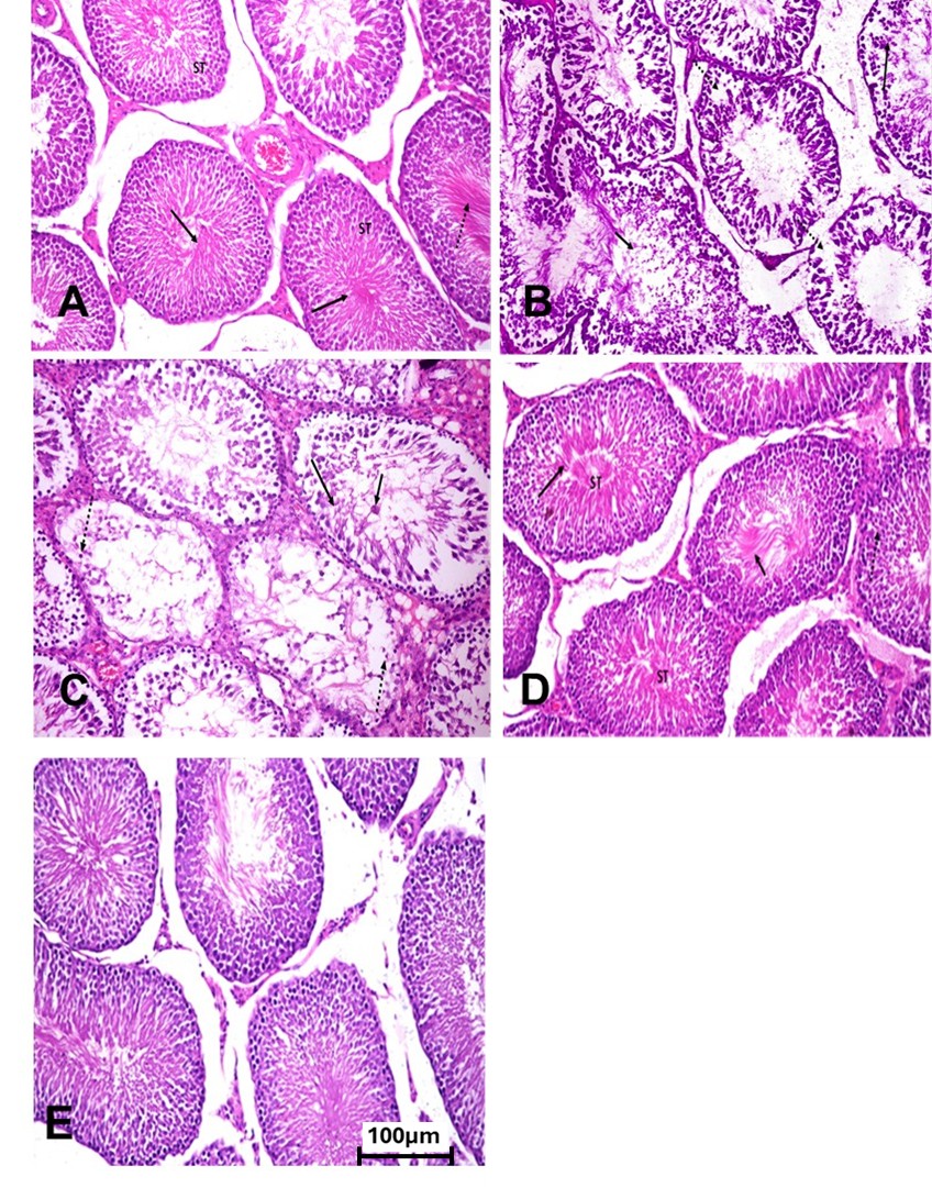
Figure 1. Histological changes in testicular tissue of control, fluoxetine-administered, Ashwagandha-treated, and combined treatment groups in adult male rats
Testis of control rats showing a normal histological structure of the seminiferous tubules (ST) with active spermatogenesis (arrow); note the presence of active sperms (dotted arrow) in the tubular lumens. (B) Testis of FLX-administered rats showing defective spermatogenesis in most of the seminiferous tubules, with degenerative and necrotic changes in the spermatogonial cells (arrow), nuclear pyknosis (dotted arrow), and attempts at spermatid giant cell formation (short arrow). (C) Testis of FLX-administered rats showing defective spermatogenesis in most of the seminiferous tubules, with degenerative and necrotic changes in spermatogonial cells (arrow), loss of some cells (dotted arrow), and necrotic debris in the lumens (short arrow). (D) Testis of ASH-treated rats showing normal histological structure of the seminiferous tubules, intact layers of spermatogonial cells (dotted arrow), active spermatogenesis (arrow), and active sperms (short arrow) in the tubular lumens. (E) Testis of FLX-administered rats treated with Ashwagandha showing well-organized spermatogonial cell layers (arrow) and active spermatogenesis (dotted arrow). (Hematoxylin and Eosin stain, X400) (scale bar 100µm).
Discussion
Fluoxetine is a selective serotonin reuptake inhibitor widely prescribed for the treatment of neurological disorders such as depression and anxiety [26].
In the present study, rats administrated a dosage of 10 mg/kg FLX for eight weeks showed significant reduction in serum level of FSH, LH and testosterone hormones compared to control group (table 1). LH stimulates the production of testosterone by Leydig cells [27]; thus, The reduction in testosterone correlates with change of LH production, as indicated by the positive association between testosterone and LH concentrations observed in the present study. The physiological significance of the LH-FSH correlation stems from its distinct effects on spermatogenesis. Gonadotrophins help spermatogenesis at two stages: spermatogonial maturation and spermiation. Separately, FSH maintains pachytene spermatocytes, while LH promotes conversion to spherical spermatids [28].
Our findings were supported by Bataineh & Daradka 2007[29] and Sakr et al. 2013 [26]. who demonstrate that long-term oral administration of FLX resulted in a significant decrease in testosterone, FSH and LH levels in adult male rats. Furthermore, Tanrikut, et al. (2010) find that the treatment with paroxetine for 5 weeks reduced level of serum testosterone [30]. On other hand our study contradicts Julanov`s study who demonstrat that treatment with FLX dose 150 mg kg-1 for 28 days increase FSH , LH [31]. Also de Oliveira et al. (2013); Gouvêa, et al. (2008) and Vieira et al. (2013) reported that their no significance difference in testosterone level and Leydig cells [32-34]
There is increasing evidence that the functionality of steroidogenic tissues may be affected in conditions of prolonged oxidative stress with ROS production. A number of experiments with rat primary testicular Leydig cells demonstrated that levels of steroidogenic enzymes decreased with increasing animal age along with Ts output in parallel with diminished levels of antioxidants [35].
Omura & Morohashi (1995) reported that ROS such as superoxide anions and H2O2 could inhibit testicular steroidogenesis [36].
Oxidative stress is known as elevated level of ROS that damage proteins, lipids, cell membranes, and genetic material [37]. Lipid peroxidation, a process where ROS primarily target membrane lipids, It is also known that spermatozoa and testicular tissues are particularly vulnerable to lipid peroxidation and ROS assault. The high concentration of polyunsaturated fatty acids in sperm membranes is thought to be the cause of testicular tissues' susceptibility to oxidation [38].
In the present study, rats administered a dosage of 10 mg/kg FLX for 8 weeks showed a significant reduction in GSH, SOD, and GST levels, along with a significant increase in MDA and NO levels compared to the control group (Table 2). The elevated MDA and NO levels, combined with the reduction in antioxidant parameters, indicate lipid peroxidation.
Many authors explained the mechanism by which fluoxetine induces testicular tissue toxicity. Inkielewicz-Stępniak (2011) showed that fluoxetine induces lipid peroxidation leading to free-radical release, which causes membrane disorganization and subsequent decreases in membrane fluidity, and finally extensive tissue damage [39].
Our results agree with Sakr, et al (2015) who demonstrate that oral administration of FLX resulted in a significant reductions in serum GSH , SOD and a significant increase in MDA concentrations [40]. On the contrary Erdemir et al. (2014) reported that that SSRIs have a negative effect on testicular tissues [41].
Spermatogenesis is a complex process in testes that forms mature male germ cells through mitosis, meiosis, and spermiogenesis. It is vulnerable to insults from genetics, environment, diet, and heavy metals, with oxidative stress affecting fertility potential and sperm dysfunction [42].
Treatment with FLX related to male infertility in the form of poor semen quality such as decreased sperms viability and motility [43]. The obtained data showed that FLX administrated group showed a highly significant decrease in seminal fructose level, and count, motility and significant increase in abnormal forms of the sperms compared to control group.
Our results indicate defective spermatogenesis, which is explained by decrease in testosterone level in FLX group, and the increase in MDA, NO.
Our data supported by Chen, et al. (2025) who demonstrated that long-term fluoxetine administration, exhibited significantly reduced mating and fertility indices, decreased sperm count and motility, and increased sperm deformities compared to the control group [44].
Studies with Alzahrani (2012); Bataineh & Daradka (2007) and Sakr et al. (2015) reported that treatment with FLX lead to significance decrease in sperm count and motility and significance increase in abnormality [29,40,45].
Regard to significance decrease in semen fructose; the secretion of the seminal vesicles contains fructose other simple sugars, amino acids, ascorbic acid, and prostaglandins. Fructose is the primary nutrient source for sperm in the semen. Seminal vesicular secretion is important for semen coagulation, sperm motility, stability of sperm chromatin, and suppression of the immune activity in the female reproductive tract [46] .
Jahromy & Moghadam (2014) recorded that sertraline administration to adult mice induced a dose-dependent decrease in sperm motility, count, viability and serum level of testosterone [47].
In human a study by Kumar et al. (2006) they demonstrated that FLX, sertraline, fluvoxamine, and citalopram negatively influence semen parameters and showed a spermicidal effect. In addition, long-term intake of FLX caused a decrease in spermatogenesis, weights of reproductive organs, and FSH levels in rats [48].
Fluoxetine damages testicular tissue causing deformation of the seminiferous tubules, degenerated spermatogenic cells with extensive cytoplasmic vacuoles, and sloughing of germ cells [49].
This study show that FLX administration group causes defective spermatogenesis in most seminiferous tubules, characterized by degenerative and necrotic changes in spermatogonial cells. Also, loss spermatogonial cell nuclear pyknosis, and attempts at spermatid giant cell formation. These findings collectively suggest that FLX can have adverse effects on male reproductive function, affecting spermatogenesis and steroidogenesis. Our finding agree with Bataineh & Daradka (2007) who reported that Long-term ingestion of fluoxetine for 60 days caused a great decrease in spermatogenesis in seminiferous tubules of the testes [29]. The previous study show that degeneration within a cell (through cell death or apoptosis) could be considered an indication of cell death [50].
W. somnifera, also known as Ashwagandha, has been used in traditional medicine for thousands of years. ASH has a variety of activities such as anti-stress, anti-inflammatory, antimicrobial, anti-cancer, anti-diabetic, anti-obesity, cardioprotective, and hypolipidemic properties. ASH exhibits a positive effect on the functioning of the endocrine system including improvement the secretory function of the thyroid gland, normalizing adrenal activity, and multidirectional improvement on functioning of the reproductive system [51] . In the current study, the histological alterations in the testes caused by FLX improved in rats treated with both FLX and ASH. Furthermore, rats administrated with FLX + ASH and ASH group revealed a significant increase in LH, FSH and TS. These results are in contract with that of Jasuja, et al. (2013) who reported that oral administration of acephate (75 mg/kg body weight/day) for 15 and 30 days caused a significant decrease in serum testosterone, follicle stimulating hormone (FSH) and luteinizing hormone (LH) concentration when compared with control group [52]. Serum testosterone, LH and FSH concentration were increased in ASH group. The mechanism of ASH effect on the reproductive system is not known entirely yet, but this mechanism is proposed to be linked to the antioxidative features and ability to improve the hormonal balance of LH, FSH, and testosterone and improve detoxification process [5].
Our results indicated that co-administration of ASH caused an increase in the antioxidant enzyme, GSH, GST and SOD while a decrease in the lipid peroxidation marker, MDA and NO. This indicates that ASH extract reacts with peroxyl radicals including the inhibition of LPO chain propagation [53]. Reduced level of LPO in ASH-treated animals is indicative of the antioxidant nature of this plant [54]. Our results agree with Jasuja et al. (2013) who stated that the level of LPO was reduced whereas GSH content, SOD and catalase activity were elevated after treatment with ASH at the dose level of 100 mg/kg body weight/day [52]. Furthermore, in several studies investigating the effects of ASH on semen quality in infertile males, enzymatic antioxidant activity was determined by measuring LPO and protein carbonyl groups in seminal plasma, which indirectly depicts the enzymatic activity of SOD and catalase. Compared with pretreatment, the amount of LPO was found to decrease after administration of ASH [55-57], which is most likely attributed to a synergistic relationship between the increase in the enzymatic activity of SOD and catalase and the innate antioxidant activity of ASH. Therefore, compounds of the ASH extract are able to donate electrons and halt the destructive chain reaction of free radicals, thereby lowering the overall ROS burden [58]. Therefore, it is reasonable to conclude that ASH root extract has been found to possess potent antioxidant activity; which is achieved by supplementing co-factors necessary for the proper functioning of antioxidant enzymes. This ability to decrease ROS and oxidative stress in spermatozoa can be attributed to two mechanisms.
Regard to semen parameter ASH-administrated group revealed significance increase sperm count and motility and significance decrease in abnormality. Our finding agree with Bhargavan, et al. (2015) and Kumar et al. (2015). They described the effects of ASH root extract on reversal of male fertility in arsenic and alcohol-induced testicular impairments respectively in rats. They observed significant augmentation of sperm count and motility after oral administration of ASH at a dose of either 100 mg/kg for 30 days or 200 mg/kg for 28 days [59,60]. Moreover, sperm morphological abnormality was found to be reduced in ASH-treated rats. The roots of ASH contain alkaloids, withanolides, flavonoids, and reducing sugars [61]. Over 20 active compounds, such as withaferin A, sitoindosides, withanosides, choline, and beta-sitosterol, have been identified [62]. These constituents likely contribute to ASH effects on semen, although the specific spermatogenic or steroidogenic roles of these compounds remain unclear [55].
Conclusions
ASH treatment significantly protected testicular tissue from injury caused by FLX. The biochemical and histopathological changes were markedly improved by the herbal extract of ASH. These protective effects of ASH therapy may be attributed to its potent antioxidant properties. It has the potential to be used as an adjunct therapy to mitigate the adverse effects of FLX in patients.
Ethical Considerations
All procedures involving animals were conducted in accordance with institutional and national guidelines for the care and use of laboratory animals. The experimental protocol was approved by the Institutional Ethics Committee of Faculty of Science, Al-Azhar University, Assiut branch. Every effort was taken to minimize animal suffering during the study.
Conflict of Interests
The authors declare no conflicts of interest.
Funding
This research received no specific grant from any funding agency in the public, commercial, or not-for-profit sectors.
Acknowledgement
The authors would like to thank the staff of the Faculty of Science, Al-Azhar University, Assiut branch, Egypt for their support and assistance during the experimental procedures.
Authors' Contributions
[Amira Siddiq]: Experiment design, data analysis, laboratory analysis, and manuscript writing.
[Alaa M. H. El-Bitar]: Animal handling, data collection, and statistics.
[Abdelbaset M. A. Abdelreheem]: Data interpretation and manuscript revision.
References
- Aggarwal A, Jethani S, Rohatagi R, Kalra J. Premature ejaculation - dose and duration dependent effect of fluoxetine: a histological study on seminal vesicle of albino rats. J Clin Diagn Res. 2014;8(9):AC14-6. [DOI: 10.7860/JCDR/2014/10084.4912] [PMID: 25386416]
- Ahmad MK, Mahdi AA, Shukla KK, Islam N, Rajender S, Madhukar D, et al. Withania somnifera improves semen quality by regulating reproductive hormone levels and oxidative stress in seminal plasma of infertile males. Fertil Steril. 2010;94(3):989-96. [DOI: 10.1016/j.fertnstert.2009.04.046] [PMID: 19501822]
- Alzahrani HA. Sister chromatid exchanges and sperm abnormalities produced by antidepressant drug fluoxetine in mouse treated in vivo. Eur Rev Med Pharmacol Sci. 2012;16(15):2154-61. [PMID: 23280034]
- Anwer T, Sharma M, Pillai KK, Khan G. Protective effect of Withania somnifera against oxidative stress and pancreatic beta-cell damage in type 2 diabetic rats. Acta Pol Pharm. 2012;69(6):1095-101. [PMID: 23285670]
- Ashrafizadeh M, Ahmadi Z, Samarghandian S, Mohammadinejad R, Yaribeygi H, Sathyapalan T, et al. MicroRNA-mediated regulation of Nrf2 signaling pathway: implications in disease therapy and protection against oxidative stress. Life Sci. 2020;244:117329. [DOI: 10.1016/j.lfs.2020.117329] [PMID: 31954747]
- Babu SR, Sadhnani MD, Swarna M, Padmavathi P, Reddy PP. Evaluation of FSH, LH and testosterone levels in different subgroups of infertile males. Indian J Clin Biochem. 2004;19(1):45-9. [DOI: 10.1007/BF02872388] [PMID: 23105425]
- Bataineh HN, Daradka T. Effects of long-term use of fluoxetine on fertility parameters in adult male rats. Neuro Endocrinol Lett. 2007;28(3):321-5. [PMID: 17627270]
- Beutler E, Kelly Bm. The effect of sodium nitrite on red cell GSH. Experientia. 1963;19:96-7. [DOI: 10.1007/BF02148042] [PMID: 13967892]
- Bhargavan D, Deepa B, Shetty H, Krishna AP. The protective effect of Withania somnifiera against oxidative damage caused by ethanol in the testes of adult male rats. Int J Bas Clin Pharm. 2015;4(6):1104-9. [DOI: 10.18203/2319-2003.ijbcp20151340]
- Chen Y, Xiong Y, Zhu L, Gu L, Liu Y. Long-term oral fluoxetine leads to reduced male reproductive function in mice and gradual recovery after discontinuation. Repord Toxicol. 2025;132:108840. [DOI: 10.1016/j.reprotox.2025.108840] [PMID: 39826770]
- Cunningham CW. Is congruence between data partitions a reliable predictor of phylogenetic accuracy? Empirically testing an iterative procedure for choosing among phylogenetic methods. Syst Biol. 1997;46(3):464-78. [DOI: 10.1093/sysbio/46.3.464] [PMID: 11975330]
- Bisht S, Faiq M, Tolahunase M, Dada R. Oxidative stress and male infertility. Nat Rev Urol. 2017;14(8):470-85. [DOI: 10.1038/nrurol.2017.69] [PMID: 28508879]
- De Oliveira WM, de Sá IR, de Torres SM, de Morais RN, Andrade AM, Maia FC, et al. Perinatal exposure to fluoxetine via placenta and lactation inhibits the testicular development in male rat offspring. Syst Biol Reprod Med. 2013;59(5):244-50. [DOI: 10.3109/19396368.2013.796021] [PMID: 23651434]
- Devi PU. Withania somnifera dunal (Ashwagandha): potential plant source of a promising drug for cancer chemotherapy and radiosensitization. Indian J Exp Biol. 1996;34(10):927-32. [PMID: 9055640]
- Eacker SM, Agrawal N, Qian K, Dichek HL, Gong EY, Lee K, et al. Hormonal regulation of testicular steroid and cholesterol homeostasis. Mol Endocrinol. 2008;22(3):623-35. [DOI: 10.1210/me.2006-0534] [PMID: 18032697]
- El-Demerdash FM. Lipid peroxidation, oxidative stress and acetylcholinesterase in rat brain exposed to organophosphate and pyrethroid insecticides. Food Chem Toxicol. 2011;49(6):1346-52. [DOI: 10.1016/j.fct.2011.03.018] [PMID: 21419823]
- Soliman M, Mahmoud B, Kefafy M, Yassien R, El-Roghy E. Effect of antidepressant drug (fluoxetine) on the testes of adult male albino rats and the possible protective role of omega-3. Menoufia Med J. 2017;30(4):1135. [DOI: 10.4103/mmj.mmj_521_17]
- El Sayed Y, Ali N, Elshazly A, Salim R, Baioumy B, El Noury H. The role of Vitamin D on fluoxetine induced testicular toxicity in adult male rats. Mansoura J Forensic Med Clin Toxicol. 2021;29(2):41-56. [DOI: 10.21608/mjfmct.2021.69367.1030]
- Erdemir F, Atilgan D, Firat F, Markoc F, Parlaktas BS, Sogut E. The effect of sertraline, paroxetine, fluoxetine and escitalopram on testicular tissue and oxidative stress parameters in rats. Int Braz J Urol. 2014;40(1):100-8. [DOI: 10.1590/S1677-5538.IBJU.2014.01.15] [PMID: 24642156]
- Esterbauer H, Cheeseman KH. [42] Determination of aldehydic lipid peroxidation products: malonaldehyde and 4-hydroxynonenal. InMethods enzymol. 1990;186:407-21. [DOI: 10.1016/0076-6879(90)86134-H]
- Filipiak-Szok A, Kurzawa M, Szłyk E, Twarużek M, Błajet-Kosicka A, Grajewski J. Determination of mycotoxins, alkaloids, phytochemicals, antioxidants and cytotoxicity in Asiatic ginseng (Ashwagandha, Dong quai, Panax ginseng). Chem Zvesti. 2017;71(6):1073-82. [DOI: 10.1007/s11696-016-0028-0] [PMID: 28553006]
- Ganzera M, Choudhary MI, Khan IA. Quantitative HPLC analysis of withanolides in Withania somnifera. Fitoterapia. 2003;74(1-2):68-76. [DOI: 10.1016/s0367-326x(02)00325-8] [PMID: 12628397]
- Gonzales GF. Function of seminal vesicles and their role on male fertility. Asian J Androl. 2001;3(4):251-8. [PMID: 11753468]
- Gouvêa TS, Morimoto HK, de Faria MJ, Moreira EG, Gerardin DC. Maternal exposure to the antidepressant fluoxetine impairs sexual motivation in adult male mice. Pharmacol Biochem Behav. 2008;90(3):416-9. [DOI: 10.1016/j.pbb.2008.03.025] [PMID: 18457868]
- Habig WH, Pabst MJ, Jakoby WB. Glutathione S-transferases. The first enzymatic step in mercapturic acid formation. J Biol Chem. 1974;249(22):7130-9. [PMID: 4436300]
- Hajizadeh Z, Mehranjani MS, Najafi G, Shariatzadeh SM, Jalali AS. Black grape seed extract modulates fluoxetine-induced oxidative stress and cytotoxicity in the mouse testis. Jundishapur J Nat Pharm Prod. 2016;11(2):e27512. [DOI: 10.17795/jjnpp-27512]
- Hamed MA, Ekundina VO, Akhigbe RE. Psychoactive drugs and male fertility: impacts and mechanisms. Reprod Biol Endocrinol. 2023;21(1):69. [DOI: 10.1186/s12958-023-01098-2] [PMID: 37507788]
- Inkielewicz-Stępniak I. Impact of fluoxetine on liver damage in rats. Pharmacol Rep. 2011;63(2):441-7. [DOI: 10.1016/s1734-1140(11)70510-2] [PMID: 21602599]
- Jahromy MH, Moghadam AA. Effects of sertraline on sperm motility, number and viability and its relation to blood levels of testosterone, FSH and LH in adult male mice. Adv Sexual Med. 2014;4(2):17-24. [DOI: 10.4236/asm.2014.42004]
- Jasuja ND, Sharma P, Joshi SC. Ameliorating effect of Withania somnifera on acephate administered male albino rats. African J Pharm Pharmacol. 2013;7(23):1554-9. [DOI: 10.5897/AJPP12.1332]
- Julanov MN, Alimbekova ME, Usmangaliyeva SS, Turganbayeva GE, Koibagarov KU, Tagayev OO, et al. Impact of fluoxetine on testosterone levels and spermatogenic cell production of male albino mice. Caspian J Environ Sci. 2023;21(5):1135-41. [DOI: 10.22124/CJES.2023.7401]
- Kulisic T, Radonic A, Katalinic V, Milos M. Use of different methods for testing antioxidative activity of oregano essential oil. Food chem. 2004;85(4):633-40. [DOI: 10.1016/j.foodchem.2003.07.024]
- Kumar A, Kumar R, Rahman MS, Iqubal MA, Anand G, Niraj PK, et al. Phytoremedial effect of Withania somnifera against arsenic-induced testicular toxicity in Charles Foster rats. Avicenna J Phytomed. 2015;5(4):355-64. [PMID: 26445714]
- Kumar VS, Sharma VL, Tiwari P, Singh D, Maikhuri JP, Gupta G, et al. The spermicidal and antitrichomonas activities of SSRI antidepressants. Bioorg Med Chem Lett. 2006;16(9):2509-12. [DOI: 10.1016/j.bmcl.2006.01.078] [PMID: 16464584]
- Laurence DR, Bacharach AL. Evaluation of drug activities: pharmacometrics. Elsevier. 2013. [LINK]
- Mahdi AA, Shukla KK, Ahmad MK, Rajender S, Shankhwar SN, Singh V, et al. Withania somnifera improves semen quality in stress-related male fertility. Evid Based Complement Alternat Med. 2011;2011:576962. [DOI: 10.1093/ecam/nep138] [PMID: 19789214]
- Matthiesson KL, McLachlan RI, O'Donnell L, Frydenberg M, Robertson DM, Stanton PG, et al. The relative roles of follicle-stimulating hormone and luteinizing hormone in maintaining spermatogonial maturation and spermiation in normal men. J Clin Endocrinol Metab. 2006;91(10):3962-9. [DOI: 10.1210/jc.2006-1145] [PMID: 16895950]
- Midzak AS, Chen H, Papadopoulos V, Zirkin BR. Leydig cell aging and the mechanisms of reduced testosterone synthesis. Mol Cell Endocrinol. 2009;299(1):23-31. [DOI: 10.1016/j.mce.2008.07.016] [PMID: 18761053]
- Mikulska P, Malinowska M, Ignacyk M, Szustowski P, Nowak J, Pesta K, et al. Ashwagandha (Withania somnifera)-current research on the health-promoting activities: a narrative review. Pharm. 2023;15(4):1057. [DOI: 10.3390/pharmaceutics15041057] [PMID: 37111543]
- Mishra LC, Singh BB, Dagenais S. Scientific basis for the therapeutic use of Withania somnifera (ashwagandha): a review. Altern Med Rev. 2000;5(4):334-46. [PMID: 10956379]
- Misra HP, Fridovich I. The role of superoxide anion in the autoxidation of epinephrine and a simple assay for superoxide dismutase. J Biol Chem. 1972;247(10):3170-5. [PMID: 4623845]
- Mohamed Kamel GA, Harahsheh E, Hussein S. Mechanisms underlying the hepatoprotective effect of silymarin on fluoxetine-induced liver injury in rats: the implication of peroxisome proliferator–activated receptor-gamma (PPAR-γ). Comp Clin Pathol. 2022;31(4):689-98. [DOI: 10.1007/s00580-022-03369-7]
- Mohammadi F, Nikzad H, Taherian A, Amini Mj, Salehi M. Effects of herbal medicine on male infertility. 2013;10(4):3-16. [LINK]
- Chen, Y., Xiong, Y., Zhu, L., Gu, L., & Liu, Y. (2025). Long-term oral fluoxetine leads to reduced male reproductive function in mice and gradual recovery after discontinuation. Reproductive Toxicology, 132, 108840 .[DOI: 10.1016/j.reprotox.2025.108840] [PMID: 39826770]
- Moradi M, Hashemian MA, Douhandeh E, Peysokhan M, Hashemian AH, Faramarzi A. The protective role of melatonin in citalopram-induced reproductive toxicity via modulating nitro-oxidative stress and apoptosis in male mice. Reprod Toxicol. 2023;118:108368. [DOI: 10.1016/j.reprotox.2023.108368] [PMID: 36966901]
- Morakinyo AO, Iranloye BO, Daramola AO, Adegoke OA. Antifertility effect of calcium channel blockers on male rats: association with oxidative stress. Adv Med Sci. 2011;56(1):95-105. [DOI: 10.2478/v10039-011-0018-y] [PMID: 21596665]
- Nasimi Doost Azgomi R, Zomorrodi A, Nazemyieh H, Fazljou SMB, Sadeghi Bazargani H, Nejatbakhsh F, et al. Effects of Withania somnifera on reproductive system: a systematic review of the available evidence. Biomed Res Int. 2018;2018:4076430. [DOI: 10.1155/2018/4076430] [PMID: 29670898]
- Newman DJ, Cragg GM. Natural products as sources of new drugs over the nearly four decades from 01/1981 to 09/2019. J Nat Prod. 2020;83(3):770-803. [DOI: 10.1021/acs.jnatprod.9b01285] [PMID: 32162523]
- Omura T, Morohashi K. Gene regulation of steroidogenesis. J Steroid Biochem Mol Biol. 1995;53(1-6):19-25. [DOI: 10.1016/0960-0760(95)00036-y] [PMID: 7626452]
- Padmanabhan S, Tripathi DN, Vikram A, Ramarao P, Jena GB. Cytotoxic and genotoxic effects of methotrexate in germ cells of male Swiss mice. Mutat Res. 2008;655(1-2):59-67. [DOI: 10.1016/j.mrgentox.2008.07.003] [PMID: 18678279]
- Sakr HF, Abbas AM, Elsamanoudy AZ, Ghoneim FM. Effect of fluoxetine and resveratrol on testicular functions and oxidative stress in a rat model of chronic mild stress-induced depression. J Physiol Pharmacol. 2015;66(4):515-27. [PMID: 26348076]
- Sakr SA, Mahran HA, El-Deeb MM. Ameliorative effect of curcumin on fluoxetine-induced reproductive toxicity and oxidative stress in male albino rats. Oxid Antioxid Med Sci. 2013;2(1):29-35. [DOI: 10.5455/oams.140213.or.027]
- Samplaski MK, Nangia AK. Adverse effects of common medications on male fertility. Nat Rev Urol. 2015;12(7):401-13. [DOI: 10.1038/nrurol.2015.145] [PMID: 26101108]
- Seed J, Chapin RE, Clegg ED, Dostal LA, Foote RH, Hurtt ME, et al. Methods for assessing sperm motility, morphology, and counts in the rat, rabbit, and dog: a consensus report. ILSI Risk Science Institute Expert Working Group on Sperm Evaluation. Reprod Toxicol. 1996;10(3):237-44. [DOI: 10.1016/0890-6238(96)00028-7] [PMID: 8738562]
- Sengupta P, Agarwal A, Pogrebetskaya M, Roychoudhury S, Durairajanayagam D, Henkel R. Role of Withania somnifera (Ashwagandha) in the management of male infertility. Reprod Biomed Online. 2018;36(3):311-26. [DOI: 10.1016/j.rbmo.2017.11.007] [PMID: 29277366]
- Sharef AY, Aziz FM, Adham AN. The protective effect of Fumaria officinalis against the testicular toxicity of fluoxetine in rat. Zanco J Med Sci. 2020;24(1):117-31. [DOI: 10.15218/zjms.2020.015]
- Sharma P, Ghanghas P, Kaushal N, Kaur J, Kaur P. Epigenetics and oxidative stress: A twin-edged sword in spermatogenesis. Andrologia. 2019;51(11):e13432. [DOI: 10.1111/and.13432] [PMID: 31583745]
- Sharma R, Sharma P, Bhardwaj R. Adaptogens: new age healing gems for physical wellbeing. Am J Multidiscip Res Dev. 2021;3(10):26-35. [LINK]
- Shukla KK, Mahdi AA, Mishra V, Rajender S, Sankhwar SN, Patel D, et al. Withania somnifera improves semen quality by combating oxidative stress and cell death and improving essential metal concentrations. Reprod Biomed Online. 2011;22(5):421-7. [DOI: 10.1016/j.rbmo.2011.01.010] [PMID: 21388887]
- Singh A, Duggal S, Singh H, Singh J, Katekhaye S. Withanolides: Phytoconstituents with significant pharmacological activities. Int J Green Pharm. 2010;4(4). [DOI: 10.22377/ijgp.v4i4.152]
- Sofikitis N, Giotitsas N, Tsounapi P, Baltogiannis D, Giannakis D, Pardalidis N. Hormonal regulation of spermatogenesis and spermiogenesis. J Steroid Biochem Mol Biol. 2008;109(3-5):323-30. [DOI: 10.1016/j.jsbmb.2008.03.004] [PMID: 18400489]
- Zochedh, A., Sundaram, S., Chandran, K., Sukumaran, S., Viswanathan, T. M., Sultan, A. B., & Kathiresan, T. (2025). An In Silico Approach to Explicate the Binding Ability, Stability, and Reactivity of Bioactive Compounds from Withania somnifera as an ATP7A Regulator against Menke's Disease. [LINK]
- Tanrikut C, Feldman AS, Altemus M, Paduch DA, Schlegel PN. Adverse effect of paroxetine on sperm. Fertil Steril. 2010;94(3):1021-6. [DOI: 10.1016/j.fertnstert.2009.04.039] [PMID: 19515367]
- Vieira ML, Hamada RY, Gonzaga NI, Bacchi AD, Barbieri M, Moreira EG, et al. Could maternal exposure to the antidepressants fluoxetine and St. John's Wort induce long-term reproductive effects on male rats? Reprod Toxicol. 2013;35:102-7. [DOI: 10.1016/j.reprotox.2012.07.006] [PMID: 22824787]
- Wiciński M, Fajkiel-Madajczyk A, Kurant Z, Kurant D, Gryczka K, Falkowski M, et al. Can ashwagandha benefit the endocrine system?-a review. Int J Mol Sci. 2023;24(22):16513. [DOI: 10.3390/ijms242216513] [PMID: 38003702]
- Wyrobek AJ, Gordon LA, Burkhart JG, Francis MW, Kapp RW Jr, Letz G, et al. An evaluation of the mouse sperm morphology test and other sperm tests in nonhuman mammals. a report of the u.s. environmental protection agency gene-tox program. Mutat Res. 1983;115(1):1-72. [DOI: 10.1016/0165-1110(83)90014-3] [PMID: 6835246]