
Demes Chornelia Martantiningtyas1,2*  , Grace Puspasari2, July Ivone3, Stella Tinia Hasianna4, Imelda Imelda5, Larissa Larissa6, Hartini Hartini5, Wenny Wati3, Eti Nurwening Sholikhah7, Hanggoro Tri Rinonce7
2 PhD Candidate in Biomedical Sciences, Faculty of Medicine, Public Health and Nursing, Universitas Gadjah Mada, Yogyakarta, Indonesia
3 Department of Public Health, Faculty of Medicine, Maranatha Christian University, Bandung, Indonesia.
4 Department of Physiology, Faculty of Medicine, Maranatha Christian University, Bandung, Indonesia.
5 Department of Histology, Faculty of Medicine, Maranatha Christian University, Bandung, Indonesia.
6 Department of Anatomical Pathology, Faculty of Medicine, Maranatha Christian University, Bandung, Indonesia.
7 Department of Anatomical Pathology, Faculty of Medicine, Public Health and Nursing, Dr Sardjito Hospital, Universitas Gadjah Mada, Yogyakarta, Indonesia.


Abstract
Background: Microplastics (MPs) are increasingly recognized as environmental toxicants capable of accumulating in renal tissue. Experimental studies suggest that MPs may aggravate renal ischemia–reperfusion injury (IRI); however, the strength of evidence varies, and mechanistic conclusions often rely on indirect data.
Methods: This narrative review synthesized experimental evidence on microplastic-induced renal toxicity and renal ischemia-reperfusion injury, with emphasis on oxidative stress, inflammation, mitochondrial dysfunction, endothelial injury, and related mechanistic pathways.
Results: Direct evidence from rodent IRI models indicates that prior MP exposure worsens tubular damage, increases oxidative stress markers, and enhances inflammatory activation during reperfusion. These findings support a contributory role for MPs in amplifying reactive oxygen species (ROS) generation, mitochondrial dysfunction, and downstream inflammatory pathways during IRI. In contrast, additional mechanisms frequently discussed in MP nephrotoxicity—such as ferroptosis, endothelial remodelling, epigenetic alterations, and co-contaminant–mediated toxicity—are largely based on non-ischemic kidney models or extra-renal systems. Distinguishing mechanistic pathways supported by direct evidence from those that remain hypothetical is essential for clarifying how MPs may influence IRI severity.
Conclusion: Current literature indicates that MPs exacerbate renal IRI primarily through oxidative and inflammatory mechanisms, while other pathways remain plausible but unproven. Further research using dedicated MP–IRI models is needed to validate these mechanisms and guide targeted therapeutic interventions.
Keywords: Inflammation; Kidney toxicity; Microplastics; Oxidative stress; Renal ischemia–reperfusion injury
Introduction
Microplastics (MPs), defined as plastic particles smaller than 5 mm, have become pervasive environmental contaminants with increasing relevance to human health. Exposure occurs primarily through ingestion and inhalation, and experimental studies demonstrate that MPs can translocate across epithelial barriers and accumulate in several organs, including the kidney [1,2,3]. Since renal ischemia–reperfusion injury (IRI) is highly sensitive to baseline redox and inflammatory status, pre-existing MP-associated renal stress may act as a modifier of injury severity during reperfusion.
The kidney is particularly vulnerable to MP-associated toxicity because of its high perfusion rate and continuous filtration of circulating particles. Findings from non-ischemic experimental models show that MPs induce oxidative stress, mitochondrial dysfunction, inflammation, and early fibrotic signalling [4-7]. However, most of these mechanisms arise from chronic exposure systems that do not involve ischemic stress, and therefore cannot be directly extrapolated to IRI. As a result, mechanistic conclusions in the literature often merge pathways demonstrated under IRI conditions with mechanisms inferred from non-ischemic or extra-renal models, complicating the interpretation of causal relevance.
Renal IRI is a major cause of acute kidney injury and plays an important role in the transition toward chronic kidney disease [8]. A limited but growing body of evidence indicates that MPs exacerbate IRI by intensifying oxidative and inflammatory responses and worsening tubular and endothelial injury [3,9,10]. In contrast, several other mechanistic themes frequently discussed in MP toxicology—such as ferroptosis, epigenetic modification, and broader immune remodelling—remain supported mainly by indirect observations and have not been validated in MP–IRI contexts.
Given these inconsistencies, the present review aimed to provide a focused and critical synthesis of current evidence. Specifically, we evaluated mechanistic pathways by which MPs influence renal IRI, distinguished mechanisms supported by direct experimental data from those that remain hypothetical, and identified areas where further research is required. This distinction is essential for clarifying the true biological relevance of MP exposure in the setting of IRI and for guiding future experimental and translational work. To improve mechanistic clarity, this review applies a mechanistic hierarchy that distinguishes (i) pathways supported by direct experimental evidence in combined MP–IRI renal models from (ii) mechanisms inferred from non-ischemic renal injury or extra-renal MP studies. This separation is used to reduce redundancy across sections, avoid overgeneralization, and define specific gaps and priorities for future hypothesis-driven research.
Microplastic Exposure and Accumulation in the Kidney
The MPs have been detected in drinking water, seafood, table salt, indoor air, and atmospheric fallout, illustrating the extent of human exposure [3,10]. After ingestion or inhalation, MPs can translocate across biological barriers such as the intestinal epithelium and pulmonary capillaries. Once they enter systemic circulation, these particles can distribute to multiple organs, including the liver, spleen, and kidney (Figure 1). Their small size, physicochemical stability, and capacity to adsorb chemical additives and environmental pollutants allow MPs to persist within tissues and partially evade immune clearance mechanisms.
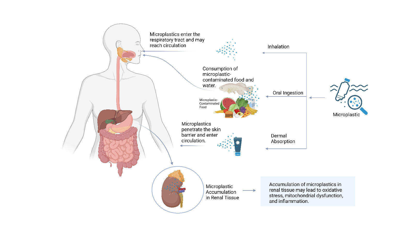
Figure 1. Major routes of MP exposure and renal accumulation. MPs enter via ingestion, inhalation, and dermal contact, translocate into the systemic circulation, and accumulate in renal tissue, where they can induce oxidative stress, mitochondrial dysfunction, inflammation, and barrier instability [20,39].
The kidney is particularly susceptible to MP deposition because of its high perfusion rate and continuous filtration of circulating substances. Animal studies demonstrate that polystyrene MPs accumulate in both the renal cortex and medulla following repeated oral exposure, resulting in reduced glomerular filtration, impaired tubular reabsorption, and structural abnormalities, including glomerular shrinkage, vacuolar degeneration, tubular apoptosis, and interstitial fibrosis [11,12]. Importantly, these pathological changes are observed in the absence of ischemic challenge, indicating that MPs alone are sufficient to induce biologically meaningful renal stress rather than acting as inert particles.
In vitro studies support these in vivo observations. Human embryonic kidney and proximal tubular epithelial cells exposed to polystyrene MPs exhibit reduced proliferation, morphological stress responses, and increased intracellular reactive oxygen species generation [13]. Transcriptomic analyses further reveal downregulation of antioxidant defence genes, such as SOD2 and CAT, accompanied by altered metabolic signalling, reflected by reduced expression of glycolytic regulators, including GAPDH. Together, these findings confirm that MPs directly disrupt renal cellular homeostasis at the molecular and metabolic levels, even under non-ischemic conditions.
Several stress-response pathways have been implicated in MP-associated renal toxicity, including oxidative imbalance, mitochondrial and endoplasmic reticulum stress, lysosomal dysfunction, and activation of inflammatory signalling [12,14-17]. MPs can also serve as carriers for environmental co-contaminants such as cadmium and polycyclic aromatic hydrocarbons, which further intensify oxidative and inflammatory injury. However, it is critical to note that most mechanistic insights at this level originate from chronic exposure models that do not involve ischemia–reperfusion injury. As such, these pathways should be interpreted as defining baseline renal vulnerability rather than direct evidence of MP-mediated modulation of ischemic injury.
Recent high-resolution approaches provide additional context for these baseline effects. A single-cell transcriptomic study demonstrated that combined exposure to MPs and a high-fat diet exacerbates extracellular matrix disorganization, activates reactive oxygen species–related oncogenic pathways, and shifts immune cell populations toward inflammatory and profibrotic phenotypes [16,18-21]. While these findings do not directly model ischemia–reperfusion injury, they highlight how MP exposure can reshape the renal microenvironment, potentially lowering the threshold for maladaptive responses to subsequent ischemic stress.
Taken together, these findings indicate that MPs accumulate within renal tissue and induce a state of epithelial, endothelial, and metabolic instability characterized by oxidative stress and inflammatory activation. These alterations overlap with recognized vulnerability nodes in renal ischemia–reperfusion injury and therefore provide a biologically plausible foundation for interaction between MP exposure and ischemic susceptibility. However, causal linkage and pathway prioritization require validation in experimental models that explicitly integrate both MP exposure and ischemia–reperfusion injury.
Pathophysiology of Renal Ischemia-Reperfusion Injury
Renal IRI is a major cause of acute kidney injury and an important driver of progression toward chronic kidney disease [24]. The injury is initiated by an interruption of renal blood flow, followed by restoration of perfusion. Although reperfusion is essential for tissue survival, the sudden reintroduction of oxygen and nutrients paradoxically exacerbates cellular and molecular damage.
During the ischemic phase, oxygen deprivation leads to rapid depletion of adenosine triphosphate, disruption of ion transport, cytoskeletal instability, and cellular swelling [24]. These metabolic disturbances compromise the integrity of tubular epithelial and vascular endothelial cells, creating a heightened state of vulnerability before reperfusion. At this stage, injury is largely driven by energetic failure rather than overt inflammation.
Upon reperfusion, the abrupt influx of oxygen triggers a burst of reactive oxygen species production [25]. This oxidative surge damages lipids, proteins, and nucleic acids, and acts as a central amplifier of injury. Reactive oxygen species activate inflammatory signalling pathways, including NF-κB and the NLRP3 inflammasome, promoting the recruitment and activation of neutrophils, macrophages, and T lymphocytes within the renal parenchyma [22,26]. The magnitude and persistence of this inflammatory response are key determinants of whether renal injury resolves or progresses toward maladaptive repair.
Endothelial dysfunction represents another defining feature of ischemia–reperfusion injury. Activated endothelial cells increase the expression of adhesion molecules such as ICAM-1 and VCAM-1[27,28], facilitating leukocyte adhesion and transmigration. These changes promote microvascular congestion, increased vascular permeability, and regional hypoxia despite restored macroscopic blood flow, a phenomenon commonly referred to as the no-reflow state. As a result, reperfusion remains heterogeneous, and focal hypoxic injury persists even after systemic perfusion is re-established.
Tubular epithelial cells are particularly susceptible during reperfusion. Necrosis and apoptosis disrupt nephron architecture and impair reabsorptive and secretory functions. Injured tubular cells release damage-associated molecular patterns, which further amplify inflammatory signalling and immune cell recruitment [24]. Although partial structural and functional recovery may occur, repair is often incomplete. Persistent fibroblast activation, capillary rarefaction, and excessive extracellular matrix deposition contribute to interstitial fibrosis and accelerate the transition from acute kidney injury to chronic kidney disease [23,24].
Recent work has highlighted the contribution of regulated cell death pathways to renal IRI. Ferroptosis, an iron-dependent form of lipid peroxidation–driven cell death, has emerged as a significant mediator of tubular injury during ischemia–reperfusion [24]. This pathway is mechanistically distinct from apoptosis and necrosis and is tightly linked to oxidative imbalance and mitochondrial dysfunction. The recognition of ferroptosis expands the mechanistic landscape of IRI and suggests additional therapeutic targets beyond classical anti-inflammatory or antioxidant strategies.
Overall, renal ischemia–reperfusion injury reflects the convergence of energetic failure, oxidative stress, inflammatory amplification, endothelial instability, and maladaptive repair processes. This mechanistic framework provides the reference against which the impact of MP exposure is evaluated in the following section, allowing discrimination between pathways that directly amplify canonical IRI mechanisms and those inferred from related but non-ischemic injury models.
Mechanistic Interplay between MPs and Renal Ischemia-Reperfusion Injury
The MPs introduce a constellation of biochemical and structural disturbances within the kidney that may lower the threshold at which ischemia–reperfusion injury becomes clinically significant. Although direct studies combining MP exposure with renal ischemia–reperfusion remain limited, experimental models consistently demonstrate oxidative, inflammatory, and endothelial vulnerabilities following MP exposure that overlap with canonical drivers of ischemic injury [11,12,13]. To improve mechanistic clarity, the pathways discussed below are interpreted using a hierarchical framework that distinguishes mechanisms supported by direct MP–IRI renal evidence from those extrapolated from non-ischemic renal or extra-renal systems.
Within this framework, “direct evidence” refers to studies that explicitly assess renal outcomes following ischemia–reperfusion in the context of prior MP exposure, whereas “indirect evidence” refers to mechanistic pathways inferred from non-ischemic kidney injury models or extra-renal MP studies that remain biologically plausible but unvalidated in MP–IRI settings.
Direct Evidence Linking MPs to Exacerbated Renal IRI
Experimental studies indicate that MP exposure establishes a renal redox environment that is less resilient to ischemic stress and subsequent reperfusion. Kidneys exposed to MPs exhibit reduced antioxidant enzyme activity, increased mitochondrial electron leakage, and impaired mitochondrial membrane potential. These alterations shift the pre-ischemic baseline toward oxidative instability rather than inducing overt injury in isolation. When oxygen is reintroduced during reperfusion, this primed state generates an exaggerated burst of reactive oxygen species, accelerating lipid peroxidation and oxidative damage to both nuclear and mitochondrial DNA. Loss of mitochondrial integrity further compromises tubular epithelial energy homeostasis, amplifying susceptibility to reperfusion-associated stress. Collectively, these findings indicate that MPs amplify canonical oxidative mechanisms of ischemia–reperfusion injury rather than introducing entirely novel injury pathways. These direct mechanisms are summarized schematically in Figure 2.
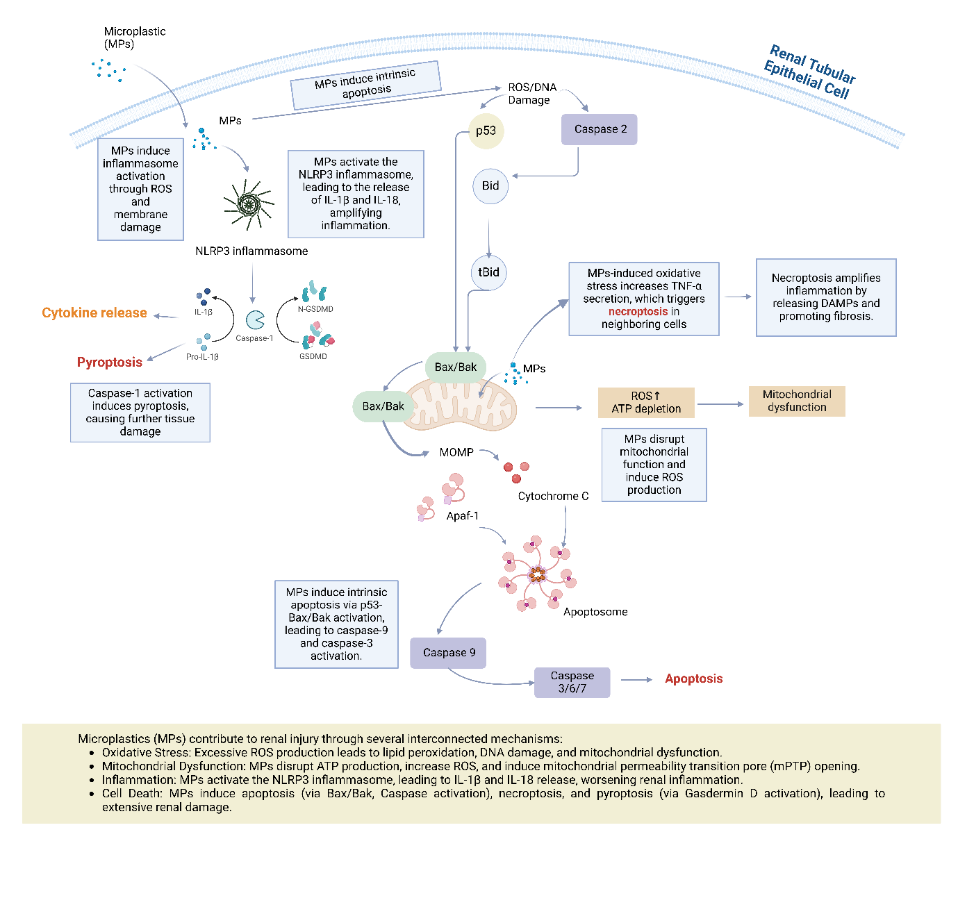
Figure 2. Mechanistic pathways of MP-induced renal injury in ischemia-reperfusion injury. MPs contribute to renal injury through multiple interconnected mechanisms, including oxidative stress, mitochondrial dysfunction, inflammation, pyroptosis [41], apoptosis [31], and necroptosis [24]. MPs trigger excessive ROS production, impair mitochondrial function, and activate inflammatory pathways, such as the NLRP3 inflammasome. Additionally, MPs induce cell death through apoptosis (via Bax/Bak activation and caspase pathways) and necroptosis, exacerbating renal damage. These processes collectively worsen renal IRI and contribute to fibrosis and long-term kidney dysfunction. [10,32] .
The MPs also intensify inflammatory activation during reperfusion. Prior exposure increases the expression of pattern recognition receptors within tubular and interstitial compartments. Upon reperfusion-induced immune cell recruitment, these receptors facilitate rapid activation of NF-κB and caspase-1 signalling, promoting early maturation of interleukin-1β and interleukin-18. The resulting pyroptotic cell death releases damage-associated molecular patterns that extend inflammatory injury beyond the originally ischemic region. These observations support the concept that MPs function as inflammatory primers, lowering the activation threshold of innate immune pathways during reperfusion.
Endothelial dysfunction represents another defining axis of direct interaction. Microplastic exposure disrupts endothelial tight junction integrity, reduces nitric oxide bioavailability, and increases expression of adhesion molecules such as ICAM-1 and VCAM-1. During reperfusion, these endothelial abnormalities promote leukocyte adhesion, microvascular congestion, and regions of incomplete capillary reperfusion, consistent with the no-reflow phenomenon. Although direct clinical evidence linking MP burden to post-ischemic vascular dysfunction is currently lacking, the mechanistic overlap observed in experimental models supports biological plausibility rather than causal inference.
The influence of MPs extends into the post-injury repair phase. MP-exposed kidneys show enhanced activation of transforming growth factor-β and Smad signalling, promoting fibroblast activation and excessive extracellular matrix deposition. Tubular regeneration becomes less efficient, and the transition from acute injury to recovery is delayed, accelerating progression toward maladaptive interstitial fibrosis. However, existing studies do not clearly distinguish whether this fibrotic acceleration is driven predominantly by persistent inflammation, sustained microvascular dysfunction, or direct effects of MPs on fibroblast biology. This unresolved distinction represents a critical mechanistic gap.
Despite these convergent findings, the direct MP–IRI evidence base remains constrained by heterogeneity in particle size, polymer composition, exposure duration, and timing of outcome assessment during reperfusion. These variables likely influence the dominant injury phenotype and currently limit pathway-specific attribution across studies.
Indirect or Extrapolated Mechanisms Relevant to the MP–IRI Axis
Several additional mechanisms relevant to ischemia–reperfusion injury have been implicated in MP toxicity but are supported primarily by indirect evidence. MPs induce mitochondrial injury, lysosomal destabilization, and endoplasmic reticulum stress in intestinal, hepatic, and pulmonary models. Since these processes are central determinants of ischemia–reperfusion susceptibility, their presence suggests mechanistic convergence rather than confirmed contribution in the kidney.
The MPs also modulate intracellular signalling pathways, including PI3K–Akt, MAPK, and interleukin-17 networks, which regulate tubular survival, inflammatory magnitude, and repair dynamics during ischemic injury. However, none of these pathways have been temporally mapped in combined MP–IRI renal models, leaving unresolved whether MPs alter the timing, magnitude, or cell-type specificity of these responses during reperfusion.
Immune remodelling observed in extra-renal tissues may also have indirect relevance. MP exposure shifts macrophage polarization, enhances effector T-cell infiltration, and alters cytokine profiles in intestinal and hepatic systems. These immune phenotypes resemble those associated with maladaptive repair following renal ischemia, particularly the persistence of pro-inflammatory macrophage states that impair tubular regeneration. Nevertheless, extrapolation to the kidney remains speculative until renal-specific immune profiling is performed under combined exposure conditions.
The MPs also impair epithelial barrier stability and tight junction integrity in gut and lung models, suggesting that similar effects on renal tubular epithelium could exacerbate post-ischemic leakiness, damage-associated molecular pattern release, and inflammatory recruitment. At present, this analogy remains biologically plausible but experimentally unverified.
Ferroptosis and co-contaminant interactions represent another layer of extrapolated mechanisms. MPs promote iron-dependent lipid peroxidation and ferroptotic signalling in intestinal and hepatic cells. Ferroptosis is now recognized as a key contributor to tubular injury in ischemia–reperfusion models. Environmental toxicants adsorbed onto MP surfaces, including cadmium, lead, and polycyclic aromatic hydrocarbons, further intensify oxidative and mitochondrial injury. However, reported ferroptotic responses vary across cell types, underscoring the importance of renal-specific validation before ferroptosis-targeted interventions can be rationally applied in MP-exposed ischemic kidneys.
Taken together, these indirect mechanisms highlight areas of biological plausibility and mechanistic convergence rather than established causality. Distinguishing which of these pathways meaningfully contribute to MP-mediated sensitization of ischemia–reperfusion injury remains a central challenge. Resolving this distinction is essential to prevent indiscriminate pathway borrowing from non-ischemic models and to ensure rational prioritization of therapeutic targets.
Therapeutic Strategies and Future Directions
Therapeutic strategies for MP-exacerbated ischemia-reperfusion injury remain conceptually grounded, as most interventions have been evaluated either in isolated MP exposure models or in classical ischemia-reperfusion settings rather than in systems that combine both insults. However, several mechanistic intersections provide plausible entry points for protection. Antioxidants with mitochondrial activity, including N acetylcysteine, MitoQ, and vitamin E, reduce MP-associated oxidative stress and help restore mitochondrial stability (Table 1). These mechanisms parallel protective effects observed in renal ischemia-reperfusion injury, suggesting that antioxidants with mitochondrial specificity may offer benefit when both exposures coexist [37,45,48]. However, existing studies do not determine whether these agents reverse early MP-induced mitochondrial vulnerability or act predominantly during the reperfusion-associated oxidative burst. This distinction is therapeutically relevant, as interventions that normalize pre-ischemic vulnerability may confer broader protection than those targeting late-stage oxidative damage alone.
Table 1. Representative therapeutic strategies targeting microplastic-exacerbated renal IRI
Targeted Mechanism | Representative Agents | Mechanistic Rationale | References |
|---|---|---|---|
Oxidative stress | N‐acetylcysteine, MitoQ, Vitamin E | Restores redox balance, scavenges ROS, stabilizes mitochondrial function | |
Inflammation | MCC950, IL-1RA, NF-κB inhibitors | Suppresses inflammasome activation and cytokine maturation | |
Endothelial Dysfunction | Statins, nitric oxide donors, ACE inhibitors | Enhances nitric oxide bioavailability and reduces endothelial activation | |
Fibrosis | Pirfenidone, Bardoxolone methyl | Inhibits TGF-β/Smad signaling and fibroblast activation | |
Ferroptosis | Ferrostatin-1, Liproxstatin-1, Deferoxamine | Limits iron-dependent lipid peroxidation and tubular ferroptotic death |
Anti-inflammatory approaches also show mechanistic promise. Inhibitors of inflammasome activation, such as MCC950 and interleukin-1 receptor antagonists, reduce NLRP3-driven cytokine maturation and may mitigate the amplified inflammatory readiness observed in MP-primed kidneys [46,47]. Modulation of NF- κB signaling further represents a rational target because MP exposure increases pattern recognition receptor expression and lowers the threshold for inflammatory activation during reperfusion. Despite this mechanistic overlap, no studies have directly evaluated whether early inhibition of innate immune sensing pathways prevents pyroptotic amplification or merely attenuates cytokine magnitude during reperfusion. Addressing this temporal uncertainty will be essential for defining optimal intervention windows.
Endothelial protection is another important direction. Statins, nitric oxide donors, and angiotensin converting enzyme inhibitors enhance endothelial stability, improve nitric oxide bioavailability, and reduce adhesion molecule expression [49,50,51]. Given that MPs disrupt tight junctions and promote upregulation of ICAM-1 and VCAM-1, these agents may be particularly valuable for limiting microvascular congestion and incomplete reperfusion. Since endothelial dysfunction appears to be an early and persistent consequence of MP exposure, vascular-targeted interventions may offer a wider therapeutic window than strategies focused solely on tubular epithelium. However, it remains unknown whether MP-associated microvascular particle retention can be reversed pharmacologically once established.
Antifibrotic agents warrant attention as well. MPs accelerate transforming growth factor beta signalling and fibroblast activation, processes intimately linked to maladaptive repair after ischemia-reperfusion. Some compounds, such as pirfenidone and bardoxolone methyl, inhibit transforming growth factor beta-mediated collagen deposition and have demonstrated benefit in renal fibrosis models [43,52]. Nevertheless, their effectiveness in MP-exposed ischemic kidneys has not been tested, and the optimal timing of antifibrotic intervention remains undefined. Whether early administration during the injury-to-repair transition provides greater benefit than late-stage treatment represents a critical unanswered question.
Ferroptosis inhibition is an emerging area of interest. The MPs promote lipid peroxidation in several tissues, and ferroptosis represents a major form of regulated cell death in IRI. Ferroptosis inhibitors, including ferrostatin-1, liproxstatin-1, and deferoxamine, reduce iron-dependent oxidative injury and support tubular viability in multiple models [53,54]. However, it remains unclear whether MP-associated ferroptotic vulnerability precedes ischemic insult or develops primarily during reperfusion. Clarifying this temporal profile will be essential for determining whether ferroptosis-targeted therapies should be administered prophylactically or during the reperfusion phase.
Taken together, the therapeutic landscape reveals several mechanistic intersections between MP toxicity and IRI. However, the lack of integrated MP ischemia-reperfusion models remains the most significant limitation to translating these concepts into targeted interventions. Future studies must incorporate controlled co-exposure designs that evaluate timing, dose, particle size, and biological compartmentalization to determine which pathways constitute true therapeutic leverage points. Work that includes clinically relevant endpoints such as renal perfusion dynamics, delayed functional recovery, and long-term fibrotic progression will be essential for determining translational value.
Clinical Implications
The MP exposure may hold clinical relevance because chronic accumulation in renal tissue can create a physiological state that is more vulnerable to ischemic stress. Patients undergoing major surgery, septic shock, cardiac arrest, or kidney transplantation are repeatedly exposed to ischemia–reperfusion events, and outcomes in these settings are strongly influenced by baseline mitochondrial integrity, endothelial function, and inflammatory tone. From a mechanistic perspective, MP-associated oxidative instability, endothelial fragility, and inflammatory priming overlap with key determinants of ischemia–reperfusion severity.
Although direct clinical evidence linking MP burden to ischemia–reperfusion outcomes is currently unavailable, experimental data suggest that environmental MP exposure could function as an unrecognized modifier of renal risk rather than a primary cause of injury. This concept is particularly relevant in populations with repeated or unavoidable ischemic insults, such as transplant recipients, critically ill patients, and individuals with cardiovascular instability.
Importantly, these implications should be interpreted as hypothesis-generating rather than predictive. Future clinical studies integrating exposure assessment, biomonitoring of MP burden, and renal functional outcomes will be required to determine whether MP exposure contributes meaningfully to perioperative acute kidney injury, delayed graft function, or impaired long-term recovery.
Conclusions
Current evidence suggests that MPs exacerbate renal ischemia–reperfusion injury mainly by amplifying oxidative stress, inflammatory activation, endothelial dysfunction, and maladaptive repair processes, thereby intensifying established ischemic injury pathways rather than introducing novel mechanisms. This review distinguishes pathways supported by direct renal MP–IRI evidence from those extrapolated from non-ischemic or extra-renal models, highlighting oxidative and inflammatory priming as the most consistently validated mechanisms. Other proposed pathways, including ferroptosis modulation, immune remodelling, and co-contaminant interactions, remain biologically plausible but insufficiently validated in combined exposure settings. Future studies should prioritize integrated MP–ischemia–reperfusion models with defined temporal resolution, dose, and particle-size stratification, and clinically relevant endpoints to establish causality and inform targeted therapeutic strategies.
Conflict of Interests
There are no conflicts to declare.
Funding
This work was supported by the SKEMA Tambahan Research Grant (Hibah SKEMA Tambahan Tahun 2021) from the Research and Community Service Institute (LPPM), Maranatha Christian University, Indonesia.
References
- Li Y, Tao L, Wang Q, Wang F, Li G, Song M. Potential health impact of microplastics: a review of environmental distribution, human exposure, and toxic effects. Environ Health. 2023;1(4):249-57. [DOI: 10.1021/envhealth.3c00052] [PMID: 39474495]
- Mohamed Nor NH, Kooi M, Diepens NJ, Koelmans AA. Lifetime accumulation of microplastic in children and adults. Environ Sci Technol. 2021;55(8):5084-96. [DOI: 10.1021/acs.est.0c07384] [PMID: 33724830]
- La Porta E, Exacoustos O, Lugani F, Angeletti A, Chiarenza DS, Bigatti C, et al. Microplastics and kidneys: an update on the evidence for deposition of plastic microparticles in human organs, tissues and fluids and renal toxicity concern. Int J Mol Sci. 2023;24(18):14391. [DOI: 10.3390/ijms241814391] [PMID: 37762695]
- Hussain KA, Romanova S, Okur I, Zhang D, Kuebler J, Huang X, et al. Assessing the release of microplastics and nanoplastics from plastic containers and reusable food pouches: implications for human health. Environ Sci Technol. 2023;57(26):9782-92. [DOI: 10.1021/acs.est.3c01942] [PMID: 37343248]
- Liang Y, Liu D, Zhan J, Liu X, Li P, Ma X, et al. Polystyrene microplastics induce kidney injury via gut barrier dysfunction and C5a/C5aR pathway activation. Environ Pollut. 2024;342:122909. [DOI: 10.1016/j.envpol.2023.122909] [PMID: 38036092]
- Lu H, Hou L, Zhang Y, Guo T, Wang Y, Xing M. Polystyrene microplastics mediate cell cycle arrest, apoptosis, and autophagy in the G2/M phase through ROS in grass carp kidney cells. Environ Toxicol. 2024;39(4):1923-35. [DOI: 10.1002/tox.24068] [PMID: 38064284]
- Luo WQ, Cao MT, Sun CX, Wang JJ, Gao MX, He XR, et al. Size-dependent internalization of polystyrene microplastics as a key factor in macrophages and systemic toxicity. J Hazard Mater. 2025;490:137701. [DOI: 10.1016/j.jhazmat.2025.137701] [PMID: 40020305]
- Bonventre JV, Yang L. Cellular pathophysiology of ischemic acute kidney injury. J Clin Invest. 2011;121(11):4210-21. [DOI: 10.1172/JCI45161] [PMID: 22045571]
- Lee SE, Kim DY, Jeong TS, Park YS. Micro- and nano-plastic-induced adverse health effects on lungs and kidneys linked to oxidative stress and inflammation. Life (Basel). 2025;15(3):392. [DOI: 10.3390/life15030392] [PMID: 40141737]
- Kuang Q, Gao L, Feng L, Xiong X, Yang J, Zhang W, et al. Toxicological effects of microplastics in renal ischemia-reperfusion injury. Environ Toxicol. 2024;39(4):2350-62. [DOI: 10.1002/tox.24115] [PMID: 38156432]
- Wang YL, Lee YH, Hsu YH, Chiu IJ, Huang CC, Huang CC, et al. The kidney-related effects of polystyrene microplastics on human kidney proximal tubular epithelial cells HK-2 and male C57BL/6 mice. Environ Health Perspect. 2021;129(5):57003. [DOI: 10.1289/EHP7612] [PMID: 33956507]
- Wang W, Guan J, Feng Y, Nie L, Xu Y, Xu H, et al. Polystyrene microplastics induced nephrotoxicity associated with oxidative stress, inflammation, and endoplasmic reticulum stress in juvenile rats. Front Nutr. 2023;9:1059660. [DOI: 10.3389/fnut.2022.1059660] [PMID: 36687698]
- Goodman KE, Hua T, Sang QA. Effects of polystyrene microplastics on human kidney and liver cell morphology, cellular proliferation, and metabolism. ACS Omega. 2022;7(38):34136-53. [DOI: 10.1021/acsomega.2c03453] [PMID: 36188270]
- Wang YL, Huang CC, Zheng CM, Liu WC, Lee YH, Chiu HW. Polystyrene microplastic-induced extracellular vesicles cause kidney-related effects in the crosstalk between tubular cells and fibroblasts. Ecotoxicol Environ Saf. 2024;273:116098. [DOI: 10.1016/j.ecoenv.2024.116098] [PMID: 38368757]
- Xu M, Chen J, Gao L, Cai S, Dong H. Microplastic exposure induces HSP90α secretion and aggravates asthmatic airway remodeling via PI3K-Akt-mTOR pathway. Ecotoxicol Environ Saf. 2025;291:117828. [DOI: 10.1016/j.ecoenv.2025.117828] [PMID: 39923560]
- Xu W, Ye S, Liu W, Guo H, Zhang L, Wei S, et al. Single-cell RNA-seq analysis decodes the kidney microenvironment induced by polystyrene microplastics in mice receiving a high-fat diet. J Nanobiotechnol. 2024;22(1):13. [DOI: 10.1186/s12951-023-02266-7] [PMID: 38167034]
- Zhu Z, Liao R, Shi Y, Li J, Cao J, Liao B, et al. Polystyrene nanoplastics induce apoptosis of human kidney proximal tubular epithelial cells via oxidative stress and MAPK signaling pathways. Environ Sci Pollut Res Int. 2023;30(51):110579-89. [DOI: 10.1007/s11356-023-30155-x] [PMID: 37792190]
- Verzola D, Rumeo N, Alberti S, Loiacono F, La Maestra S, Passalacqua M, et al. Coexposure to microplastic and bisphenol a exhacerbates damage to human kidney proximal tubular cells. Heliyon. 2024;10(20):e39426. [DOI: 10.1016/j.heliyon.2024.e39426] [PMID: 39498083]
- Wen Y, Cai J, Zhang H, Li Y, Yu M, Liu J, et al. The Potential mechanisms involved in the disruption of spermatogenesis in mice by nanoplastics and microplastics. Biomed. 2024;12(8):1714. [DOI: 10.3390/biomedicines12081714]
- Xiong X, Gao L, Chen C, Zhu K, Luo P, Li L. The microplastics exposure induce the kidney injury in mice revealed by RNA-seq. Ecotoxicol Environ Saf. 2023;256:114821. [DOI: 10.1016/j.ecoenv.2023.114821] [PMID: 36989554]
- Xiong Y, Chen Z, Xiang H, Liu Y, Wang Y. Polystyrene microplastics disrupt adrenal steroid synthesis in male mice via mitochondrial dysfunction. Ecotoxicol Environ Saf. 2025;290:117528. [DOI: 10.1016/j.ecoenv.2024.117528] [PMID: 39674024]
- Zhang Y, Men J, Yin K, Zhang Y, Yang J, Li X, et al. Activation of gut metabolite ACSL4/LPCAT3 by microplastics in drinking water mediates ferroptosis via gut-kidney axis. Commun Biol. 2025;8(1):211. [DOI: 10.1038/s42003-025-07641-8] [PMID: 39930042]
- Polichnowski AJ, Griffin KA, Licea-Vargas H, Lan R, Picken MM, Long J, et al. Pathophysiology of unilateral ischemia-reperfusion injury: importance of renal counterbalance and implications for the AKI-CKD transition. Am J Physiol Renal Physiol. 2020;318(5):1086-99. [DOI: 10.1152/ajprenal.00590.2019] [PMID: 32174143]
- Li C, Yu Y, Zhu S, Hu Y, Ling X, Xu L, et al. The emerging role of regulated cell death in ischemia and reperfusion-induced acute kidney injury: current evidence and future perspectives. Cell Death Discov. 2024;10(1):216. [DOI: 10.1038/s41420-024-01979-4] [PMID: 38704372]
- Granger DN, Kvietys PR. Reperfusion injury and reactive oxygen species: the evolution of a concept. Redox Biol. 2015;6:524-51. [DOI: 10.1016/j.redox.2015.08.020] [PMID: 26484802]
- Ogurlu B, Hamelink TL, Van Tricht IM, Leuvenink HGD, De Borst MH, Moers C, et al. Utilizing pathophysiological concepts of ischemia-reperfusion injury to design renoprotective strategies and therapeutic interventions for normothermic ex vivo kidney perfusion. Am J Transplant. 2024;24(7):1110-26. [DOI: 10.1016/j.ajt.2024.01.001] [PMID: 38184242]
- Akhtar AM, Schneider JE, Chapman SJ, Jefferson A, Digby JE, Mankia K, et al. In vivo quantification of VCAM-1 expression in renal ischemia reperfusion injury using non-invasive magnetic resonance molecular imaging. Plos One. 2010;5(9):e12800. [DOI: 10.1371/journal.pone.0012800] [PMID: 20877722]
- Burne MJ, Elghandour A, Haq M, Saba SR, Norman J, Condon T, et al. IL-1 and TNF independent pathways mediate ICAM-1/VCAM-1 up-regulation in ischemia reperfusion injury. J Leukoc Biol. 2001;70(2):192-8. [PMID: 11493610]
- Kadac-Czapska K, Ośko J, Knez E, Grembecka M. Microplastics and oxidative stress-current problems and prospects. Antioxidants. 2024;13(5):579. [DOI: 10.3390/antiox13050579] [PMID: 38790684]
- Liu T, Hou B, Wang Z, Yang Y. Polystyrene microplastics induce mitochondrial damage in mouse GC-2 cells. Ecotoxicol Environ Saf. 2022;237:113520. [DOI: 10.1016/j.ecoenv.2022.113520] [PMID: 35489138]
- Mai NTQ, Batjargal U, Kim WS, Kim JH, Park JW, Kwak IS, et al. Microplastic induces mitochondrial pathway mediated cellular apoptosis in mussel (Mytilus galloprovincialis) via inhibition of the AKT and ERK signaling pathway. Cell Death Discov. 2023;9(1):442. [DOI: 10.1038/s41420-023-01740-3] [PMID: 38057300]
- Zou H, Chen Y, Qu H, Sun J, Wang T, Ma Y, et al. Microplastics exacerbate cadmium-induced kidney injury by enhancing oxidative stress, autophagy, apoptosis, and fibrosis. Int J Mol Sci. 2022;23(22):14411. [DOI: 10.3390/ijms232214411] [PMID: 36430889]
- Zhou B, Zhang A, Wang Y, Feng S, Xue Q, Liu Z, et al. Microplastics induce human kidney development retardation through ATP-mediated glucose metabolism rewiring. J Hazard Mater. 2025;486:137002. [DOI: 10.1016/j.jhazmat.2024.137002] [PMID: 39754872]
- Feng L, Chen C, Xiong X, Wang X, Li X, Kuang Q, et al. PS-MPs promotes the progression of inflammation and fibrosis in diabetic nephropathy through NLRP3/Caspase-1 and TGF-β1/Smad2/3 signaling pathways. Ecotoxicol Environ Saf. 2024;273:116102. [DOI: 10.1016/j.ecoenv.2024.116102] [PMID: 38382346]
- Yang W, Jannatun N, Zeng Y, Liu T, Zhang G, Chen C, et al. Impacts of microplastics on immunity. Front Toxicol. 2022;4:956885. [DOI: 10.3389/ftox.2022.956885] [PMID: 36238600]
- Cheng Y, Yang Y, Bai L, Cui J. Microplastics: an often-overlooked issue in the transition from chronic inflammation to cancer. J Transl Med. 2024;22(1):959. [DOI: 10.1186/s12967-024-05731-5] [PMID: 39438955]
- Wang X, Jia Z, Zhou X, Su L, Wang M, Wang T, et al. Nanoplastic-induced vascular endothelial injury and coagulation dysfunction in mice. Sci Total Environ. 2023;865:161271. [DOI: 10.1016/j.scitotenv.2022.161271] [PMID: 36587662]
- Lee SE, Yoon HK, Kim DY, Jeong TS, Park YS. An emerging role of micro- and nanoplastics in vascular diseases. Life (Basel). 2024;14(2):255. [DOI: 10.3390/life14020255] [PMID: 38398764]
- Kehinde SA, Olajide AT, Ogunsanya ST, Fatokun TP, Olulana DI, Olabiyi FA, et al. Polyethylene microplastics disrupt renal function, mitochondrial bioenergetics, redox homeostasis, and histoarchitecture in Wistar rats. Sci Rep. 2025;15(1):41120. [DOI: 10.1038/s41598-025-24887-8] [PMID: 41266508]
- Wang H, Lei X, Zhang Z, Ommati MM, Tang Z, Yuan J. Chronic exposure of bisphenol-A impairs cognitive function and disrupts hippocampal insulin signaling pathway in male mice. Toxicol. 2022;472:153192. [DOI: 10.1016/j.tox.2022.153192] [PMID: 35489422]
- Li S, Gu X, Zhang M, Jiang Q, Xu T. Di (2-ethylhexyl) phthalate and polystyrene microplastics co-exposure caused oxidative stress to activate NF-κB/NLRP3 pathway aggravated pyroptosis and inflammation in mouse kidney. Sci Total Environ. 2024;926:171817. [DOI: 10.1016/j.scitotenv.2024.171817] [PMID: 38513858]
- Rojas-Rivera J, Ortiz A, Egido J. Antioxidants in kidney diseases: the impact of bardoxolone methyl. Int J Nephrol. 2012;2012:321714. [DOI: 10.1155/2012/321714] [PMID: 22701794]
- Huang R, Fu P, Ma L. Kidney fibrosis: from mechanisms to therapeutic medicines. Signal Transduct Target Ther. 2023;8(1):129. [DOI: 10.1038/s41392-023-01379-7] [PMID: 36932062]
- Xiong X, Gao L, Chen C, Zhu K, Luo P, Li L. The microplastics exposure induce the kidney injury in mice revealed by RNA-seq. Ecotoxicol Environ Saf. 2023;256:114821. [DOI: 10.1016/j.ecoenv.2023.114821]. [PMID:36989554].
- Dare AJ, Bolton EA, Pettigrew GJ, Bradley JA, Saeb-Parsy K, Murphy MP. Protection against renal ischemia–reperfusion injury in vivo by the mitochondria targeted antioxidant MitoQ. Redox Biology. 2015;5:163-8. [DOI: 10.1016/j.redox.2015.04.008]
- Zhang C, Zhu X, Li L, Ma T, Shi M, Yang Y, et al. A small molecule inhibitor MCC950 ameliorates kidney injury in diabetic nephropathy by inhibiting NLRP3 inflammasome activation. Diabetes Metab Syndr Obes. 2019;12:1297-1309. [DOI: 10.2147/DMSO.S199802] [PMID: 31447572]
- Corcoran SE, Halai R, Cooper MA. pharmacological inhibition of the nod-like receptor family pyrin domain containing 3 inflammasome with MCC950. Pharmacol Rev. 2021;73(3):968-1000. [DOI: 10.1124/pharmrev.120.000171] [PMID: 34117094]
- Small DM, Sanchez WY, Roy SF, Morais C, Brooks HL, Coombes JS, et al. N-acetyl-cysteine increases cellular dysfunction in progressive chronic kidney damage after acute kidney injury by dampening endogenous antioxidant responses. Am J Physiol-Renal Physiol. 2018;314(5):956-68. [DOI: 10.1152/ajprenal.00057.2017]
- García-Menéndez E, Marques Vidas M, Alique M, Carracedo J, de Sequera P, Corchete E, et al. Statins and antiplatelet agents are associated with changes in the circulatory markers of endothelial dysfunction in chronic kidney disease. Nefrologia. 2019;39(3):287-93. [DOI: 10.1016/j.nefro.2018.11.001] [PMID: 30732927]
- Su JB. Vascular endothelial dysfunction and pharmacological treatment. World J Cardiol. 2015;7(11):719-41. [DOI: 10.4330/wjc.v7.i11.719] [PMID: 26635921]
- Ruszkowski P, Masajtis-Zagajewska A, Nowicki M. Effects of combined statin and ACE inhibitor therapy on endothelial function and blood pressure in essential hypertension - a randomized double-blind, placebo controlled crossover study. J Renin Angiotensin Aldosterone Syst. 2019;20(3):1470320319868890. [DOI: 10.1177/1470320319868890] [PMID: 31486700]
- Lahane GP, Dhar A, Bhat A. Therapeutic approaches and novel antifibrotic agents in renal fibrosis: a comprehensive review. J Biochem Mol Toxicol. 2024;38(8):e23795. [DOI: 10.1002/jbt.23795] [PMID: 39132761]
- Miotto G, Rossetto M, Di Paolo ML, Orian L, Venerando R, Roveri A, et al. Insight into the mechanism of ferroptosis inhibition by ferrostatin-1. Redox Biol. 2020;28:101328. [DOI: 10.1016/j.redox.2019.101328] [PMID: 31574461]
- Jin R, Dai Y, Wang Z, Hu Q, Zhang C, Gao H, et al. Unraveling ferroptosis: a new frontier in combating renal fibrosis and CKD progression. Biology. 2024;14(1):12. [DOI: 10.3390/biology14010012] [PMID: 39857243]