



Abstract
Background: Thiamine deficiency heightens tissue sensitivity to toxins by increasing oxidative stress and promoting inflammation. Various toxins deplete glutathione (GSH) levels, which are essential for cellular signaling, particularly within the NF-κB pathway that regulates responses to harmful substances. Maintaining balance in the NF-κB pathway and GSH is vital for toxin protection. We examined rat liver and heart tissues exposed to carbon tetrachloride (CCl4).
Methods: A total of 40 rats were allocated into four experimental groups: group 1 (control), group 2 (control supplemented with thiamine), group 3 (OSR-untreated), and group 4 (OSR supplemented with thiamine). The rats in the thiamine-treated groups were given 180 mg/L daily for a period of two weeks through their drinking water. Oxidative stress was induced by injecting 1 ml/kg of CCl4 intraperitoneally. Levels of GSH, GSSG, and NF-κB expression were assessed, along with indicators of liver and heart function. Liver tissue was analyzed under a microscope by a pathologist.
Results: Thiamine improved hepatic and cardiac function, reduced inflammation, and enhanced the antioxidant capacity of the body, liver, and heart against oxidative stress. Additionally, it downregulated NF-κB activity in both the liver and heart and prevented hepatic damage and fat accumulation, with effects reaching statistical significance (p<0.001).
Conclusion: Thiamine protected the liver and heart against oxidative stress-induced damage by enhancing antioxidant capacity and reducing NF-κB signaling and inflammation. This protection was achieved through boosting the body's ability to fight oxidative stress and lowering biomolecule oxidation. Downregulation of NF-κB levels and enhancement of GSH metabolism appears to be crucial ways in which thiamine safeguards both the liver and the heart.
Keywords: Heart, Liver, Nuclear factor- kβ, Oxidative stress, Reduced glutathione, Thiamine
Introduction
Exogenous and endogenous irritants can induce both short- and long-term tissue damage. Oxidative stress causes cell death through two primary mechanisms: by enhancing the calcium levels inside cells and by harming important molecules [1]. Over time, these free radicals harm the body by damaging parts of cells [2]. Additionally, various toxins can damage tissues by reducing glutathione (GSH), a key molecule that regulates important processes, such as the nuclear factor-kappa B (NF-κB) pathway, which plays a major role in the way tissues react to harmful substances. Changing this pathway and improving GSH levels are important ways to reduce damage from harmful chemicals. When GSH levels drop in the liver and heart, it can lead to liver and heart diseases [3, 4].
Looking into natural ways to prevent oxidative stress and inflammation is important to reduce their harmful effects and avoid problems when medicines are used together [5, 6]. Thiamine, also known as vitamin B1, is a strong antioxidant and plays a key role in how the body uses energy. When thiamine is deficient, tissues become more sensitive to harmful substances due to increased oxidative stress and inflammation [7, 8]. Oxidative stress may cause tissue damage by lowering the levels of thiamine-related chemicals and the enzymes that support them [9]. Moreover, thiamine deficiency can exacerbate oxidative stress. In this review, we examine how thiamine affects NF-κB signaling in the liver and heart, as well as markers of oxidative stress and inflammation, using a rat model exposed to oxidative stress (OSR). We also assessed how thiamine treatment influences markers indicative of hepatic and cardiac function.
Materials and Methods
Materials
Buying top-quality reagents from Sigma in the United States, Merck in Germany, and Yekta Tajhiz Azma in Iran.
Study design
The rats were obtained from the animal lab at the Royan Institute in Tehran, Iran. After spending two weeks getting used to their new environment, the rats were split into four groups: group 1 (control), group 2 (control supplemented with thiamine), group 3 (OSR untreated), group 4 (OSR supplemented with thiamine).
Oxidative stress was induced by giving the rats a single dose of 1 ml per kilogram of a mixture containing CCl4 and olive oil on day 15. The rats in the treated groups were given 180 mg/L daily for a period of two weeks through their drinking water. Rats in the thiamine-treated groups were provided with water containing an additional 0.18 g of thiamine per liter for a duration of two weeks. Acute liver damage is induced by a single dose of CCl4 at 1 ml/kg. Inducing liver fibrosis and cirrhosis in rodents using CCl4 is a commonly used and widely acknowledged experimental method for researching these conditions [10]. The decision to use this particular treatment dose was based on its superior antioxidant and anti-inflammatory properties as indicated by our recent investigation [11]. Following the guidance from earlier sources and research, we selected a singular dose, which is typically preferred when the most effective dose is known, while multiple doses are commonly employed when the ideal dose is uncertain. All groups of rats had an equal total weight, and the amount of water they consumed was proportional to their weight. Therefore, introducing the treatment into their water source avoided conflicts in scheduling and lessened stress [12]. The research was approved by the Ethics Committee of Ardabil University of Medical Sciences (IR.ARUMS.AEC.1401.046). Blood samples were collected after the rats were anesthetized using cardiac puncture. The tissues were immediately taken, weighed, and then mixed to create homogenates.
Measuring the biochemical profile
We tested the activity of several enzymes, including total creatine kinase (CK), CK-MB, alanine transaminase (ALT), aspartate transaminase (AST), alkaline phosphatase (ALP), and gamma-glutamyl transpeptidase (GGT), using commercial kits that rely on photometric methods. We also measured the levels of total bilirubin (T-Bil), total serum protein, and albumin (Alb). Total globulin (Glo) was calculated by subtracting albumin from total protein, and the Alb/Glo ratio was determined. We also calculated the liver weight index (LWI). The levels of cardiac troponin I in the rat serum were measured using a CUSABIO ELISA kit provided by Wuhan Huamei Biotech Co., China.
Assessment of Oxidative Stress, Antioxidant, and Inflammatory Markers in Serum, Liver, and Heart Homogenates
Advanced oxidation protein products (AOPP), malondialdehyde (MDA), and ferric ion-reducing antioxidant power (FRAP) were measured using spectrophotometric methods [13]. Total glutathione (GSH) and oxidized glutathione (GSSG) were quantified through reverse-phase-high-performance liquid chromatography at 215 nm, and the GSH/GSSG ratio was calculated [14]. The activities of (PON) [12], catalase (CAT) [13], and myeloperoxidase (MPO) [14]were determined using kinetic methods. Interleukin-1β (IL-1β) was measured using a CUSABIO ELISA kit from Wuhan Huamei Biotech Co, China.
Gene expression of hepatic NF-κB
Total RNA was extracted from heart and liver tissues using TRIzol reagent (Invitrogen, USA). The quantity and quality of the RNA were assessed using a Nanodrop spectrophotometer at 260 nm, and the 260/280 ratio was checked to ensure good quality. Complementary DNA (cDNA) was synthesized through reverse transcription following the manufacturer's instructions. Quantitative reverse transcription PCR (qRT-PCR) was performed using a standard SYBR Green PCR kit (Toyobo, Japan), and the specific gene amplification was carried out on an ABI 7300 system (Applied Biosystems, Germany). The results of gene expression were normalized using glyceraldehyde-3-phosphate dehydrogenase (GAPDH) as the reference gene. The following primer sequences were used:
NF-κB:5´-CCTGTCTGCACCTGTTCCAA-3´ (forward)
3´ACTCCTGGGTCTGTGTTGTT-5´ (reverse)
GAPDH: 5´-CTGGACAGGGTATACAGGGTTAG -3´ (forward)
3´-ACTGGTGCCGTTTATGCCTTG-5´ (reverse)
The gene expression ratio of NF-κB to GAPDH was calculated using the 2-ΔΔCT method.
Pathological study
Liver sections were prepared by fixing, embedding in paraffin, and staining with hematoxylin and eosin (H&E) for examination [15].
Statistical Analysis
A multivariate analysis of variance (MANOVA) was carried out, along with Turkey’s post hoc test, to compare various factors. A P-value < 0.05 was considered statistically significant. All statistical analyses were conducted using SPSS (version 16).
Results
Table 1 shows signs of liver problems (e.g., enzyme activity), T-Bill levels, liver weight index (LWI), and the protein levels in the blood. It also includes signs of heart problems, such as creatine kinase (CK) and CK-MB activity, troponin-I levels, and the weight of the rat groups. The OSR group had the highest levels of enzymes, T-Bill, and LWI. On the other hand, this group had the lowest levels of total protein, albumin (Alb), globulin (Glo), and the ratio of Alb to Glo. The OSR rats also showed higher levels of heart-related markers. Thiamine administration improved some of these parsameters in the OSR (thiamine) group; however, it did not fully restore the levels to those observed in the control group, except for LWI and the Alb/Glo ratio, which were statistically significant (p<0.001).
Table 1. Effect of thiamine (B1) on hepatic function parameters, body weight, and liver weight index in normal (N) and CCl4-induced oxidative stress rats (OSR)
Parameter | Groups (n=7 rats in each group) | |||
|---|---|---|---|---|
N | N (Thiamine) | OSR | OSR (Thiamine) | |
Alanine transaminase, ALT (U/L) | 21.35± 1.19 | 19.84 ± 1.07 | 54.63± 3.29 * | 37.01 ±2.08 *, # |
Aspartate transaminase, AST (U/L) | 28.42 ± 1.53 | 27.82 ± 1.61 | 98.25 ± 5.90 * | 55.04 ± 3.52 *, # |
Alkaline phosphatase (U/L) | 95.72± 5.60 | 98.38 ± 5.33 | 169.44 ± 8.07 * | 120.24±7.63*, # |
γ-glutamyl transpeptidase (U/L) | 22.35 ± 0.94 | 23.87 ± 0.88 | 36.02 ± 2.58 * | 26.18 ±1.30*, # |
Total protein (g/dl) | 6.97 ± 0.36 | 7.06 ± 0.38 | 5.49 ± 0.21* | 6.37 ± 0.32 *, # |
Albumin (g/dl) | 3.88± 0.19 | 3.91 ± 0.22 | 2.64 ± 0.19 * | 3.50 ± 0.17*, # |
Globulins (g/dl) | 3.09± 0.16 | 3.15 ± 0.13 | 2.85 ± 0.15 * | 2.87 ± 0.19 *, # |
Albumin/Globulins | 1.25± 0.16 | 1.24 ± 0.13 | 0.92 ± 0.15 * | 1.21 ± 0.19 # |
Total bilirubin | 0.39± 0.03 | 0.37 ± 0.02 | 1.02 ± 0.17 * | 0.51 ± 0.06*, # |
Liver weight index (%) | 3.66± 0.24 | 3.69 ± 0.26 | 2.96 ± 0.20 * | 3.73 ± 0.28 # |
Body weight (g) | 298.29± 16.47 | 307.20± 16.95 | 250.41± 14.73 * | 300.26 ± 17.59 # |
* Indicates significant difference with the N group (p<0.001)
# Indicates significant difference with the OSR group (p<0.001)
Figures 1a to 1d present the liver tissue changes in different groups of rats. The liver from the normal group had healthy, hexagonal liver cells arranged in layers (Figure 1a). However, the liver of the OSR group showed several harmful changes, such as cell death (marked with a triangle), scarring (large arrow), fat buildup (stars), empty spaces inside cells (circles), immune cell buildup (arrows), and cells with two nuclei (cross). Thiamine helped reduce these harmful changes (Figure 1e).
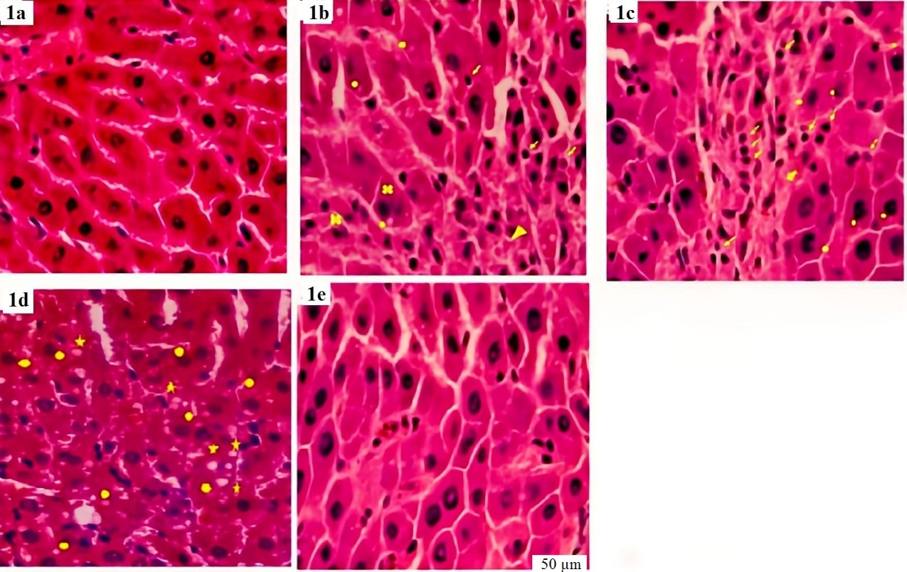
Figure 1. Histopathologic views (stained by hematoxylin and eosin and original magnification ×400) of the liver in the normal, oxidative stress rat model group (OSR), and the treated one
The hepatocytes of the N and N (Thiamine) groups were polygonal in shape and arrayed in plates (Fig-1a).
Necrosis (triangle, Fig.1b), fibrosis (huge arrow, Fig.1c), fatty change (stars, Fig.1d), cytoplasmic vacuolization (circles, Figs 1c and 1d), infiltration (arrows, Figs 1b and 1c), and binucleated hepatocytes (cross, Fig 1b) were observed in the liver of the OSR group.
Thiamine prevented CCL4-induced histopathological changes in the rat’s liver of the OSR (Thiamine) group (Fig 1e).
Tables 2 and 3 compare different markers, including antioxidant, oxidative stress, and inflammatory, measured in both blood and tissue samples (heart and liver). The treated groups showed significantly lower levels of oxidative stress and inflammation, and higher levels of antioxidant markers, with all differences being highly significant (p<0.001).
Figure 2 indicates the ratio of NF-κB to GAPDH in liver and heart tissues from the treated and untreated N and OSR groups. The ratio of NF-κB to GAPDH was much lower in the thiamine-treated N and OSR groups compared to the untreated OSR group, and this difference was statistically significant (p<0.001).
Table 2. Comparison of antioxidant profile (total glutathione, GSH/GSSG, CAT, PON, and FRAP) in the serum and tissue homogenates of normal, CCL4, and treated ones
Parameter | Sample | N | N (Thiamine) | CCL4 | CCL4 (Thiamine) |
|---|---|---|---|---|---|
Total glutathione | Serum (µmol/L) | 239.62± 15.47 | 257.11 ± 18.50* | 68.52± 4.21*, # | 219.33 ± 14.96*, # |
Liver (nmol/mg protein) | 600.35 ±32.84 | 644.62± 36.01* | 379.58 ± 19.63 * | 492.05 ± 25.47*, # | |
GSH/GSSG | Serum | 8.61 ±0.53 | 9.03± 0.57* | 0.918± 0.06 * | 6.49 ± 0.47*, # |
Liver | 10.19± 0.66 | 11.45± 0.74* | 1.06.06± 0.08* | 7.57 ± 0.52 *, # | |
CAT | Serum (U/mg protein) | 143.19 ±9.30 | 158.29±10.61* | 103.26± 6.08 * | 119.71 ± 7.54 *, # |
Liver (U/mg protein) | 307.25± 16.01 | 349.76± 17.45* | 99.47± 5.28* | 221.52± 13.80 *, # | |
PON | Serum (U/mg protein) | 126.62 ±8.07 | 142.44±9.63* | 68.79± 4.02 * | 91.51 ± 4.35*, # |
Liver (U/mg protein) | 229.92± 13.45 | 261.03± 14.36* | 140.17± 801* | 188.54 ± 9.73 *, # | |
FRAP | Serum (µmol/L) | 1108.31 ±79.25 | 1191.72± 84.30* | 498.07± 28.40 * | 932.65 ± 49.38 *, # |
Liver (nmol/g tissue) | 7.41± 0.43 | 8.39± 0.59* | 4.04± 0.21* | 6.16± 0.37*, # |
Table 3. Effect of thiamine on oxidative stress and inflammatory markers in sera, liver, and heart homogenates of all groups
Parameter | Sample | N | N (Thiamine) | CCL4 | CCL4 (Thiamine) |
|---|---|---|---|---|---|
MDA | Serum (nmol/L) | 8.46 ±0.45 | 7.81± 0.38* | 23.05± 1.36 * | 15.38 ± 0.87 *, # |
Liver (nmol/g tissue) | 11.54± 0.63 | 8.61± 0.51* | 50.26± 3.09* | 23.20 ± 1.88 *, # | |
AOPP | Serum (µmol/L) | 8.94±0.43 | 6.25± 0.31* | 34.82± 2.04 * | 20.53 ± 1.37 *, # |
Liver (nmol/g tissue) | 12.08± 0.56 | 11.79± 0.51* | 66.42± 4.05* | 54.80± 2.92 *, # | |
IL-1β | Serum (Pg./mg protein) | 289.55± 15.06 | 241.63± 29.23* | 710.36± 38.91* | 453.72 ± 31.52 *, # |
Liver (Pg./mg protein) | 114.22 ±7.02 | 83.99± 5.15* | 314.67± 16.42* | 191.75 ± 11.98 *, # | |
MPO | Serum (U/mg protein) | 0.42 ±0.06 | 0.28± 0.03* | 5.93± 0.37 * | 1.99 ± 0.21 *, # |
Liver (U/mg protein) | 0.75± 0.08 | 0.53± 0.08* | 3.84± 0.43* | 1.40 ± 0.17*, # |
* Indicates significant difference with the N group (p<0.001)
# Indicates significant difference with the CCl4group (p<0.001)
MDA, malondialdehyde; AOPP, advanced oxidation protein products; IL-1β, interleukine-1β; MPO, myeloperoxidase
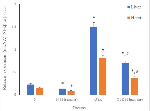
Figure 2. Comparison of relative hepatic and cardiac nuclear factor-kβ (NF-kβ) to glysealdehide -3- phosphate dehydrogenase (GAPDH), in normal (N) and oxidative stress rat model group (OSR)
* Indicates a significant difference with N group (p <0.001)
# Indicates a significant difference with ROS group (p<0.001
NF-κB/ACTB; Nuclear factor κB/ Glyceraldehyde-3-phosphate dehydrogenase
Discussion
In this study, exposure to CCl4 caused oxidative stress, leading to acute liver damage and heart muscle issues. This was shown through higher levels of dysfunction markers and changes in tissue samples, which were related to more free radicals and increased activity of NF-κB. Thiamine reduced these harmful changes and improved both hepatic and cardiac function by boosting the body's capacity to manage free radicals, especially through enhancement of the GSH levels.
The OSR group showed signs of liver and heart damage. The CCl4 caused more inflammation and oxidative stress in both the blood and tissues. It also raised the levels of lipid-protein markers in the blood and tissue samples (Table 2), and lowered the body's ability to fight oxidative stress
(Table 3). This confirms that CCl4 causes oxidative stress. Glutathione (GSH) plays a key role in breaking down the harmful chemicals made by CCl4. Thiamine treatment helped reduce oxidative stress by increasing total glutathione levels, the ratio of GSH to GSSG, and the activity of antioxidant enzymes in both normal and OSR groups (Table 2). A higher GSH/GSSG ratio facilitates the elimination of free radicals [16]. Our findings are consistent with two studies showing that thiamine deficiency induces oxidative stress in animal models [17] and in lab-grown liver cells [18] by reducing GSH and antioxidant enzyme levels, while increasing MDA and GSSG. This suggests that thiamine improves the body's antioxidant capacity. In our study, the OSR group exhibited higher levels of NF-βB, IL-1β, and MPO activity. Research has shown that antioxidants such as curcumin, lycopene and L-carnitine can boost the activity of antioxidant enzymes in models of liver damage caused by CCl4 [19, 20]. Thiamine helped reduce inflammation by lowering NF-κB activity (Fig. 2), IL-1β levels, and MPO activity (Table 3). Recent research has shown that thiamine has antioxidant and anti-inflammatory effects in diabetic patients [21] and in models of long-term inflammation [22]. A recent study indicated that thiamine might serve as a successful preventative remedy for liver injury in endotoxemia in mice. This is associated with its capacity to control Gal-3, improving the inflammatory reaction and halting galactose metabolism [23].
The harmful effects of CCl4 on the liver were shown through various signs, such as cell death, fat accumulation, cell swelling, and cells with two nuclei (Figs. 1b -1d). These harmful effects were also seen through higher levels of certain enzymes (transaminases, ALP, and GGT) and increased T-Bill levels in the OSR group, along with lower levels of total protein, albumin, globulin, and the albumin/globulin ratio. The lower liver weight index in the OSR group (Table 1) indicates that CCl4 caused liver damage, which is consistent with findings from previous studies [24, 25]. The treatment showed a protective effect on the liver by helping to reduce the liver weight index (Table 1).
Similar research has demonstrated the significant influence of oxidative stress on liver damage and the efficiency of antioxidants such as a combination of curcumin with vitamin E [26]. Over 40 years ago, it was found that a diet lacking thiamine made rats more sensitive to harmful substances (e.g., CCl4), by causing more oxidative stress and inflammation [8]. Additionally, Cells with two nuclei or more are signs of liver damage caused by oxidative stress. Their absence in the group that received thiamine in the OSR study shows that thiamine has antioxidant and liver-protecting properties. In this study, thiamine prevented liver scarring and cell death (Fig. 1e) and improved liver function (Table 1) by reducing activation of the NF-κB pathway and inflammation, and by increasing antioxidant levels both in the body and in the liver (Table 2) in the OSR (Thiamine) group. The lower activity of the NF-κB pathway and higher antioxidant levels in the treated N and OSR groups support the protective effect of thiamine on the liver. These results agree with studies showing that stopping the NF-κB pathway helps reduce liver damage in mice [27]. Oxidative stress caused by CCl4 also harms the heart by increasing heart NF-κB activity (Fig. 2) and lowering antioxidant levels (Table 2). This heart damage is supported by higher levels of heart dysfunction markers (Table 1). Here, we present the new discovery of thiamine's ability to protect the heart from oxidative Stress. Our findings are supported by the literature, which demonstrates that thiamine plays a critical role in the liver's ability to detoxify harmful compounds and produce energy. A reduction in thiamine levels can lead to a decline in the liver's efficiency, potentially leading to conditions such as fatty liver disease or hepatic encephalopathy [28]. Furthermore, our results are consistent with previous research suggesting that antioxidants, including polyphenolic substances such as curcumin, quercetin, kaempferol, can attenuate CCl4-induced liver injury by inhibiting the NF-κB pathway and enhancing antioxidant enzymes function [19, 20]. The study faced some limitations, with the most notable one being the inability to analyze cardiac tissue and assess additional antioxidant enzymes because of financial restrictions.
Conclusions
Based on results obtained, Thiamine protected the liver and heart from oxidative stress-induced damage by enhancing antioxidant capacity and attenuating NF-κB signaling and inflammation. This protective effect was achieved through the augmentation of the body's ability to combat oxidative stress. Thiamine also shielded liver cells from death and decreased the buildup of liver fat caused by CCl4. Decreased NF-κB activation, elevated GSH levels, and reduced biomolecule oxidation appear to be crucial mechanisms through which thiamine safeguards both the liver and heart.
Conflicts of Interest
The authors declare no conflicts of interest.
Funding
This research was financially supported by Ardabil University of Medical Sciences.
Acknowledgement
The results explained in this paper were part of the Sana Irani's thesis. The authors would like to thank Ardabil University of Medical Sciences for their financial support.
Compliance with Ethical Guidelines
This study was approved by the Ethics Committee of Ardebil University of Medical Sciences (IR.ARUMS.AEC.1401.046).
Authors' Contributions
Writing original draft: Sina Mahdavifard
Project administration, Formal analysis, Data curation, and methodology: Sina Mahdavifard & Sana Irani
Conceptualization, Review & editing, Supervision, Conceptualization: Sina Mahdavifard
References
- Ermak G, Davies KJ. Calcium and oxidative stress: from cell signaling to cell death. Mol Immunol. 2002;38(10):713-21. [DOI: 10.1016/S0161-5890(01)00108-0]
- Gonçalves RV, Costa AMA. Oxidative stress and tissue repair: mechanism biomarkers and therapeutics. Oxid Med Cell Longev. 2021;2021:6204096-99. [DOI: 10.1155/2021/6204096] [PMID: 33728020]
- Hentze H, Gantner F, Kolb SA, Wendel A. Depletion of hepatic glutathione prevents death receptor-dependent apoptotic and necrotic liver injury in mice. Am J Pathol. 2000;156(6):2045-56. [DOI: 10.1016/S0002-9440(10)65076-6]
- Tan M, Yin Y, Ma X, Zhang J, Pan W, Tan M, et al. Glutathione system enhancement for cardiac protection: pharmacological options against oxidative stress and ferroptosis. Cell Death Dis. 2023;14(2):131. [DOI: 10.1038/s41419-023-05645-y] [PMID: 36792890]
- Pan X, Ma X, Jiang Y, Wen J, Yang L, Chen D, et al. A comprehensive review of natural products against liver fibrosis: flavonoids quinones lignans phenols and acids. Evid Based Complement Alternat Med. 2020;2020:7171498. [DOI: 10.1155/2020/7171498]
- Mahdavifard S NM. Effect of linalool on the activity of glyoxalase-I and diverse glycation products in rats with type 2 diabetes. J Mazandaran Univ Med Sci. 2020;30(186):24-33. [LINK]
- Cassiano LMG, Oliveira MS, Pioline J, Salim ACM, Coimbra RS. Neuroinflammation regulates the balance between hippocampal neuron death and neurogenesis in an ex vivo model of thiamine deficiency. J Neuroinflammation. 2022;19. [DOI: 10.1186/s12974-022-02624-6] [PMID: 36376954]
- Moore L. Enhanced hepatotoxicity and inhibition of liver endoplasmic reticulum calcium pump by ccl4 in rats fed a thiamine deficient diet. Life Sci. 1983;32(7):741-5. [DOI: 10.1016/0024-3205(83)90307-7] [PMID: 6298546]
- Mrowicka M, Mrowicki J, Dragan G, Majsterek I. The importance of thiamine (vitamin b1) in humans. 2023;43(10). [DOI: 10.1042/BSR20230374]
- Ritesh KR, Suganya A, Dileepkumar HV, Rajashekar Y, Shivanandappa T. A single acute hepatotoxic dose of ccl(4) causes oxidative stress in the rat brain. Toxicol Rep. 2015;2:891-5. [DOI: 10.1016/j.toxrep.2015.05.012]
- Mahdavifard S, Malekzadeh HR. Symbiotic anti-oxidant anti-glycation and anti-inflammatory qualities of a combination of thiamine and niacin protected type-2 diabetic male rats against both macro and micro-vascular complications. Iran J Basic Med Sci. 2025;28(1):98-104. [DOI: 10.22038/ijbms.2024.77553.16771] [PMID: 39877627]
- Kim J LT, Yang M. Environmental exposure to lead (pb) and variations in its susceptibility. J Environ Sci Heal Part C Environ Carcinog Ecotoxicol Rev 2014;32:159–85. [DOI: 10.1080/10590501.2014.907461] [PMID: 24875442]
- Fernandes RP TM, Tonin FG, Lima CG, Pugine SM, Munekata PE, Lorenzo JM, et al. Evaluation of antioxidant capacity of 13 plant extracts by three different methods: cluster analyses applied for selection of the natural extracts with higher antioxidant capacity to replace synthetic antioxidant in lamb burgers. J Food Sci Technol. 2016;53(1):451-60. [DOI: 10.1007/s13197-015-1994-x] [PMID: 26787964]
- Begic A, Djuric A, Gobeljic B, Stevanovic I, Lukic V, Stanojevic I, et al. The simple isocratic hplc—uv method for the simultaneous determination of reduced and oxidized glutathione in animal tissue. Acta Chromatographica. 2017;29(1):67-84. [DOI: 10.1556/1326.2017.29.1.5]
- Kavita S, Neerja R, Khushboo G. Saurabh S. Probable benefits of green tea with genetic implications. J Oral Maxillofac Pathol. 2017;21(1):107-14. [DOI: 10.4103/0973-029X.203758] [PMID: 28479696]
- Muri J KM. Redox regulation of immunometabolism. Nat Rev Immunol. 2021;16:363-81. [DOI: 10.1038/s41577-020-00478-8] [PMID: 33340021]
- Chauhan A SN, Bubber P. Thiamine deficiency induced dietary disparity promotes oxidative stress and neurodegeneration. Indian J Clin Biochem. 2018;33(4):422-8. [DOI: 10.1007/s12291-017-0690-1] [PMID: 30319188]
Shangari N, Mehta R, O'Brien P J. Hepatocyte susceptibility to glyoxal is dependent on cell thiamin content. Chem Biol Interact. 2007;165(2):146-54. [DOI: 10.1016/j.cbi.2006.11.009]- Harsh Kumar NKb, Sumeet Kapoor , Eugenie Nepovimova d, Chirag Chopra, Tejas Suthar DaU, et al. Dietary antioxidants in mitigating carbon tetrachloride-induced hepatoxicity: an integrative review of preclinical insights. Journal of functional food. 2025;133:177020. [DOI: 10.1016/j.jff.2025.107020]
- Shahid A SS, Salamat M, Shafiq S, Ali ZTA. Ameliorative effects of lycopene and l-carnitine on cci4-Induced liver fibrosis rat model. Food Sci Nutr. 2026;14:71689. [DOI: 10.1002/fsn3.71689] [PMID: 41971612]
- Amirani E, Aghadavod E, Shafabakhsh R, Asemi Z, Tabassi Z, Panahandeh I, et al. Anti-inflammatory and antioxidative effects of thiamin supplements in patients with gestational diabetes mellitus. J Matern Fetal Neonatal Med. 2022;35(11):2085-90. [DOI: 10.1080/14767058.2020.1779212]
- Menezes RR, Godin AM, Rodrigues FF, Coura GME, Melo ISF, Brito AMS, et al. Thiamine and riboflavin inhibit production of cytokines and increase the anti-inflammatory activity of a corticosteroid in a chronic model of inflammation induced by complete freund's adjuvant. Pharmacol Rep. 2017;69(5):1036-43. [DOI: 10.1016/j.pharep.2017.04.011]
- Li T, Bai J, Du Y, Tan P, Zheng T, Chen Y, et al. Thiamine pretreatment improves endotoxemia-related liver injury and cholestatic complications by regulating galactose metabolism and inhibiting macrophage activation. Int Immunopharmacol. 2022;108:108892. [DOI: 10.1016/j.intimp.2022.108892]
- Johra FT, Hossain S, Jain P, Bristy AT, Emran T, Ahmed R, et al. Amelioration of cci(4)-induced oxidative stress and hepatotoxicity by ganoderma iucidum in long evans rats. Sci Rep. 2023;13(1):9909. [DOI: 10.1038/s41598-023-35228-y] [PMID: 37336915]
- Lamia SS, Emran T, Rikta JK, Chowdhury NI, Sarker M, Jain P, et al. Coenzyme q10 and silymarin reduce cci(4)-induced oxidative stress and liver and kidney injury in ovariectomized rats-implications for protective therapy in chronic liver and kidney diseases. Pathophysiology. 2021;28(1):50-63. [DOI: 10.3390/pathophysiology28010005]
- Venkatanarayana G SG, Sivajyothi P, Indira P. Protective effects of curcumin and vitamin e on carbon tetrachloride-induced nephrotoxicity in rats. EXCLI J. 2012;11:641-50. [PMID: 27847452]
- Zhang L, Liu C, Yin L, Huang C, Fan S. Mangiferin relieves cci4-induced liver fibrosis in mice. Sci Rep. 2023;13(1):4172. [DOI : 10.1038/s41598-023-30582-3] [PMID: 36914687]
- Butterworth RF. Thiamine deficiency-related brain dysfunction in chronic liver failure. Metab Brain Dis. 2009;24(1):189-96. [LINK]