



Abstract
Background: Bisphenol A is a widely used chemical in various industries and has cytotoxic and genotoxic effects. Nicorandil is a drug for the treatment of ischemic heart disease with useful antioxidant and tissue-protective properties. In the present study, nicorandil nanoliposomes coated with tocopheryl polyethylene glycol succinate-1000 were developed to enhance its antioxidant properties, and their protective effects were evaluated against bisphenol A-induced cytotoxicity.
Methods: Nanoliposomes were prepared by the film hydration method, and the characteristics of the particles, loading efficiency, and release profile were assessed. Human umbilical vein endothelial cells (HUVEC) were exposed to bisphenol A (450 µM) after 24-hr pretreatment with nicorandil or nicorandil-nanoliposomes (1-100 µM). Cell viability (MTT assay), intracellular reactive oxygen species (ROS) levels, total antioxidant capacity (TAC), and glutathione content were evaluated.
Results: Nanoliposomes were obtained with proper particle characteristics and a loading efficiency of 76%. The IC50 of bisphenol A was calculated as about 450 µM. Pretreatment of cells with nicorandil and nicorandil-nanoliposomes significantly reduced intracellular ROS levels and increased TAC but could not elevate cellular glutathione content. Nicorandil-nanoliposomes led to a greater decrease in ROS and increase in TAC than did nicorandil.
Conclusion: The findings indicate the cytoprotective and antioxidant effects of nicorandil nanoliposomes coated with tocopheryl polyethylene glycol succinate against cytotoxicity induced by bisphenol A.
Keywords: Bisphenol A, Cytotoxicity Tests, Endothelial cells, Liposomes, Nicorandil
Introduction
Bisphenol A (BPA) is a chemical widely used across industries, especially in the production of plastics and resins, and in products such as plastic containers, water bottles, toys, and medical supplies [1]. Human exposure to BPA can occur through exposure to food and beverages, as well as through inhalation and skin contact. Bisphenol A has been found in amniotic fluid, blood, milk, sweat, and urine of people [2]. The association between exposure to BPA and various diseases, such as fetal damage and malformations, impaired fertility, complications in the central nervous system, cardiovascular toxicity, neuroendocrine disorders, and cytotoxic, genotoxic, and carcinogenic effects, has been confirmed in various papers [3, 6]. Induction of cytotoxicity due to inflammation and oxidative stress, and increased levels of reactive oxygen species (ROS) following exposure to BPA, have been reported in many in vitro and in vivo studies [7, 8].
Nicorandil (NIC) is a nicotinamide ester and a vasodilator drug that is effective in the prevention and treatment of ischemic heart diseases by releasing nitric oxide (NO) [9]. This drug is chemically unstable and sensitive to moisture, heat, and mechanical stress during manufacturing. Moreover, NIC has antioxidant and anti-apoptotic properties and has been shown to protect myocardial tissue from oxidative stress-induced damage [10, 11]. Studies have shown that NIC has beneficial and protective effects against tissue damage caused by toxic compounds and drugs. The protective effects of NIC against acetaminophen-induced liver damage have been demonstrated through maintaining NO balance, antioxidant and anti-inflammatory effects, and reducing apoptotic responses [12]. In another study, the effect of long-term use of NIC was investigated on endothelial function, systemic inflammatory markers, and oxidative stress in patients with coronary artery disease, and the results demonstrated that treatment with NIC was associated with improvement in endothelial function and reduction in oxidative damage and systemic inflammation [13]. Given the reported antioxidant and tissue-protective effects of NIC, this study investigated the effect of NIC nanoliposomes (NIC-NL) in protecting vascular endothelial cells against BPA-induced toxicity.
Nanoliposomes have received great attention as novel drug delivery systems in modern drug delivery. These biocompatible nanostructures can act as effective drug carriers, directing drugs to targeted tissues and reducing their side effects. These nanostructures can help address problems such as low solubility and insufficient stability of drugs, and can increase the duration of drug persistence in the body [14,15]. Tocopheryl polyethylene glycol succinate (TPGS), a water-soluble derivative of vitamin E, is considered a novel compound for the production of nanoparticles with high drug entrapment efficiency and desirable physicochemical and pharmaceutical properties. TPGS is a well-known surfactant in nano-carrier science and is approved as a safe supplement and drug solubilizer. In addition, TPGS has antioxidant properties that can protect cells from damage caused by oxidative stress [17, 16]. Considering the protective effects of NIC, the present study aimed to prepare nanoliposomes containing this drug and coat them with TPGS in order to increase the stability and half-life of the drug and enhance its antioxidant effects, and to investigate the protective effects of NIC-NL on BPA-induced cell toxicity in an endothelial cell culture model.
Materials and Methods
Preparation of NIC-NL
The film hydration method was used to prepare nanoliposomes [18]. For this purpose, 30 mg of phosphatidyl choline, 6 mg of cholesterol, and 2 mg of NIC were dissolved in a mixture of chloroform-methanol (1:2) using sonication. Then, 7.8 molar% TPGS (relative to the weight of phosphatidylcholine) was added to the previous mixture and stirred until a clear solution was obtained. Then, methanol and chloroform were removed using a rotary evaporator, leaving a thin, uniform film. In the next step, the resulting film was hydrated with 5 mL of phosphate buffer (pH 7.4) at 60°C for 2 h, and the particle size was reduced using a sonicator probe to obtain nanoparticulate liposomes.
Physical Characterization of NIC-NL
Using a Malvern Zeta Sizer, the average particle size, particle size distribution, zeta potential, and polydispersity index (PDI) of the nanoparticles were evaluated [19]. In addition, the saturation solubility of NIC and NIC-NL was calculated by adding the excess amounts of the drugs in phosphate buffer containing 0.5% tween until they no longer dissolved. After separating the supernatant, the absorbance was spectrophotometrically detected at 261 nm [20].
Entrapment Efficiency
To check the amount of drug loaded in nanoliposomes, 1 mL of the hydrated solution in phosphate buffer of drug-free nanoliposomes and drug-loaded nanoliposomes was centrifuged, the absorbance of the supernatant was read at 261 nm, and the loading efficiency was calculated based on the difference between the total and free drug concentrations.
In vitro Drug Release Profile of NIC-NL
The release rate of NIC from liposomes was determined using the dialysis technique in phosphate buffer (pH 7.4) medium containing Tween (0.5%). The dialysis bags were filled with a suspension of NPs and placed in beakers containing release medium. Drug release was investigated at 37±0.5°C for 48 h, and 1 mL of sample was collected at each time interval (0.5, 1, 2, 3, 6, 12, 24, and 48 h), and the medium was replaced with fresh medium. After centrifugation, the absorbance of the samples was recorded by a UV spectrophotometer at 261 nm, and the studies were repeated three times [21].
Cell Culture
Human umbilical vein endothelial cells (HUVEC) obtained from the Pasteur Institute of Iran were cultured in RPMI medium containing nutrient F-12, 10% fetal bovine serum, streptomycin (100 μg/mL), and penicillin (100 units/mL) in a 37°C incubator with humidified atmosphere containing 5% CO2 and 95% O2.
MTT Assay
A suspension of 5×10⁴ cells was plated in a 96-well plate. After 24 h of growth at 37°C, HUVECs were again incubated for 24 h with different concentrations (1- 200 μM) of each of the NIC alone, NIC-NL, and drug-free nanoliposomes for assessment of their possible cytotoxic effect, or with BPA (10-1500 μM) for estimation of its IC50 value [6]. After 24 h of incubation, the cells were incubated with MTT reagent for 3 h. Then, dimethyl sulfoxide (DMSO) was added, and absorbance was assessed at 540 nm by a microplate reader/spectrophotometer. The safe concentrations of NIC alone and NIC-NL (without inhibitory effect on cell viability), as well as an appropriate concentration of BPA that caused at least a 50% reduction in cell survival, were selected for further experiments.
To evaluate the protective effects of NIC-NL against BPA-induced toxicity and compare it with NIC, HUVECs were pretreated with 1-100 μM concentrations of NIC-NL or NIC for 24 h. Then, the cells were again exposed to BPA (450 μM) for 24 h, and finally, cell survival was examined using the MTT method. Cells that were not exposed to any treatment and were only supplemented with an equivalent volume of phosphate buffer were considered as controls. The experiments were repeated 3 times for each sample.
Total Antioxidant Capacity (TAC) Assay
To evaluate TAC, a special kit (Danesh Pajouh Co., Iran) and the Ferric Reducing Antioxidant Power (FRAP) method were used. This method is based on the reduction of ferric to ferrous ions in the presence of tripyridyl-triazine (TPTZ), also known as 2,4,6-tripyridyl-s-triazine. The absorbance of the Fe2+-TPTZ complex was read by a spectrophotometer at 570 nm, and the concentrations were calculated in terms of ferrous sulfate based on the corresponding standard curve [22].
Intracellular ROS Assay
For this purpose, a commercial kit (Kiazist Co., Iran) was used. In this assay, 2,7-dichlorofluorescein diacetate (DCFDA) was used to detect intracellular ROS levels by the fluorimetric method. This substance penetrates living cells, is hydrolyzed by intracellular esterase enzymes, and is then trapped inside the cell. It then exhibits fluorescence upon oxidation by ROS [22].
Glutathione Assay
Intracellular glutathione content was determined using a standard kit (Kiazist Co., Iran) and based on the reaction of free thiol groups with Ellman's solution, which produces a yellow color. The procedure was performed according to the manufacturer's instructions, and the absorbance of the complex resulting from the reaction of the reagent with reduced sulfhydryl was measured at 405 nm [22].
Statistical Analysis
For statistical analysis, the mean and standard deviation (SD) or standard error of the mean (SEM) were calculated. The differences between the results in the experiments were analyzed statistically by one-way ANOVA and Tukey's supplementary test using the SPSS (version 25) software, and the results were considered significant with P<0.05.
Results
The results of the size and zeta potential of nanoliposome samples without drug before and after coating with TPGS, as well as nanoliposome samples containing NIC coated with TPGS, are given in Table 1. The average saturation concentration of free NIC was 70.42±15.9 µg/mL, and the average saturation concentration of the NIC-loaded liposome sample was 226.38±18.54 µg/mL. The loading efficiency of NIC in nanoliposomes was calculated to be 76%.
The results of the study of the release behavior of NIC from nanoliposomes over 48 h are presented in Figure 1, which indicates a drug release of 49%, and the majority of the release occurred in the first 12 h.
In cell culture experiments, the effect of different concentrations of BPA on the viability of HUVEC was first investigated using the MTT method. After 24 h of incubation, BPA at concentrations of 10 to 1000 μM induced cell death, with an IC50 of 453.08 μM (Figure 2).
Table 1. Physicochemical characteristics of nanoparticles
PDI | Zeta Potential | Z-average Size | |
|---|---|---|---|
0.544±0.027 | 16.60±1.35 mv | 191.24±4.52 nm | NL |
0.330±0.034 | 8.30±0.13 mv | 111.51±3.73 nm | NL-TPGS |
0.249±0.041 | 5.21±0.11 mv | 87.32±2.85 nm | NIC-NL-TPGS |
Values are expressed as mean±SD.
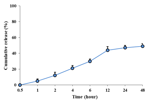
Figure 1. In vitro release profile of NIC nanoliposomes in phosphate buffer medium containing tween (0.5%) at 37°C and a pH of 7.4 (n=3).
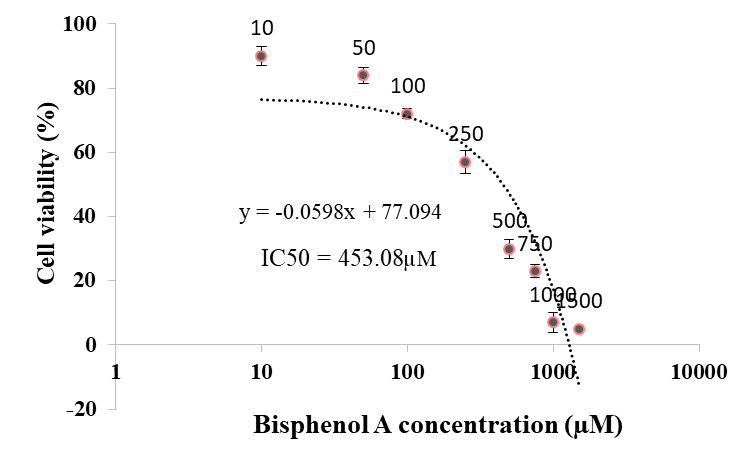
Figure 2. Viability percentage of HUVEC evaluated by MTT assay after 24 h incubation with different concentrations of bisphenol A. Data represent mean±SEM (n=3).
Therefore, in subsequent experiments, a concentration of 450 μM BPA was used to induce toxicity in HUVEC.
The possible toxicity of NIC, drug-free nanoliposomes, and NIC-NL toward HUVEC cells was also evaluated based on the MTT assay. As indicated in Figure 3, there was no inhibitory impact on the survival rate of cells after 24-hour exposure to the concentrations of 1-100 μM of the aforementioned compounds. Only NIC and NIC-NL at a concentration of 200 μM caused a significant decrease in HUVEC cell viability (P<0.05). Therefore, the concentrations of 1 to 100 μM were used in other experiments.
Figure 4 shows the cytoprotective effects of different concentrations of NIC and NIC-NL on HUVECs exposed to 450 μM BPA for 24 h, as assessed by the MTT method. Pretreatment of cells with NIC at 25-100 μM and NIC-NL at 5-100 μM significantly reduced BPA-induced cytotoxicity and maintained HUVEC cell survival.
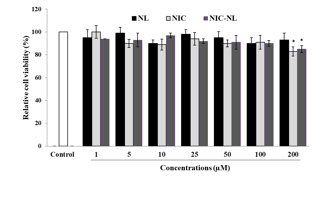
Figure 3. Viability percentage of HUVEC evaluated by MTT assay after 24 h incubation with different concentrations of nicorandil (NIC), drug-free nanoliposomes (NL), and nicorandil nanoliposomes (NIC-NL). Data represent mean±SEM (n=3). *P<0.001 versus control (untreated cells).
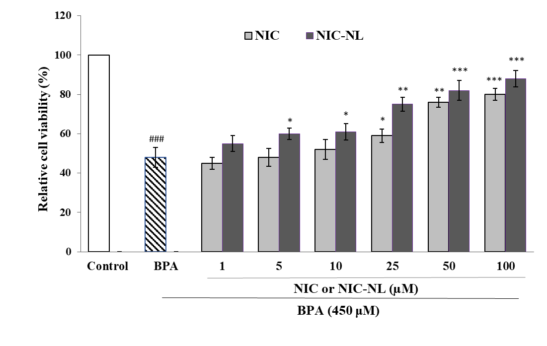
Figure 4. Viability percentage of HUVEC evaluated by MTT assay after 24 h pretreatment with nicorandil (NIC) or nicorandil nanoliposomes (NIC-NL) in bisphenol A-induced toxicity. Data represent mean±SEM (n=3). ###P<0.001 versus control (untreated cells); *P<0.05, **P<0.01 and ***p<0.001versus BPA.
Figure 5 indicates the Impact of NIC and NIC-NL on TAC in HUVEC under BPA-induced toxicity conditions. Incubation of cells with BPA significantly decreased TAC compared to control cells (P<0.001). The results indicated that pretreatment of cells with NIC at concentrations of 25-100 μM and pretreatment with NIC-NL at 5-100 μM significantly increased TAC under BPA-induced cytotoxicity. Moreover, NIC-NL at a concentration of 100 μM increased TAC more effectively compared to 100 μM of NIC (P<0.05).
The effect of NIC and NIC-NL on the amount of ROS produced in HUVEC during a 24-h exposure to BPA (450 μM) is demonstrated in Figure 5. Exposure to BPA increased intracellular ROS levels by approximately 2-fold compared to the control group (p<0.001). Pretreatment with NIC and NIC-NL at 1-100 μM significantly reduced intracellular ROS levels, bringing them close to normal levels. In addition, NIC-NL at a concentration of 100 μM was more effective in reducing ROS levels compared to 100 μM of NIC (P<0.05).
Figure 6 indicates the effect of different concentrations of NIC and NIC-NL on the glutathione content of HUVEC under BPA-induced cytotoxicity. Accordingly, none of the drugs were able to prevent the decrease in glutathione levels caused by BPA in the cells.
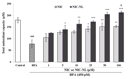
Figure 5. Total antioxidant capacity in HUVEC evaluated as FRAP value after 24 h pretreatment with nicorandil (NIC) or nicorandil nanoliposomes (NIC-NL) in bisphenol A-induced toxicity. Data represent mean±SEM (n=3). ###P<0.001 versus control (untreated cells); *P<0.05, **P<0.01 and ***P<0.001versus BPA; &P<0.05 versus NIC.
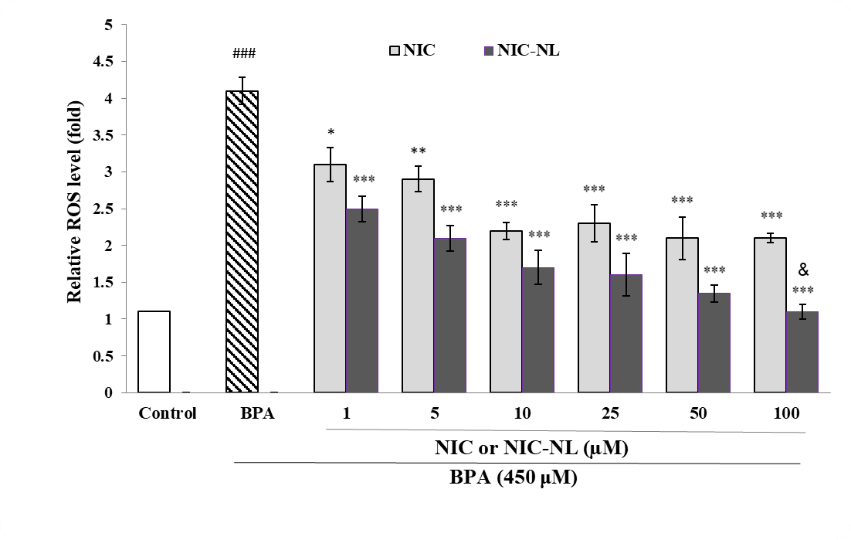
Figure 6. Intracellular ROS level in HUVEC evaluated by fluorescence intensity after 24 h pretreatment with nicorandil (NIC) or nicorandil nanoliposomes (NIC-NL) in bisphenol A (BPA)-induced toxicity. Data represent mean±SEM (n=3). ###P<0.001 versus control (untreated cells); *P<0.05, **P<0.01 and ***P<0.001versus BPA; &P<0.05 versus NIC.
Discussion
In the present study, BPA at 453.08 μM caused a 50% decrease in HUVEC survival. Many studies have shown that BPA can cause toxicity in various cells. In a study conducted by Russo et al., the IC50 of BPA in HUVEC, as assessed by the MTT assay, was reported to be 300 μM [6]. Çinar et al. exposed neuronal cells to 100 μM of BPA to induce cytotoxicity and observed that cell viability was reduced by approximately 60% in the MTT assay [23]. Various mechanisms have been proposed for BPA-induced cytotoxicity. Data have indicated that BPA can cause cell death by causing oxidative stress and inducing apoptosis. By activating the TRPV4 (Transient receptor potential vanilloid 4) signaling pathway, BPA leads to the accumulation of calcium and zinc ions in mitochondria, which subsequently results in the disruption of the mitochondrial membrane potential, ROS production, and lipid peroxidation. These changes also stimulate apoptosis through the activation of various caspase enzymes and affect the expression of genes involved in apoptosis, such as p53, Bax, and Bcl-2 [6, 23].
In addition, it has been reported that BPA activates inflammatory pathways in vascular cells and leads to an increase in inflammatory cytokines such as interleukins (IL)1-beta and IL-8, monocyte chemoattractant protein-1 (MCP-1), and intercellular adhesion molecules, such as ICAM (intercellular adhesion molecule), VCAM (vascular cell adhesion molecule), and selectin [6].
The present investigation showed that NIC and NIC-NL at concentrations of 1 to 100 μM had no inhibitory effect on HUVEC cell survival. Asensio-Lopez et al. examined the effect of NIC on cardiomyocyte cell viability and found that NIC at concentrations of 10–100 μM had no inhibitory effect on normal cell growth [24]. In another study, Wei et al. exposed lymphocytes to 1 nM, 1 μM, and 1 mM concentrations of NIC and reported that 1 μM concentration of NIC could induce cytotoxic effects in lymphocytes but lower concentrations were safe [25]. In another study, NIC at a concentration of 100 μM had no inhibitory effects on the survival of pulmonary artery endothelial cells [26].
The findings of the present study indicated that NIC at concentrations of 25 to 100 μM and NIC-NL at concentrations of 5 to 100 μM significantly inhibited BPA-induced cytotoxicity in HUVEC. In a study by Wang et al., pretreatment of pulmonary artery endothelial cells with NIC (100 μM) protected the cells under hypoxic conditions by increasing endothelial nitric oxide synthase (eNOS) expression and decreasing nuclear factor κB (NF-κB) expression. In addition, NIC inhibited the expression of caspase enzymes and increased the expression of Bcl-2 relative to Bax, subsequently reducing apoptosis in endothelial cells [26]. The anti-apoptotic effects of NIC have been reported to protect cardiac cells in cardiomyopathy conditions by activating the PI3K/Akt (Phosphatidylinositol 3-kinase/Akt) pathway [27]. In addition, the anti-inflammatory effects of NIC have been reported, and it has been able to reduce the secretion of tumor necrosis factor (TNF) from lymphocytes through the production of NO and affecting the potassium channels [25]. It was also shown that NIC protects cells by inhibiting oxidative stress. In a study of Asensio-Lopez et al., NIC at concentrations of 10 to 100 μM protected cardiomyocytes against doxorubicin-induced toxicity [24].
The present research revealed that NIC-NL enhanced the protective effects of NIC in PBA-induced toxicity, such that NIC-NL increased cell viability in HUVEC at lower concentrations compared to NIC. In a similar study on nitroglycerin, which acts similarly to NIC, nitroglycerin nanoliposomes were readily taken up by endothelial cells and demonstrated 70 times greater therapeutic and anti-inflammatory efficacy than nitroglycerin [28]. In another study, chitosan nanoparticles loaded with nitroglycerin showed stronger renal protective effects than free nitroglycerin in ischemia-reperfusion injury [29].
In this study, exposure of endothelial cells to BPA resulted in a significant increase in intracellular ROS levels, a severe decrease in TAC, and a decrease in glutathione content in HUVEC. Oxidative stress has been proposed as a key mechanism in BPA-induced cytotoxicity. In the study of Ebrahimi et al., exposure of normal gingival cells to BPA (100 μM) increased intracellular ROS levels by about 4-fold, lipid peroxides by about 2-fold, and decreased glutathione content by about 5-fold [30]. BPA can reduce TAC in cells and tissues by reducing antioxidant compounds, such as glutathione, and antioxidant enzymes, including superoxide dismutase, catalase, and glutathione peroxidase [31, 32].
The results of the present study demonstrated that pretreatment of HUVEC cells with NIC and NIC-NL significantly reduced intracellular ROS levels and increased TAC levels, but failed to increase cellular glutathione content. The antioxidant effects of NIC have been reported in several studies. The NIC at concentrations of 10 to 100 μM dose-dependently protected cardiomyocyte cells against doxorubicin-induced toxicity by counteracting oxidative stress through reducing ROS levels and lipid peroxidation in the study of Asensio-López et al. They indicated that activation of ATP-sensitive potassium channels by NIC causes mitochondrial depolarization and subsequent inhibition of the mitochondrial NADPH oxidase enzyme, which plays a major role in ROS production [24]. NIC can also play a role in inhibiting oxidative stress by inhibiting the NF-κB signaling pathway [26].
In our study, NIC and NIC-NL failed to provide a beneficial effect in increasing cellular glutathione content. It is important to note that following high plasma concentrations of nitroglycerin, contradictory effects have been reported, such as increased mitochondrial oxidative stress through the production of superoxide and peroxynitrite radicals, as well as depletion of sulfhydryl stores, which are among the mechanisms proposed in the development of tolerance to nitrites. Nitroglycerin non-selectively reacts with thiol-containing compounds to produce dinitrate metabolites. It directly oxidizes sulfhydryl groups on several cellular proteins, including aldehyde dehydrogenase and glutathione transferase, and tolerance to this drug subsequently develops by reducing redox signaling [33].
In addition, the results of the present study indicated that NIC-NL were more effective in reducing ROS and increasing TAC compared to free NIC. In a recent study, it was found that chitosan nanoparticles loaded with nitroglycerin had significantly stronger effects on reducing total oxidative status and increasing TAC in kidney tissue of rats with ischemia/reperfusion-induced renal injury compared with free nitroglycerin [29]. In a study conducted by Ardakani et al., nanoliposomes containing 0.07 μM nitroglycerin produced an efficacy equivalent to that of nitroglycerin at a dose of 5 μM, meaning that the efficiency of nanoliposomes in producing NO, anti-inflammatory effects, and preventing mitochondrial superoxide radical formation was 70 times greater than that of free nitroglycerin [28]. In the present work, NIC-NL coated with TPGS was used. The liposomal nanoparticle form of NIC can both improve drug stability and reduce the need for repeated doses by slowly releasing the drug. The preparation of TPGS-coated NIC-NL allows for the use of lower doses of this drug, thereby helping reduce its side effects. In addition, by enhancing the antioxidant effects of NIC, it protects cells against damage caused by oxidative stress [34].
Conclusions
The findings of the present study indicated that TPGS-coated NIC-NL has cytoprotective and antioxidant effects in endothelial cells, increasing cell survival and TAC and reducing intracellular ROS production under BPA-induced toxicity. Further studies are needed to reveal the precise molecular mechanisms of the beneficial effects of this drug and its nanoliposomes.
Conflicts of Interest
The authors declare no conflicts of interest with any entities.
Funding
This study was financially supported by the Vice Chancellor for Research and Technology, Isfahan University of Medical Sciences, Isfahan, Iran (Grant No. 3402521).
Compliance with Ethical Guidelines
This study received approval from the Institutional Research Ethics Committee of Isfahan University of Medical Sciences (ID: IR.MUI.AEC.1402.043).
Authors' Contributions
All authors equally contributed to preparing this article.
Data Access and Responsibility
The authors confirm that this paper contains original work and accept full responsibility for its content.
References
- Rykowska I, Wasiak W. Properties, threats, and methods of analysis of bisphenol A and its derivatives. Acta Chromatograph. 2006;16:7-27. [LINK]
- Vandenberg LN, Hauser R, Marcus M, Olea N, Welshons WV. Human exposure to bisphenol A (BPA). Reprod Toxicol. 2007;24(2):139-77. [DOI: 10.1016/j.reprotox.2007.07.010] [PMID: 17825522]
- Tarafdar A, Sirohi R, Balakumaran PA, Reshmy R, Madhavan A, Sindhu R, et al. The hazardous threat of Bisphenol A: Toxicity, detection and remediation. J Hazard Mater. 2022;423:127097. [DOI: 10.1016/j.jhazmat.2021.127097] [PMID: 34488101]
- Cooper BL, Posnack NG. Characteristics of bisphenol cardiotoxicity: impaired excitability, contractility, and relaxation. Cardiovasc Toxicol. 2022;22(3):273-80. [DOI: 10.1007/s12012-022-09719-9] [PMID: 35143014]
- Afzal G, Ahmad HI, Hussain R, Jamal A, Kiran S, Hussain T, et al. Bisphenol A induces histopathological, hematobiochemical alterations, oxidative stress, and genotoxicity in common carp (Cyprinus carpio L.). Oxid Med Cell Longev. 2022;2022:5450421. [DOI: 10.1155/2022/5450421] [PMID: 35126815]
- Russo C, Maugeri A, Albergamo A, Dugo G, Navarra M, Cirmi S. Protective effects of a red grape juice extract against bisphenol A-induced toxicity in human umbilical vein endothelial cells. Toxics. 2023;11:391. [DOI: 10.3390/toxics11040391] [PMID: 37112618]
- Ramos C, Ladeira C, Zeferino S, Dias A, Faria I, Cristovam E, et al. Cytotoxic and genotoxic effects of environmentally relevant concentrations of bisphenol A and interactions with doxorubicin. Mutat Res Genet Toxicol Environ Mutagen. 2019;838:28-36. [DOI: 10.1016/j.mrgentox.2018.11.009] [PMID: 30678825]
- Ikhlas S, Usman A, Ahmad M. In vitro study to evaluate the cytotoxicity of BPA analogues based on their oxidative and genotoxic potential using human peripheral blood cells. Toxicol in Vitro. 2019;60:229-36. [DOI: 10.1016/j.tiv.2019.06.001] [PMID: 31173877]
- Ahmed LA. Nicorandil: A drug with ongoing benefits and different mechanisms in various diseased conditions. Indian J Pharmacol. 2019;51(5):296-301. [DOI: 10.4103/ijp.IJP_298_19] [PMID: 31831918]
- Zheng J, Long M, Qin Z, Wang F, Chen Z, Li L. Nicorandil inhibits cardiomyocyte apoptosis and improves cardiac function by suppressing the HtrA2/XIAP/PARP signaling after coronary microembolization in rats. Pharmacol Res Perspect. 2021;9(1):e00699. [DOI: 10.1002/prp2.699] [PMID: 33448699]
- Sánchez-Duarte S, Márquez-Gamino S, Montoya-Pérez R, Villicana-Gómez EA, Vera-Delgado KS, Caudillo-Cisneros C, et al. Nicorandil decreases oxidative stress in slow- and fast-twitch muscle fibers of diabetic rats by improving glutathione system functioning. J Diabetes Investig. 2021;12(7):1152-61. [DOI: 10.1111/jdi.13513] [PMID: 33503290]
- El-Kashef DH, Sharawy MH. Hepatoprotective effect of nicorandil against acetaminophen-induced oxidative stress and hepatotoxicity in mice via modulating NO synthesis. Environ Sci Pollut Res Int. 2023;30(6):14253-64. [DOI: 10.1007/s11356-022-23139-w] [PMID: 36149558]
- Zong Y, Li J, Xu X, Xu X. Effects of nicorandil on systemic inflammation and oxidative stress induced by percutaneous coronary intervention in patients with coronary heart disease. J Int Med Res. 2021;49(12):3000605211058873. [DOI: 10.1177/03000605211058873] [PMID: 34871513]
- Kumar S, Dilbaghi N, Saharan R, Bhanjana G. Nanotechnology as emerging tool for enhancing solubility of poorly water-soluble drugs. Bionanosci. 2012;2:227-50. [DOI: 10.1007/s12668-012-0060-7]
- Lu H, Zhang S, Wang J, Chen Q. A review on polymer and lipid-based nanocarriers and their application to nano-pharmaceutical and food-based systems. Front Nutr. 2021;8:783831. [DOI: 10.3389/fnut.2021.783831] [PMID: 34926557]
- Sadoqi M, Lau-Cam CA, Wu SH. Investigation of the micellar properties of the tocopheryl polyethylene glycol succinate surfactants TPGS 400 and TPGS 1000 by steady-state fluorometry. J Colloid Interface Sci. 2009;333(2):585-9. [DOI: 10.1016/j.jcis.2009.01.048] [PMID: 19232633]
- Nguyen TK, Nilakantan V, Felix CC, Khanna AK, Pieper GM. Beneficial effect of alpha-tocopheryl succinate in rat cardiac transplants. J Heart Lung Transplant. 2006;25(6):707-15. [DOI: 10.1016/j.healun.2006.02.007] [PMID: 16730577]
- Song T, Wang H, Liu Y, Cai R, Yang D, Xiong Y. TPGS-modified long-circulating liposomes loading Ziyuglycoside I for enhanced therapy of myelosuppression. Int J Nanomed. 2021;16:6281-95. [DOI: 10.2147/IJN.S326629] [PMID: 34548791]
- Danaei MR, Dehghankhold M, Ataei S, Hasanzadeh Davarani F, Javanmard R, et al. Impact of particle size and polydispersity index on the clinical applications of lipidic nanocarrier systems. Pharmaceutics. 2018;10(2):57. [DOI: 10.3390/pharmaceutics10020057] [PMID: 29783687]
- Dayani L, Taheri A, Taymouri S, Najafi RB, Esmaeilian F. Preparation and in vitro evaluation of Vancomycin-loaded Montmorillonite-Sodium Alginate topical gel for wound infection. Brazil J Pharm Sci. 2022;58:e21034. [DOI: 10.1590/s2175-97902022e21034]
- Ghassami E, Varshosaz J, Mirian M, Jahanian‐Najafabadi A. HER‐2 aptamer‐targeted Ecoflex® nanoparticles loaded with docetaxel promote breast cancer cell apoptosis and anti‐metastatic effect. IET Nanobiotechnol. 2019;13(4):428-34. [DOI: 10.1049/iet-nbt.2018.5047] [PMID: 31171748]
- Safaeian L, Sadeghi-Dinani M, Ramezani A, Asghari-Varzaneh M. Impact of Opuntia ficus-indica fruit extracts on toxicity induced by doxorubicin in cardiomyocytes. Res J Pharmacogn. 2025;12(4):5–8. [DOI: 10.22127/rjp.2025.525978.2844]
- Çinar R, Nazıroğlu M. Bisphenol A induces neuronal apoptosis and oxidative stress through TRPV4 channel signaling pathways: Protective role of alpha‐lipoic acid. Environ Toxicol. 2025;40(11):1261-76. [DOI: 10.1002/tox.24541] [PMID: 40396745]
- Asensio-López MC, Soler F, Pascual-Figal D, Fernández-Belda F, Lax A. Doxorubicin-induced oxidative stress: The protective effect of nicorandil on HL-1 cardiomyocytes. PLoS One. 2017;12(2):e0172803. [DOI: 10.1371/journal.pone.0172803] [PMID: 28245258]
- Wei XM, Heywood GJ, Di Girolamo N, Thomas PS. Nicorandil inhibits the release of TNFα from a lymphocyte cell line and peripheral blood lymphocytes. Int Immunopharmacol. 2003;3(12):1581-8. [DOI: 10.1016/S1567-5769(03)00176-0] [PMID: 14555283]
- Wang H, Zuo X, Wang Q, Yu Y, Xie L, et al. Nicorandil inhibits hypoxia-induced apoptosis in human pulmonary artery endothelial cells through activation of mitoKATP and regulation of eNOS and the NF-κB pathway. Int J Mol Med. 2013;32(1):187-94. [DOI: 10.3892/ijmm.2013.1379] [PMID: 23670355]
- Zhan B, Xu Z, Zhang Y, Wan K, Deng H, Wang D, et al. Nicorandil reversed homocysteine-induced coronary microvascular dysfunction via regulating PI3K/Akt/eNOS pathway. Biomed Pharmacother. 2020;127:110121. [DOI: 10.1016/j.biopha.2020.110121] [PMID: 32407984]
- Ardekani S, Scott HA, Gupta S, Eum S, Yang X, Brunelle AR, et al. Nanoliposomal nitroglycerin exerts potent anti-inflammatory effects. Sci Rep. 2015;5(1):16258. [DOI: 10.1038/srep16258] [PMID: 26584637]
- Karimi Z, Asadi K, Ghahramani P, Gholami A. Trinitroglycerine-loaded chitosan nanoparticles attenuate renal ischemia-reperfusion injury by modulating oxidative stress. Sci Rep. 2024;14(1):32112. [DOI: 10.1038/s41598-024-83886-3] [PMID: 39738455]
- Ebrahimi R, Shokrzadeh M, Ghassemi Barghi N. Effects of melatonin on the bisphenol-A-induced cytotoxicity and genetic toxicity in colon cancer cell lines, normal gingival cell lines, and bone marrow stem cell lines. Cancer Inform. 2021;20:11769351211056295. [DOI: 10.1177/11769351211056295] [PMID: 34819716]
- Sangai NP, Patel CN, Pandya HA. Ameliorative effects of quercetin against bisphenol A-caused oxidative stress in human erythrocytes: an in vitro and in silico study. Toxicol Res. 2018;7(6):1091-9. [DOI: 10.1039/c8tx00105g] [PMID: 30542603]
- Aneta Maćczak, Monika Cyrkler, Bożena Bukowska, Jaromir Michałowicz. Bisphenol A, bisphenol S, bisphenol F and bisphenol AF induce different oxidative stress and damage in human red blood cells (in vitro study). Toxicol in Vitro. 2017;41:143-9. [DOI: 10.1016/j.tiv.2017.02.018] [PMID: 28259788]
- Tsou PS, Addanki V, Haas JA, Page NA, Fung HL. Role of glutaredoxin-mediated protein S-glutathionylation in cellular nitroglycerin tolerance. J Pharmacol Exp Ther. 2009;329(2):649-56. [DOI: 10.1124/jpet.108.149997] [PMID: 19223666]
- Leme Goto P, Cinato M, Merachli F, Vons B, Jimenez T, Marsal D, et al. In vitro and in vivo cardioprotective and metabolic efficacy of vitamin E TPGS/Apelin. J Mol Cell Cardiol. 2020;138:165-74. [DOI: 10.1016/j.yjmcc.2019.12.001] [PMID: 31836542]