



Abstract
Background: Given the propensity of standard antidiabetic medications to elicit undesirable side effects over extended periods, natural product derived from medicinal plants have emerged as a compelling area of investigation for their potential therapeutic value and reduced toxicity. The present study examined Cotoneaster manna extract for its bioactive compounds.
Methods: The in vitro cytotoxicity of the extract was assessed on HT29 colon cancer cells and HEK293 normal cells using the MTT assay. The antidiabetic potential of the extract was investigated in vivo using a streptozotocin-induced diabetic rat model. The in vivo antioxidant efficacy was investigated using a D-galactose-induced rat model of aging.
Results: The extract reduced HT29 cell viability in a concentration-dependent manner (p<0.05), while exhibiting lower toxicity toward HEK293 cells. In diabetic rats, liver histopathology demonstrated partial restoration of tissue architecture and reduced hepatocellular necrosis compared with untreated controls. Compared to the D-galactose group, the extract significantly (p<0.05) elevated catalase and superoxide dismutase activities and suppressed malondialdehyde formation in both serum and liver tissues.
Conclusion: Cotoneaster manna exhibits hypoglycemic, anticancer, and antioxidant properties, likely attributable to its phenolic and flavonoid constituents. Further molecular and histopathological studies are warranted to confirm these mechanisms.
Keywords: Antineoplastic agents, Antioxidants, Hypoglycemic agents, Liver, Plant extracts, Phytogenic
Introduction
Cancer remains a leading cause of mortality worldwide, especially in developing regions. Among various malignancies, colorectal cancer is among the most prevalent and fatal, particularly in men, accounting for substantial global mortality [1]. Conventional chemotherapeutic agents used for cancer treatment are often limited by their severe side effects and lack of selectivity for malignant cells over normal tissues. Consequently, research has increasingly focused on medicinal plant–derived bioactive compounds as safer and more sustainable therapeutic alternatives [2].
Diabetes mellitus is another common metabolic disorder affecting millions globally. According to reports, about 200 million individuals currently live with diabetes, a figure projected to reach 300 million by 2025. Given the liver's pivotal role in glucose homeostasis as the key target of insulin and glucagon, the preservation of its function is a crucial therapeutic objective in diabetes management. However, long‑term administration of synthetic antidiabetic drugs often results in adverse effects such as headache, nausea, peptic ulcers, and gastrointestinal disturbances. Therefore, the use of natural agents of plant origin has gained attention as a complementary approach with fewer side effects [3].
Manna -known locally as Shir-Khesht- is a natural exudate obtained from several species of the genus Cotoneaster (family Rosaceae). The principal constituent of Cotoneaster species is manna, which predominantly comprises sugars such as glucose and mannitol. Cotoneaster manna is also rich in flavonoids, phenolic compounds, and trace elements that collectively exert notable pharmacological activities [4, 5]. In Iranian traditional medicine, Shir-Khesht has long been used for the management of ailments such as constipation and neonatal jaundice. Manna extracted from Cotoneaster species has also demonstrated hepatoprotective, anticancer, antioxidant, antibacterial, and bilirubin‑reducing properties in experimental reports [6, 7].
Given its pharmacological significance, the current research aimed to assess the anticancer, antidiabetic, and antioxidant potential of Cotoneaster manna extract through both in vitro cytotoxicity assays on human colon cancer (HT‑29) and normal kidney (HEK‑293) cell lines, and in vivo experiments on streptozotocin‑induced diabetic and D‑galactose‑treated rat models.
Materials and Methods
Plant Material and Extraction Procedure
During the summer of 2019, samples of Cotoneaster manna were harvested from the northwestern slopes of Shah‑Jahan Peak located in North Khorasan Province, Iran. For preparation of the hydroalcoholic extract, 20 g of the finely powdered dried material was blended with a mixture of 160 mL of 70% ethanol and 40 mL of distilled water in a 500 mL vessel [8]. The suspension was continuously agitated for 24 h, then kept in darkness for a further 48 h to achieve exhaustive extraction. The resulting liquid phase was separated through Whatman filter paper, concentrated under reduced pressure, and oven‑dried at 45 °C for 24 h to yield the final dry extract [9].
MTT Cytotoxicity Assay
A stock solution for cytotoxicity testing was prepared by dissolving 300 mg of the extract powder in 2 mL of RPMI 1640 culture medium to achieve a homogeneous suspension. Human embryonic kidney (HEK‑293) and colorectal carcinoma (HT‑29) cell lines were procured from the Pasteur Institute, Tehran, Iran. To ensure optimal cell viability and proliferation, cultures were maintained in a complete growth medium comprising RPMI 1640, 10% heat-inactivated fetal bovine serum, and 1% penicillin-streptomycin. Cells were incubated under standard culture conditions at 37 °C in a humidified atmosphere containing 5% CO₂. When cell density reached nearly 70% confluence, aliquots of 5 × 10⁴ cells per well were seeded into 96‑well microplates and treated with varying concentrations of the extract (25, 50, and 75 µg/mL). Following 20 h of treatment, cellular metabolic activity was assessed by a 4-hour incubation with 10 µL of MTT (5 mg/mL in PBS). The produced formazan deposits were dissolved using 100 µL of dimethyl sulfoxide (DMSO), and optical density was recorded at 540 nm employing an ELISA microplate reader [10].
Antidiabetic Study
All experimental procedures were conducted using a cohort of 24 adult male Wistar rats (Rattus norvegicus), with individual body weights ranging from 250-300 g. All rats were housed in a controlled environment (22±2 °C, 12-h light/dark cycle) at the institutional Animal Facility and had free access to a standard diet and water. After 10 days of acclimatization, the rats were randomly assigned to three groups (n=8): 1. healthy control, 2. streptozotocin-induced diabetic, and 3. diabetic rats treated with C. manna hydroalcoholic extract (100 mg/kg b.w., i.p.). Experimental diabetes was established through intraperitoneal administration of streptozotocin (50 mg/kg body weight) for 7 consecutive days. Subsequently, the treated group received the plant extract for 21 days, whereas the control group received saline only. Diabetes was confirmed biochemically following a 12-hour fast. Blood glucose measurements obtained from the tail vein using a calibrated glucometer revealed a subset of rats with levels >300 mg/dL; these animals were classified as diabetic for the study [11].
Antioxidant Evaluation
Forty-eight adult Wistar rats were divided into four groups (n=6 per group):
1. Negative control (NC): received normal saline orally.
2. D-galactose-induced group: received D-galactose (120 mg/kg bw, s.c.) dissolved in 0.9% saline and administered once daily for 21 days, following previously described methods [12, 13].
3. Positive control (PC): received ascorbic acid (10 mg/kg, orally) [12].
4. Treatment group: received Cotoneaster manna extract (100 mg/kg bw, orally) [12].
Treatments were administered
3 times per week for 21 days [12]. Upon completion of the study, animals were humanely euthanized. This procedure involved deep anesthesia with sodium pentobarbital (i.p.), followed by terminal blood collection through cardiac puncture. The liver was immediately harvested, and sections from distinct lobes were either snap-frozen in liquid nitrogen for biochemical analysis or fixed for histological examination to assess tissue morphology.
Liver tissues were homogenized in 10 mL of 10 mM phosphate buffer (pH=7.4).
Homogenization was performed on ice for 3 cycles of 60 s. Catalase (CAT) activity was determined according to the method described by Weydert and Cullen (2010) using commercial kits (Beytime, Shanghai, China). The activity of the antioxidant enzyme CAT was measured in milli-Katal per mg protein (mK/mg), while superoxide dismutase (SOD) activity was expressed in units per mg protein (U/mg). The levels of malondialdehyde (MDA) in serum and liver were reported as nmol/mg of protein [14, 15].
Histopathological Examination
Animals were anesthetized by diethyl ether inhalation. After a longitudinal abdominal incision, the liver was excised and fixed in 10% neutral-buffered formalin (NBF). The tissues were processed by standard histological procedures, paraffin-embedded, sectioned using microtome, and stained with hematoxylin and eosin (H&E) processing and paraffin embedding were performed, and sections were cut using a microtome and stained with H&E for microscopic evaluation. The present study was carried out in strict compliance with international standards for animal welfare. Ethical approval for this research was granted by the Institutional Ethics Committee of Zabol University under reference No. IR.UOZ.REC.1401.011. Histopathological evaluation of liver sections was performed through microscopic examination.
Statistical analysis
All statistical analyses were conducted using SPSS software (version 22; IBM Corp., Armonk, NY, USA). Continuous variables are expressed as the mean ± standard deviation (SD). Statistical significance between group means was assessed by one-way ANOVA. In cases where the ANOVA indicated a significant difference (p<0.05), the Least Significant Difference post-hoc test was applied for pairwise comparisons. All reported p-values are two-tailed.
Results
Blood glucose
Our findings suggest that Cotoneaster manna extract significantly reduces blood glucose levels in diabetic rats (p<0.05). Analysis of comparison of means among groups revealed a statistically significant difference between diabetic rats treated with the plant extract and untreated diabetic rats (p<0.05). Furthermore, the mean comparison results demonstrated that blood glucose concentration in the diabetic group before treatment was significantly higher than that in diabetic rats after treatment, as shown in Figure 1.
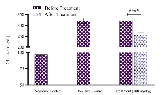 Figure 1. Variations in blood glucose levels observed among normal (Negative Control), untreated diabetic (Positive Control), and diabetic rats receiving Shir‑Khesht extract (100 mg/kg body weight). The symbol #### indicates a highly significant statistical reduction (p<0.0001) in glucose values, following extract administration compared with pre‑treatment measurements in diabetic rats.
Cell cytotoxicity evaluation using MTT Assay
Figure 2 illustrates the effect of Shir-Khesht manna extract on the viability percentage of cancer (HT‑29) and normal (HEK‑293) cells. The results of one‑way ANOVA indicated that Shir-Khesht extract markedly diminished the viability of cancer cells in a dose‑dependent fashion. Treatment with the extract at concentrations of 50 and 75 μg/mL resulted in a significant (p˂0.05) reduction in the viability of HT-29 cells compared to the untreated control. Moreover, a markedly significant variation (p˂0.0001) was observed between the mean viability of normal cells and cancer cells treated with a concentration of 75 μg/mL, indicating selective cytotoxicity of the extract toward cancer cells.
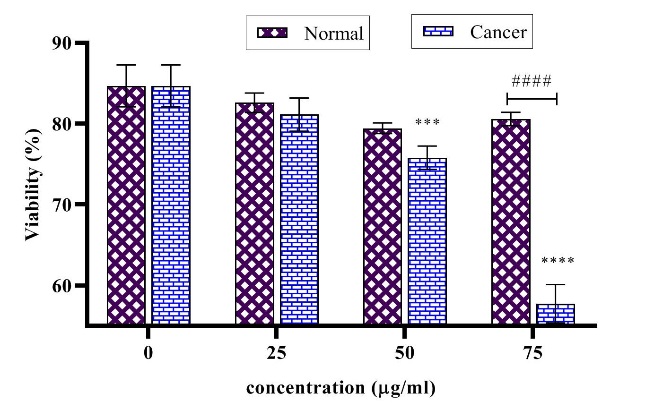
Figure 2. Effects of Shir‑Khesht manna extract on the viability of HT‑29 (colorectal cancer) and HEK‑293 (normal) cell lines at concentrations of 0, 25, 50, and 75 µg/mL. Symbols *** and **** represent statistically significant reductions in HT‑29 cell viability at 50 and 75 µg/mL, respectively, when compared with untreated control cells. The mark #### indicates a highly significant distinction (p<0.0001) between HT‑29 cells treated with 75 µg/mL and HEK‑293 cells exposed to the same concentration of the extract.
Antioxidant assessments
The antioxidant potential of the plant extract was assessed using a D‑galactose‑induced rat model. The results demonstrated a significant decrease in CAT activity in the serum and liver of D‑galactose‑treated rats compared with the negative control (NC) group, showing oxidative stress-induced aging in D‑galactose‑induced rats (Figure 3). Compared to the NC group, the D-galactose model group exhibited a significant impairment in antioxidant capacity, as evidenced by a ~70% decrease in hepatic CAT activity and a ~64% decrease in serum CAT activity. However, the catalase activity in the liver and serum samples of rats in the positive control (PC) group and the extract‑treated group was markedly (p<0.05) elevated in comparison to the D‑galactose group, therefore suggesting activation of the antioxidant defense system in rats receiving ascorbic acid and Shir-Khesht extract.
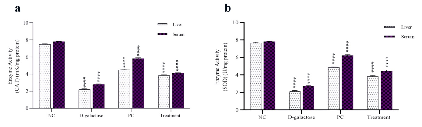
Figure 3. Catalase (a) and superoxide dismutase (b) activities measured in liver and serum samples from D‑galactose‑treated, ascorbic acid‑positive control (PC), and extract‑treated rat groups. The symbol **** denotes statistically significant (p < 0.0001) alterations in enzymatic activity across D‑galactose, PC, and experimental groups relative to the Negative Control (NC) group.
Superoxide dismutase activity in the liver and serum was significantly lower in the D-galactose group than in the NC group (p<0.05). In contrast, treatment with the extract or the positive control compound effectively restored SOD activity, resulting in levels significantly exceeding those of the diseased model (Figure 3). A pronounced increase in MDA concentration was detected in both hepatic and serum samples of the D‑galactose‑induced rats. Conversely, animals treated with either the plant extract or ascorbic acid exhibited markedly reduced MDA levels compared with the negative control group (p<0.05). These findings indicate that both treatments mitigated oxidative stress. However, the Cotoneaster extract produced an effect comparable to ascorbic acid (Figure 4).
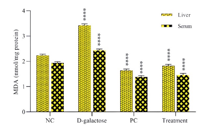
Figure 4. Comparative levels of malondialdehyde (MDA) in liver and serum samples of experimental rat groups. NC represents the Negative Control, PC refers to the Positive Control (ascorbic acid), D‑galactose denotes rats subjected to D‑galactose induction, and Treatment indicates animals administered the plant extract. Marker **** shows statistically significant variations in MDA concentrations among PC, D‑galactose, and Treatment groups when compared with the NC group (p < 0.05).
Histopathological Studies
Liver tissue samples were sectioned and stained with H&E for histological evaluation under a light microscope. The histopathological findings of the liver in healthy rats are presented in Figure 5A, showing normal hepatic architecture with clearly defined central veins and well‑arranged hepatic cords and intact sinusoids. In the untreated diabetic group, marked hepatocellular necrosis, disorganization, hyperemia, and infiltration of inflammatory cells around the central vein, as well as sinusoidal dilation, were observed (Figure 5B). In contrast, hepatic histology of extract‑administered diabetic rats revealed only slight pathological alterations and substantial recovery of hepatic architecture, accompanied by considerably less cellular degeneration than observed in the ascorbic acid control group.
Histopathological Findings
Histological evaluation of liver sections from healthy rats demonstrated normal morphology and well-organized hepatic cords, with no pathological alterations observed in this group. Induction of diabetes by streptozotocin produced marked hepatocellular necrosis, pronounced inflammatory infiltration, and disruption of hepatic sinusoidal integrity. Treatment of diabetic rats with the plant extract at a dose of 100 mg/kg body weight markedly reduced hepatocellular necrosis, attenuated local inflammation, and restored the structural integrity of hepatic sinusoids.
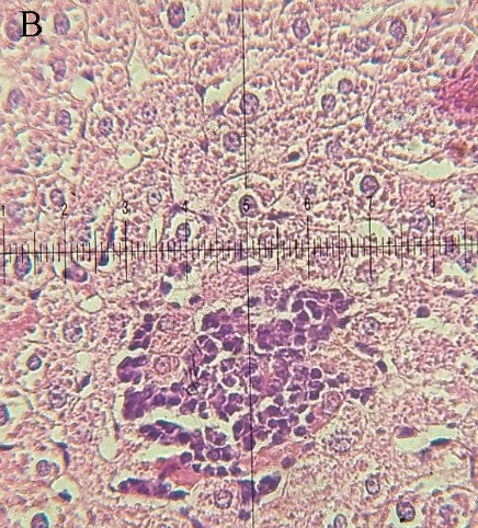
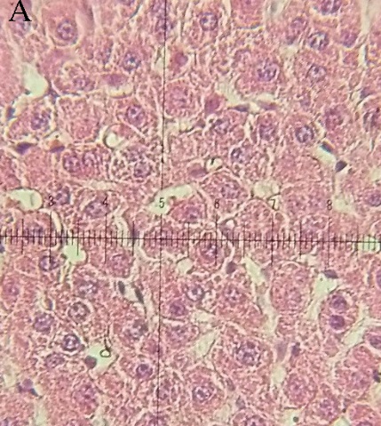
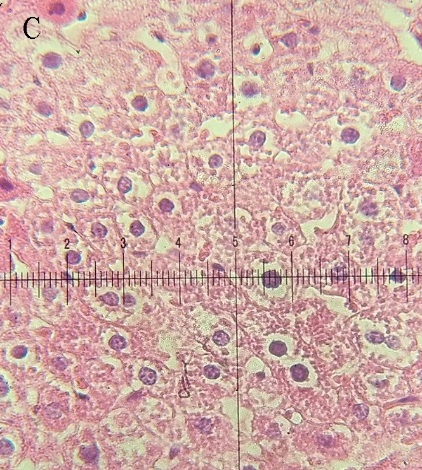
Figure 5. Representative hepatic histology of experimental rat groups. (A) Normal control group displaying intact hepatic cords and uniform hepatocyte morphology. (B) Streptozotocin‑induced diabetic group showing marked structural disorganization, hepatocellular necrosis, vascular congestion, and dense aggregation of inflammatory cells around the central vein. (C) Extract‑treated diabetic group exhibiting evident hepatocyte regeneration and restoration of hepatic architecture with minimal tissue injury (H&E staining, ×200).
Discussion
To date, the therapeutic potential of various plant‑derived compounds for the treatment of diseases such as cancer and diabetes has attracted considerable scientific attention worldwide [16]. In the present study, Shir-Khesht extract exhibited a dose‑dependent cytotoxic effect, significantly reducing the viability of colon cancer cells without affecting normal cells. Moreover, daily consumption of plants from the Artemisia genus has been reported to prevent or suppress cancer progression [17]. Previous investigations suggested that artemisinin reduces cancer cell growth via induction of apoptotic signaling [18,19]. In accordance with these observations, the Shir-Khesht extract showed potent inhibitory effects on the proliferation of colon cancer cells. Similarly, antitumor effects of Mentha species have been documented in gastric, HeLa, and HL‑60 cancer cells, where the inhibitory action of mint extracts on tumor progression is largely attributed to the presence of antioxidant flavones such as eupatilin and jaceosidin [20]. The anticancer mechanism of Shir-Khesht extract thus appears to be associated with its phenolic and flavonoid constituents, which can scavenge reactive oxygen species (ROS) and suppress oxidative-stress‑mediated damage in cancer cells.
Reactive oxygen species can damage cellular macromolecules, such as DNA, proteins, and lipids, via lipid peroxidation processes. Aging in living organisms is closely associated with oxidative stress; therefore, the development of antioxidant agents to mitigate the detrimental effects of oxidative-stress‑related aging is crucial [21]. In this experiment, a D‑galactose‑induced model was employed to evaluate the antioxidant activity of Shir-Khesht extract. Administration of D‑galactose significantly reduced serum and hepatic activities of SOD and CAT, while serum and liver MDA levels markedly increased (figures 3 and 4). These results align with earlier reports showing that long-term exposure to D‑galactose can impair antioxidant enzyme performance and weaken immune function in rat models [22]. Superoxide dismutase regulates the balance between the generation and detoxification of ROS, thereby protecting cells from free radical‑induced injury. Likewise, CAT converts hydrogen peroxide into water and oxygen, thereby mitigating oxidative stress [14]. In the present study, Shir-Khesht extract effectively restored the reduced activities of SOD and CAT in D‑galactose‑treated rats.
In a related investigation, rambutan (Nephelium lappaceum) peel extract displayed marked antioxidant activity both in vitro and in vivo; it protected HepG2 cells against H₂O₂‑induced oxidative damage and enhanced SOD activity, thereby limiting cellular apoptosis. Moreover, in a D‑galactose‑induced aging model, rambutan peel extract improved the antioxidant defense system in rats [23]. Similarly, stevia residue extract demonstrated considerable antioxidative activity in D‑galactose‑induced mice, reflected by increased SOD, CAT, and glutathione peroxidase (GPx) activities, elevated total antioxidant capacity, and reduced MDA and acetylcholinesterase levels. These effects were mediated through activation of the Akt/Nrf2/HO‑1 signaling pathway [15].
The close relationship between diabetic conditions and their associated pathological changes has been well documented in earlier reports [24, 25], and this is consistent with the histological findings obtained in the present study. The antioxidant compounds in Shir-Khesht extract appear to promote tissue regeneration in streptozotocin‑induced diabetic liver by improving antioxidant defenses and suppressing inflammatory responses. Histopathological examination of the diabetic rat liver showed disorganization of hepatocytes and distortion of the central vein.
Conclusions
The findings obtained clearly show that administering Shir‑Khesht manna extract to streptozotocin‑induced diabetic rats led to a marked decline in blood glucose, improvement of hepatic function, and attenuation of liver necrosis associated with streptozotocin toxicity. The hepatoprotective effect of Shir-Khesht extract may be attributed to the antioxidant capacity of its bioactive constituents. In both experimental models, the extract elevated the activities of SOD and CAT, thereby protecting against D-galactose-induced oxidative damage. Owing to its strong antioxidant properties, Shir-Khesht manna extract could be considered a natural anti‑aging and antioxidant agent with potential applications in the pharmaceutical and food industries.
Conflicts of Interest
The authors declare no competing interests that could have affected the outcomes or interpretation of this research.
Funding
The authors declare that no financial support was received for the present research.
Acknowledgement
This article is derived from the M.Sc. thesis, conducted at the Department of Biology, Faculty of Sciences, University of Zabol, Iran. The authors would like to express their sincere gratitude to the Faculty of Sciences and the Faculty of Veterinary at the University of Zabol for providing the necessary technical support and laboratory facilities for this study.
Compliance with Ethical Guidelines
All animal procedures were conducted in accordance with the ethical guidelines of the Ethics Code: IR.UOZ.REC.1401.011.
Authors' Contributions
All authors contributed equally to the study design, data collection, and manuscript preparation.
Data Access and Responsibility
All authors had full access to the study data and take responsibility for the integrity and accuracy of the analysis. The datasets supporting this article have been included within the manuscript and its supplementary materials. Any further raw data required to reproduce these findings can be requested from the corresponding author.
References
- Wong RS. Apoptosis in cancer: from pathogenesis to treatment. J Exp Clin Cancer Res. 2011;30(87):87. [DOI: 10.1186/1756-9966-30-87] [PMID: 21943236]
- Fock K. The epidemiology and prevention of gastric cancer. Aliment Pharmacol Ther. 2014; 40(3):250‑60. [DOI: 10.1111/apt.12814]
- Rahmatullah M, Azam NK, Khatun Z, Seraj S, Islam F, Rahman A, et al. Medicinal plants used for treatment of diabetes by the Marakh sect of the Garo tribe living in Mymensingh district, Bangladesh. Afr J Tradit Complement Altern Med. 2012;9(3):380-5. [DOI: 10.4314/ajtcam.v9i3.12] [PMID: 23983370]
- Li F, Fan Q, Li Q, Chen S, Guo W, Cui D, et al. Molecular phylogeny of Cotoneaster (Rosaceae) inferred from nuclear ITS and multiple chloroplast sequences. Plant Syst Evol. 2014;300:1533-46. [LINK]
- Les F, López V, Caprioli G, Iannarelli R, Fiorini D, Innicenti M, et al. Chemical constituents, radical scavenging activity and enzyme inhibitory capacity of fruits from Cotoneaster pannosus Franch. Food Funct. 2017;8(5):1775-84. [DOI: 10.1039/c7fo00330g]
- Azadbakht M, Pishva N, Mohammadi-Samani S, Alinejad F. Effect of manna from Cotoneaster discolor on infant jaundice (effect on blood bilirubin level). J Med Plants. 2005;4(14):36-44. [LINK]
- Amiri MS, Joharchi MR, TaghavizadehYazdi ME. Ethnomedicinal plants used to cure jaundice by traditional healers of Mashhad, Iran. Iran J Pharm Res. 2014;13(1):157-62. [DOI: 10.22037/ijpr.2014.1445] [PMID: 24734067]
- AbouZid S, Sleem A. Hepatoprotective and antioxidant activities of Tamarix nilotica flowers. Pharm Biol. 2011;49(4):392‑5. [DOI: 10.3109/13880209.2010.518971]
- Khan S, Ullah F, Mahmood T. In vitro antimicrobial and cytotoxic activity of Tamarix dioica Roxb leaves. Turk J Biol. 2013;37(3):329‑35. [DOI: 10.3906/biy‑1204‑18]
- Setayesh‑Mehr Z, Asoodeh A. The inhibitory activity of hl‑7 and HL-10 peptides from scorpion venom (Hemiscorpius lepturus) on angiotensin-converting enzyme: kinetic and docking study. Bioorg Chem. 2017;75:30‑7. [DOI: 10.1016/j.bioorg.2017.09.006]
- Gad SC. Animal models in toxicology 2nd ed. Boca Raton: CRC Press. 2006:952. [DOI: 10.1201/9781420014204]
- Okati V, Setayesh-Mehr Z, Hajinezhad MR, Esmaeilzadeh Bahabadi S. Investigation of some therapeutic characteristics of hydroalcoholic extract of Ferula gummosa. Agric Biotechnol. 2024;13(1):1-10. (Persian) [DOI: 10.22084/ab.2024.27377.1485]
- Setayesh-Mehr Z, Ghasemi LV, Asoodeh A. Evaluation of the in vivo antihypertensive effect and antioxidant activity of hl-7 and hl-10 peptide in mice. Mol Biol Rep. 2021;48(7):5571-8. [DOI: 10.1007/s11033-021-06576-7]
- Weydert CJ, Cullen JJ. Measurement of superoxide dismutase, catalase, and glutathione peroxidase in cultured cells and tissue. Nat Protoc. 2010;5(1):51‑66. [DOI: 10.1038/nprot.2009.197] [PMID: 20057381]
- Zhao L, Yang H, Xu M, Wang X, Wang C, Lian Y, et al. Stevia residue extract ameliorates oxidative stress in D-galactose-induced aging mice via Akt/Nrf2/HO-1 pathway. J Funct Foods. 2019;52:587-95. [DOI: 10.1016/j.jff.2018.11.044]
- Lee HG, Yu KA, Oh WK, Baeg TW, Oh HC, Ahn JS, et al. Inhibitory effect of jaceosidin isolated from Artemisia argyi on the function of E6 and E7 oncoproteins of hpv‑16. J Ethnopharmacol. 2005;98(3):339‑43. [DOI: 10.1016/j.jep.2005.01.054]
Lai HC, Singh NP, Sasaki T. Development of artemisinin compounds for cancer treatment. Investig New Drugs. 2013;31(1):230‑46. [DOI: 10.1007/s10637‑012‑9873‑z] [PMID: 22935909]- Emami A, Zamanitaghizadeh A, Ahi A, Mahmoodi M. Inhibitory effect of Artemisia annua extracts on gastric cancer cells via apoptosis induction. J Shahrekord Univ Med Sci. 2010;11(4):1-10. (Persian) [LINK]
- Lai H, Singh NP. Oral artemisinin prevents and delays development of dmba‑induced breast cancer in rats. Cancer Lett. 2006;231(1):43‑8. [DOI: 10.1016/j.canlet.2005.01.019]
- Seo JM, Kang HM, Son KH, Kim JH, Lee CW, Kim HM, et al. Antitumor activity of flavones isolated from Artemisia argyi. Planta Med. 2003;69(3):218-22. [DOI: 10.1055/s‑2003‑38486] [PMID: 12677524]
- Valko M, Rhodes CJ, Moncol J, Izakovic M, Mazur M. Free radicals, metals, and antioxidants in oxidative stress‑induced cancer. Chem Biol Interact. 2006;160(1):1‑40. [DOI: 10.1016/j.cbi.2005.12.009]
- Song X, Bao M, Li D, Li YM. Advanced glycation in D-galactose-induced mouse aging model. Mech Ageing Dev. 1999;108(3):239-51. [DOI: 10.1016/S0047‑6374(99)00022‑6] [PMID: 10405984]
- Zhuang Y, Ma Q, Guo Y, Sun L. Protective effects of rambutan (Nephelium lappaceum) peel phenolics on H2O2-induced oxidative damage in HepG2 cells and D-galactose-induced aging mice. Food Chem Toxicol. 2017;108(Pt B):554-62. [DOI: 10.1016/j.fct.2017.01.022] [PMID: 28137606]
- Nyblom H, Björnsson E, Simrén M, Aldenborg F, Almer S, Olsson R. The AST/ALT ratio as an indicator of cirrhosis in patients with PBC. Liver Int. 2006;26(7):840‑5. [DOI: 10.1111/j.1478‑3231.2006.01304.x]
- Assaei R, Mokarram P, Dastghaib S, Darbandi S, Darbandi M, Zal F, et al. Hypoglycemic effect of aquatic extract of Stevia in pancreas of diabetic rats: PPARγ-dependent regulation or antioxidant potential. Avicenna J Med Biotechnol. 2016;8(2):65-74. [PMID: 27141265]