Write your message
Volume 18, Issue 1 (January 2024)                   IJT 2024, 18(1): 39-44 | Back to browse issues page


XML Print


Download citation:
BibTeX | RIS | EndNote | Medlars | ProCite | Reference Manager | RefWorks
Send citation to:

Asoudeh-Fard A, Najafipour R, Salehi M, Mahmoudi M, Salahshourifar I, Eghdami A, et al . Apoptotic Effect of Phycocyanin on HT-29 Colon Cancer through Activation of Caspase Enzymes and P53 Cell Signaling Pathway. IJT 2024; 18 (1) :39-44
URL: http://ijt.arakmu.ac.ir/article-1-1253-en.html
1- INSERM U1148, Laboratory for Vascular Translation Science (LVTS), Cardiovascular Bioengineering, University Sorbonne Paris North, Paris, France.
2- Genetics Research Center, the University of Social Welfare and Rehabilitation Science, Tehran, Iran.
3- Student Research Committee, School of Medicine, Qazvin University of Medical Sciences, Qazvin, Iran.
4- Faculty of Convergent Sciences and Technologies Islamic Azad University of Science and Research Tehran Iran.
5- Assistant professor, Department of Biology, Science and Research Branch, Islamic Azad University, Tehran, Iran.
6- Department of Biochemistry, School of Medicine, Saveh Branch, Islamic Azad University, Saveh, Iran.
7- Rayan Novin Pajoohan Pras, Biotechnology Company, Biotechnology Incubator, Shiraz University of Medical Sciences, Shiraz, Iran.
8- Cellular and Molecular Research Center, Research Institute for prevention of Non-Communicable Disease, Qazvin University of Medical Sciences, Qazvin, Iran , hosseinpiry@gmail.com
Full-Text [PDF 741 kb]   (243 Downloads)     |   Abstract (HTML)  (391 Views)
Full-Text:   (69 Views)
Introduction
Colorectal cancer cells (CRC) have been responsible for 900,000 deaths in 2020 [1, 2]. New natural products for the treatment of CRC are necessary to lower the death rate in the long-term administration [3]. As a member of the phycobiliprotein (PBP) family, C-phycocyanin (C-PC) is a remarkable photosynthetic associate protein obtained from cyanobacterial and other algal species [4]. There are two available mechanisms for algal pigments to prevent the development of human tumor cells: G0/G1 cell cycle arrest and activation of the apoptotic pathway [5]. Recent studies have assessed the anti-cancer properties of C-phycocyaninin cancer cells. This compound shows good antineoplastic effects on various types of cancer cells in-vitro including lung cancer [6], ovarian cancer [7], and melanoma cancer [8]. Further, utilization of C-phycocyanin to enhance radiation therapy in colon cancer model has been evaluated previously [9]. However, the regulatory effects of C-phycocyanin on pathways associated with cancer cells, such as apoptosis, have not yet been investigated.
One of the modifications in cell physiology is partial suppression and bypassing the apoptosis  that leads to the growth of malignant cells, and tumor progression [10]. Most chemotherapeutic drugs activate the apoptotic pathway that play strategic roles in their cytotoxic activities. Moreover, dysregulation of cell cycle is strongly involved in tumor growth [11]. In this context, caspases 3, 7 amd 9 have key roles in the apoptotic pathways [12]. As an important tumor suppressor gene, p53 activates apoptosis and its mutation is effective in the late stages of colon cancer. Colorectoal cancer patients with p53 mutations have been reported to have the worst prognosis and short survival time [13]. As a transcription factor, p53 protein causes cell cycle arrest and apoptosis under cellular stress. It has been found that 40-50% of colorectoal cancer patients have p53 mutations, which is related to the progression and poor clinical outcomes [14].
In recent years, our knowledge of molecular mechanisms and the effect of gene mutations in colon cancer development has improved. Further, chemotherapy has adverse effects on normal body cells, thus it is nessesary to investigate the potential natural products that induce apoptosis with low or no side effects. Therefore, the anti-cancer effects of C-phycocyanin in human colorectal cell line (HT-29) were investigated in -vitro through molecular and cellular asssements.
Materials and Methods
Materials
The frozen cells of HT-29 colerectal cancer cell line and HUVEC origin were obtained from the National Cells Bank of Iran at the Pasteur Institute, and Stem Cell Technology Research Center of Iran (Tehran, Iran), respectively. Also, the following materials were used in our experiments during this research:
Dulbecco's Modified Eagle Medium (DMEM)  from Capricorn (Germany) (Cat. #: DMEM-HA).
Fetal bovine serum (FBS) from Gibco Life Technology, (USA) (Cat. #: 11573397).
[3-(4,5-dimethylthiazol-2-yl)-2,5-diphenyl-2H-tetrazolium bromide] (MTT) from Sigma-Aldrich (Germany) (Cat. #: M5655).
Total RNA extraction Kit of Pars Tous (Iran) (Cat. #: A101231), TRIzol® Reagent from Thermo Fisher Scientific (Waltham, USA) (Cat. #: 15596026).
Dimethyl sulfoxide (DMSO) from Merck (Germany) (Cat. #: 67-68-5).
Phycocyanin powder from Bio Green (USA) (Cat. #: FSSC22000),
Flow cytometry kit from ApoFlowEx FITC Kit, Exbico company (USA) (Cat. #: ED7044). Primers from Pishgam Co. (Tehran, Iran).
Cell Culture Conditions
The human HT-29 colerectal cancer cells were cultured at a seeding density of 104 viable cells/cm2 in DMEM culture medium, containing 10% FBS and 1% penicillin/streptomycin. They were incubated at 37°C, 5% CO2 and humidity, and passaged 15 times. The endothelial cell line, HUVEC, was isolated from an umbilical cord vein, and cultured in DMEM medium, and was passaged six times under the same condition as that of HT-29.
Phycocyanin Treatment and MTT Assay
The human HT-29 colerectal cancer cells and HUVEC cell line were seeded at 104 cells in 200μL DMEM medium per well in 96-well plates a day prior to the treatment. After incubation for 24hr, the cell line was treated in the absence or presence of phycocyanin at 12.5, 25, 50, 75, or 100 mg/ml for 3hr. Then, 10μL/well tetrazolium salt solution (MTT colorometric assay) was added to the cells and incubated for another 3hr at 37°C. Finally,the medium was discarded and DMSO was added to the wells. The absorbance of the solution from each well was measured spectrophotometrically at 540 and 570 nm, respectively [15, 16].
Gene Expresion Analyses
Using total RNA extraction Kit (Pars Tous; Tehran, Iran) based on Trizol method, the total RNA contents from both cell lines were extracted. The quality and quantity of the isolated RNA were assessed on a nanodrop spectrophotometer. One μg of normalized RNA was utilized for cDNA synthesis based on the manufacturer’s protocol for Norgen's TruScript™ First Strand cDNA Synthesis Kit (Canada). Using gene specific primers and SYBR green, the gene expressions in both untreated and Phycocyanin-treated cell lines were evaluated. The final reaction volume was 20μL per well. Each well contained Power SYBR Green PCR Master Mix (10 μL), cDNA (1 μL), and both forward and reverse primers (each 0.25 μM). The sequences of the primers are listed in Table 1.

Table 1. Primer sequences in real-time PCR
Gene Title Primer Sequence
Beta actin F: GCCAACAGAGAGAAGATGACAC
R: GTAACACCATCACCAGAGTCCA
p53 F: TGCGTGTGGAGTATTTGGATGA
R: TGGTACAGTCAGAGCCAACCTCG
Caspase-3 F: CCTCAGAGAGACATTCATGGG
R: GCAGTAGTCGCCTCTGAAGAT
Caspase-8 F: CAGGCAGGGCTCAAATTTCTG
R: TCTGCTCACTTCTTCTGAAATC
Caspase-9 F: AGTTCCCGGGTGCTGTCTAT
R: GCCATGGTCTTTCTGCTCAC

The experimental steps for PCR cycling were as follows: once incubated at 95°C for 10 minutes, the forty cycles were run over the following three steps:
Denaturation step (10 seconds at 95°C),
Annealing step (20 seconds at 52°C), and
Extension step (20 seconds at 72°C) .
Beta actin was selected as the house-keeping gene. Finally, using the Paddle method 2–ΔΔCT, the acquired cycle threshold (Ct) was analyzed [17, 18].
Flow Cytometry
To evaluate the apoptotic cells, the Annexin V-FITC test was conducted on HT-29 colorectal cancer cell lines. The cells were cultured in the presence or absence of C-PC. Briefly, after washing the cells twice with phosphate buffer saline (PBS), the cells were detached by trypsin-EDTA, then were centrifuged at 3000 rpm at 4ºC for 5-min, and the supernatant was collected. The detached cells were resuspended in binding buffer and labeled with Annexin V-FITC for 15-min. After incubation in the dark at room temperature, the cells were washed twice with Annexin-V binding buffer and fixed with cold 70% ethanol (4°C for 30-min). Then, 200μl of the same buffer was added to the cell pellets followed by adding 10μg/mL RNase, the cells were incubated at room temperature for 40-min. Finally, the cells were resuspended in propidium iodide (PI) binding buffer and the apoptosis rate was estimated.
Statistical Analyses
The statistical differences were determined by one-way analysis of variance (ANOVA) and the results were considered statistically significant at P values less than 0.05* and 0.01**. Graphs were plotted by GraphPad Prism software, version 9.4. All of the experiments were run in duplicates.
Results
Inhibition of HT-29 Cell Line
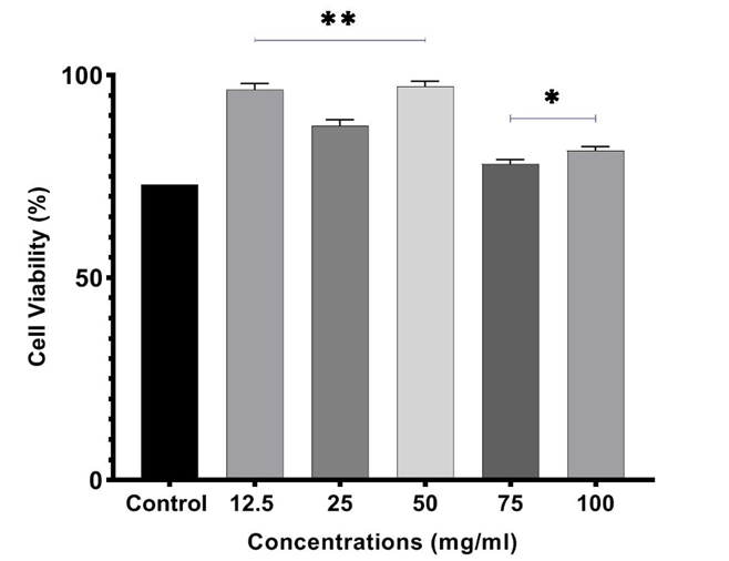
Using the MTT assay, the impact of C-PC was evaluated on the growth of HT-29 and HUVEC cells. The number of HT-29 cells gradually decreased with the rise in C-PC concentration (Figure 1). The data for the HUVEC cell line are presented in Figure 2.
The calculated value of IC50 for C-PC in HT-29 cells was 55.47 mg/mL. Moreover, the results showed a marked decrease in the colony formation in HT-29 cells after treatment with C-PC compared to that of HUVEC cells; i.e., there was no toxic effect evident on HUVEC cells. This finding points out to the effective inhibition of growth in colon cancer cells by phycocyanin.
Effects on Caspase Enzymes
In the next step, we evaluated the signaling pathways involved in apoptosis induction in caspases and p53 genes on HT-29 colon cancer cells. The result as presented in Figure 3, demonstrated that C-PC-mediated apoptosis in HT-29 cells is dependent on caspase and p53 signaling pathways. Briefly, phycocyanin can increase the expression of p53 gene and activate caspases 3, 8 and 9. The concentration of C-PC at 55.47 mg/mL prevented the growth of HT-29 cells up to 50%. This compound increases apoptosis in HT-29 cells by activating caspases and p53. The quantities of caspases and p53, as expressed in HT-29 cell line, were higher than that of the control HT-29 cells (P<0.05).


Figure 1. Assessment of HT-29 cell viability by MTT assay after 3 hr of incubation with
C-phycocyanin (12.5, 25, 50, 75 & 100 mg/ml). The cell viability of treated cells was compared with the untreated group (control) through ANOVA. * = Significant difference (P<0.05), ** = Highly significant (P<0.01**).


Figure 2. Assessments of HUVEC cell viability by MTT assay after incubation with C-phycocyanin (12.5, 25, 50, 75, and 100 mg/ml). The cell viability of treated cells was compared with the untreated group (control) through ANOVA. * = Significant difference (P<0.05), ** = Highly significant (P<0.01**).


Figure 3. Assessments of pre-apoptotic effects of C-phycocyanin in the HT-29 human colorectal cell line with qRT- PCR method.  The reactions showed increases in the expression of caspases 9, 8, 3 and p53 genes after treatment with C-phycocyanin (IC50 value). The viability of treated cells was compared with the untreated group (control) through ANOVA. ** = Highly significant difference (P<0.01).

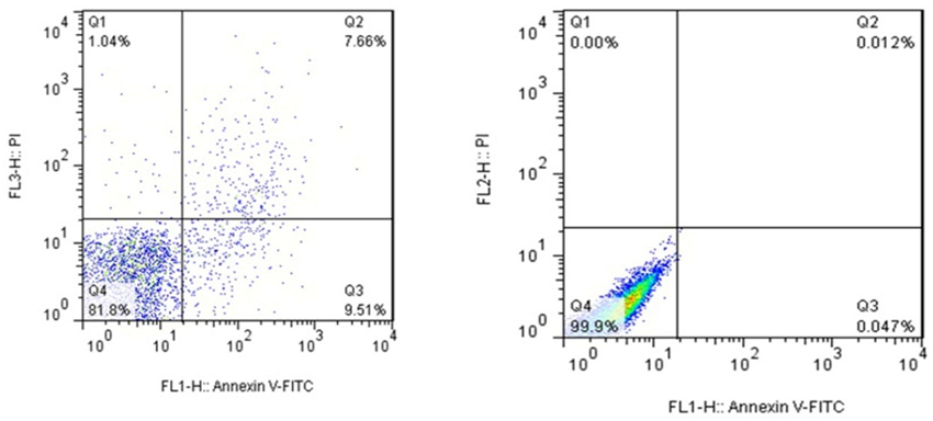
Figure 4. HT-29 cell line treated with C-phycocyanin and its pre-apoptotic effects was analyzed
by flow cytometry method.  Left Panel: HT-29 cells treated with C-phycocyanin (IC50 value). Right Panel: Control or untreated cells. Q1: Necrosis, Q2: Late apoptosis, Q3: Early apoptosis, Q4: Live cells.
The treated cells demonstrated higher levels of caspases and p53 genes. This finding indicated the up-regulation of the genes in the treated cells.
Flow Cytometry
Although C-PC has been proven to induce apoptosis in cancer cells in numerous studies [16-18], we specifically examined the effect of C-PC on the apoptosis in HT-29 cells by Annexin V. The induction of both early and late apoptosis was indicated based on the results from the C-PC-treated HT-29 cells (Figure 4).
Discussion
Colorectal cancer represents a global health challenge as the third most common malignancy. Therefore, novel therapeutic methods are urgently warranted to control this cancer. In the past decades, exploiting natural resources for cancer inhibition and treatment has become critical, and cyanobacteria hold a great potential among the essential products found in S. platensis [19].
Recently, more studies have explored the pharmacological and immunological effects of C-PC, such as photo-induced cytotoxicity [20], activation of the immune system [21], and anti-oxidative [22] and anti-inflammatory properties [23]. Besides, there is a potent anti-tumor role for C-PC in a number of in vitro and in vivo cancer cells from the blood, liver, breast, colon, and lung tissues [24-28].
Despite the encouraging results, the pharmaceutical market still lacks phycocyanin-based chemotherapy, largely due to the poorly known mechanisms of its action. In the current study, we investigated the effects of C-PC on some cellular targets and mechanisms. C-PC appears to have a key role in decreasing the expression of transcription factors, signal transducers, and pro-inflammatory cytokines. Also, this biliprotein is able to increase the expression of IL-4, a potent anti-inflammatory cytokine [29]. Further, C-PC can improve immune function by enhancing lymphocytic activities, thus increasing the body's defense against diseases. This compound is likely to become a new candidate for cancer therapy [30]. This is mainly because of the high expression of scavenger receptor-A (SR-A) expressed on the surface of tumor-associated macrophages (TAMs) and due to its affinity toward this receptor [30].
The antiproliferative and cytotoxic properties of C-PC have been shown by earlier in vitro and in vivo studies. Thangam, et al. have demonstrated the antioxidant and antiproliferative potential of C-PC against A549 and HT-29 cell lines through G0/G1 phase arrest and DNA fragmentation [5]. Also, C-PC prevents cancer cell proliferation by recruiting GAPDH from the nucleus to the plasma membrane, arresting the cell cycle at the G0/G1 phase [31, 32]. Further research has demonstrated that C-PC stops G1-phase in myeloid leukemia cells (K562), allowing them to follow the apoptotic pathway [33]. Moreover, C-PC can arrest the cells at the G2-phase and induce apoptosis in human hepatoma cell line (HepG2) [20] and human ovarian cancer cell line (SKOV-3) [7].
Phycocyanin is able to cleave polyADP-ribose polymerase 1(PARP1) and change the Bcl-2/Bax ratio by activating caspases in both apoptotic pathways.  Subhashini, et al. have shown that there is a significant decrease (49%) in proliferation, elevated apoptosis and down regulation of Bcl-2 after treatment of human chronic myelogenous leukemia cells (K562) by C-PC [24]. In another research, Ravi, et al. suggested the increased expression of p21 and decreased expression of cyclin-E in human breast cancer cell line [34]. Liao, et al. have shown that C-PC causes a stop in human pancreatic cancer cell line (PANC-1) at the G2 phase checkpoint, after which the cells undergo apoptosis. Further, these authors have demonstrated that apoptosis can be activated by phycocyanin through several pathways, such as NF-κB, PI3K/Akt/mTOR and MAPK [35].
High levels of vascular endothelial growth factor (VEGF-A), matrix metallopeptidase enzymes (MMP2 & MMP9), are the basis for metastasis, which are down regulated by C-PC through binding to VEGFR1 [29]. In addition, C-PC alters the cellular redox state and inhibits cell proliferation by targeting enzymatic and non-enzymatic antioxidants. This compound also alters the mitochondrial membrane potential by releasing cytochrome C from mitochondria during the early stages of apoptosis. The release of cytochrome C increases the production of reactive oxygen species (ROS) and ultimately activates the signaling pathways of pro-apoptosis in cancer cell lines [36, 37]. Another role for phycocyanin in cells is scavenging free radicals that prevents DNA oxidative damage in nerve cells [38].
The p53 as a tumor suppressor gene has a key role in the regulation of cell cycle, and is strictly related to tumor cell proliferation [39]. The p53 pathway plays an important role in arresting cells at the G1 and G2 checkpoints after DNA damage, thus forcing them to apoptosis [40]. Saini, et al. have shown that C-PC can stimulate the cell cycle arrest and mediate apoptosis by activating p53 gene [41]. Overall, p53 and caspase-mediated apoptosis pathways are involved in anti-cancer effects of C-PC against colon cancer HT-29 cell line. These effects were established via cellular and molecular assessments in the current study (Figures 3 & 4).
Our study showed that Phycocyanin inhibited the proliferation of tumor cells in HT-29 colerectal cancer cell line and promoted tumor cell apoptosis through p53 and caspase-mediated apoptosis pathways. Our data suggest that activation of apoptosis-p53 pathway by Phycocyanin can be a novel therapeutic candidate to overcome resistance to chemotherapy. Considering the fact that phycocyanin has not shown any cytotoxic effects on endothelial cells in-vitro, it is likely to be a promising, natural therapeutic agent for clinical use in the management of human colorectal cancer.  However, further well-designed studies are warranted on Phycocyanin.
Conflicts of Interest
The authors declare that they have no conflict of interests.
Ethical Approval
The survey was confirmed by the Ethics Committee, the University of Medical Science, Iran (Code: IR-UMS.REC.1399.545). This study did not conduct any experiments on humans or animals.
Funding
This research project was financially supported by Student Research Committee of Qazvin University of Medical Sciences.

Acknowledgments
The authors would like to appreciate Qazvin University of Medical Sciences for the generous financial support toward this study.
Authors' Contributions
HP and AP designed the study; MS, AAF, and MM performed laboratory tests and data collection. HP and IS carried out the data analyses. MM, RN and AA wrote the final draft of the manuscript. All authors reviewed and approved the final manuscript.
Type of Study: Research | Subject: Special

References
1. Rawla P, Sunkara T, Barsouk A. Epidemiology of colorectal cancer: incidence, mortality, survival, and risk factors. Prz Gastroenterol. 2019;14(2):89-103. [DOI:10.5114/pg.2018.81072] [PMID] []
2. Xi Y, Xu P. Global colorectal cancer burden in 2020 and projections to 2040. Transl Oncol. 2021;14(10):101174. [DOI:10.1016/j.tranon.2021.101174] [PMID] []
3. Jiang L, Wang Y, Yin Q, Liu G, Liu H, Huang Y, et al. Phycocyanin: A Potential Drug for Cancer Treatment. J Cancer. 2017;8(17):3416-29. [DOI:10.7150/jca.21058] [PMID] []
4. Bannu SM, Lomada D, Gulla S, Chandrasekhar T, Reddanna P, Reddy MC. Potential Therapeutic Applications of C-Phycocyanin. Curr Drug Metab. 2019;20(12):967-76. [DOI:10.2174/1389200220666191127110857] [PMID]
5. Thangam R, Suresh V, Asenath Princy W, Rajkumar M, Senthilkumar N, Gunasekaran P, et al. C-Phycocyanin from Oscillatoria tenuis exhibited an antioxidant and in vitro antiproliferative activity through induction of apoptosis and G0/G1 cell cycle arrest. Food Chem. 2013;140(1-2):262-72. [DOI:10.1016/j.foodchem.2013.02.060]
6. Hao S, Li S, Wang J, Zhao L, Yan Y, Wu T, et al. C-Phycocyanin Suppresses the In Vitro Proliferation and Migration of Non-Small-Cell Lung Cancer Cells through Reduction of RIPK1/NF-kappaB Activity. Mar Drugs. 2019;17(6). [DOI:10.3390/md17060362] [PMID]
7. Ying J, Wang J, Ji H, Lin C, Pan R, Zhou L, et al. Transcriptome analysis of phycocyanin inhibitory effects on SKOV-3 cell proliferation. Gene. 2016;585(1):58-64. [DOI:10.1016/j.gene.2016.03.023] [PMID]
8. Hao S, Li S, Wang J, Zhao L, Zhang C, Huang W, et al. Phycocyanin Reduces Proliferation of Melanoma Cells through Downregulating GRB2/ERK Signaling. J Agric Food Chem. 2018;66(41):10921-9. [DOI:10.1021/acs.jafc.8b03495] [PMID]
9. Kefayat A, Ghahremani F, Safavi A, Hajiaghababa A, Moshtaghian J. C-phycocyanin: a natural product with radiosensitizing property for enhancement of colon cancer radiation therapy efficacy through inhibition of COX-2 expression. Sci Rep. 2019;9(1):19161. [DOI:10.1038/s41598-019-55605-w] [PMID] []
10. Hanahan D, Weinberg RA. The hallmarks of cancer. Cell. 2000;100(1):57-70. [DOI:10.1016/S0092-8674(00)81683-9] [PMID]
11. Ghafelehbashi R, Farshbafnadi M, Aghdam NS, Amiri S, Salehi M, Razi S. Nanoimmunoengineering strategies in cancer diagnosis and therapy. Clin Transl Oncol. 2023;25(1):78-90. [DOI:10.1007/s12094-022-02935-3] [PMID]
12. Walczak H, Bouchon A, Stahl H, Krammer PH. Tumor necrosis factor-related apoptosis-inducing ligand retains its apoptosis-inducing capacity on Bcl-2- or Bcl-xL-overexpressing chemotherapy-resistant tumor cells. Cancer Res. 2000;60(11):3051-7.
13. Russo A, Bazan V, Iacopetta B, Kerr D, Soussi T, Gebbia N, et al. The TP53 colorectal cancer international collaborative study on the prognostic and predictive significance of p53 mutation: influence of tumor site, type of mutation, and adjuvant treatment. J Clin Oncol. 2005;23(30):7518-28. [DOI:10.1200/JCO.2005.00.471] [PMID]
14. Takayama T, Miyanishi K, Hayashi T, Sato Y, Niitsu Y. Colorectal cancer: genetics of development and metastasis. J Gastroenterol. 2006;41(3):185-92. [DOI:10.1007/s00535-006-1801-6] [PMID]
15. Radagdam S, Asoudeh-Fard A, Karimi MA, Faridvand Y, Gholinejad Z, Gerayesh Nejad S. Calcitriol modulates cholesteryl ester transfer protein (CETP) levels and lipid profile in hypercholesterolemic male rabbits: A pilot study. Int J Vitam Nutr Res. 2021;91(3-4):212-6. [DOI:10.1024/0300-9831/a000613] [PMID]
16. Salehi M, Piri H, Farasat A, Pakbin B, Gheibi N. Activation of apoptosis and G0/G1 cell cycle arrest along with inhibition of melanogenesis by humic acid and fulvic acid: BAX/BCL-2 and Tyr genes expression and evaluation of nanomechanical properties in A375 human melanoma cell line. Iran J Basic Med Sci. 2022;25(4):489-96.
17. Livak KJ, Schmittgen TD. Analysis of relative gene expression data using real-time quantitative PCR and the 2(-Delta Delta C(T)) Method. Methods. 2001;25(4):402-8. [DOI:10.1006/meth.2001.1262] [PMID]
18. Eide HA, Halvorsen AR, Bjaanaes MM, Piri H, Holm R, Solberg S, et al. The MYCN-HMGA2-CDKN2A pathway in non-small cell lung carcinoma--differences in histological subtypes. BMC Cancer. 2016;16:71. [DOI:10.1186/s12885-016-2104-9] [PMID] []
19. ElFar OA, Billa N, Lim HR, Chew KW, Cheah WY, Munawaroh HSH, et al. Advances in delivery methods of Arthrospira platensis (spirulina) for enhanced therapeutic outcomes. Bioengineered. 2022;13(6):14681-718. [DOI:10.1080/21655979.2022.2100863] [PMID]
20. Wang CY, Wang X, Wang Y, Zhou T, Bai Y, Li YC, et al. Photosensitization of phycocyanin extracted from Microcystis in human hepatocellular carcinoma cells: implication of mitochondria-dependent apoptosis. J Photochem Photobiol B. 2012;117:70-9. [DOI:10.1016/j.jphotobiol.2012.09.001] [PMID]
21. Cian RE, Lopez-Posadas R, Drago SR, de Medina FS, Martinez-Augustin O. Immunomodulatory properties of the protein fraction from Phorphyra columbina. J Agric Food Chem. 2012;60(33):8146-54. [DOI:10.1021/jf300928j] [PMID]
22. Pleonsil P, Soogarun S, Suwanwong Y. Anti-oxidant activity of holo- and apo-c-phycocyanin and their protective effects on human erythrocytes. Int J Biol Macromol. 2013;60:393-8. [DOI:10.1016/j.ijbiomac.2013.06.016] [PMID]
23. Zhu C, Ling Q, Cai Z, Wang Y, Zhang Y, Hoffmann PR, et al. Selenium-Containing Phycocyanin from Se-Enriched Spirulina platensis Reduces Inflammation in Dextran Sulfate Sodium-Induced Colitis by Inhibiting NF-kappaB Activation. J Agric Food Chem. 2016;64(24):5060-70. [DOI:10.1021/acs.jafc.6b01308] [PMID]
24. Subhashini J, Mahipal SV, Reddy MC, Mallikarjuna Reddy M, Rachamallu A, Reddanna P. Molecular mechanisms in C-Phycocyanin induced apoptosis in human chronic myeloid leukemia cell line-K562. Biochem Pharmacol. 2004;68(3):453-62. [DOI:10.1016/j.bcp.2004.02.025] [PMID]
25. Roy KR, Arunasree KM, Reddy NP, Dheeraj B, Reddy GV, Reddanna P. Alteration of mitochondrial membrane potential by Spirulina platensis C-phycocyanin induces apoptosis in the doxorubicinresistant human hepatocellular-carcinoma cell line HepG2. Biotechnol Appl Biochem. 2007;47(Pt 3):159-67. [DOI:10.1042/BA20060206] [PMID]
26. Chen T, Wong YS. In vitro antioxidant and antiproliferative activities of selenium-containing phycocyanin from selenium-enriched Spirulina platensis. J Agric Food Chem. 2008;56(12):4352-8. [DOI:10.1021/jf073399k] [PMID]
27. Lu W, Yu P, Li J. Induction of apoptosis in human colon carcinoma COLO 205 cells by the recombinant alpha subunit of C-phycocyanin. Biotechnol Lett. 2011;33(3):637-44. [DOI:10.1007/s10529-010-0464-9] [PMID]
28. Bingula R, Dupuis C, Pichon C, Berthon JY, Filaire M, Pigeon L, et al. Study of the Effects of Betaine and/or C-Phycocyanin on the Growth of Lung Cancer A549 Cells In Vitro and In Vivo. J Oncol. 2016;2016:8162952. [DOI:10.1155/2016/8162952] [PMID] []
29. Saini MK, Sanyal SN. Targeting angiogenic pathway for chemoprevention of experimental colon cancer using C-phycocyanin as cyclooxygenase-2 inhibitor. Biochem Cell Biol. 2014;92(3):206-18. [DOI:10.1139/bcb-2014-0016] [PMID]
30. Wan DH, Zheng BY, Ke MR, Duan JY, Zheng YQ, Yeh CK, et al. C-Phycocyanin as a tumour-associated macrophage-targeted photosensitiser and a vehicle of phthalocyanine for enhanced photodynamic therapy. Chem Commun (Camb). 2017;53(29):4112-5. [DOI:10.1039/C6CC09541K] [PMID]
31. Wang H, Liu Y, Gao X, Carter CL, Liu ZR. The recombinant beta subunit of C-phycocyanin inhibits cell proliferation and induces apoptosis. Cancer Lett. 2007;247(1):150-8. [DOI:10.1016/j.canlet.2006.04.002] [PMID]
32. Gupta NK, Gupta KP. Effects of C-Phycocyanin on the representative genes of tumor development in mouse skin exposed to 12-O-tetradecanoyl-phorbol-13-acetate. Environ Toxicol Pharmacol. 2012;34(3):941-8. [DOI:10.1016/j.etap.2012.08.001] [PMID]
33. Liu Y, Xu L, Cheng N, Lin L, Zhang C. Inhibitory effect of phycocyanin from Spirulina platensis on the growth of human leukemia K562 cells. Journal of Applied Phycology. 2000;12(2):125-30. [DOI:10.1023/A:1008132210772]
34. Ravi M, Tentu S, Baskar G, Rohan Prasad S, Raghavan S, Jayaprakash P, et al. Molecular mechanism of anti-cancer activity of phycocyanin in triple-negative breast cancer cells. BMC Cancer. 2015;15:768. [DOI:10.1186/s12885-015-1784-x] [PMID] []
35. Liao G, Gao B, Gao Y, Yang X, Cheng X, Ou Y. Phycocyanin Inhibits Tumorigenic Potential of Pancreatic Cancer Cells: Role of Apoptosis and Autophagy. Sci Rep. 2016;6:34564. [DOI:10.1038/srep34564] [PMID] []
36. Pardhasaradhi BV, Ali AM, Kumari AL, Reddanna P, Khar A. Phycocyanin-mediated apoptosis in AK-5 tumor cells involves down-regulation of Bcl-2 and generation of ROS. Mol Cancer Ther. 2003;2(11):1165-70.
37. Pan R, Lu R, Zhang Y, Zhu M, Zhu W, Yang R, et al. Spirulina phycocyanin induces differential protein expression and apoptosis in SKOV-3 cells. Int J Biol Macromol. 2015;81:951-9. [DOI:10.1016/j.ijbiomac.2015.09.039] [PMID]
38. Rimbau V, Camins A, Pubill D, Sureda FX, Romay C, Gonzalez R, et al. C-phycocyanin protects cerebellar granule cells from low potassium/serum deprivation-induced apoptosis. Naunyn Schmiedebergs Arch Pharmacol. 2001;364(2):96-104. [DOI:10.1007/s002100100437]
39. Bates S, Vousden KH. Mechanisms of p53-mediated apoptosis. Cell Mol Life Sci. 1999;55(1):28-37. [DOI:10.1007/s000180050267] [PMID]
40. Taylor WR, Stark GR. Regulation of the G2/M transition by p53. Oncogene. 2001;20(15):1803-15. [DOI:10.1038/sj.onc.1204252] [PMID]
41. Saini MK, Sanyal SN. Cell cycle regulation and apoptotic cell death in experimental colon carcinogenesis: intervening with cyclooxygenase-2 inhibitors. Nutr Cancer. 2015;67(4):620-36. [DOI:10.1080/01635581.2015.1015743]

Add your comments about this article : Your username or Email:
CAPTCHA

Send email to the article author


Rights and permissions
Creative Commons License This work is licensed under a Creative Commons Attribution-NonCommercial 4.0 International License.

© 2024 CC BY-NC 4.0 | Iranian Journal of Toxicology

Designed & Developed by : Yektaweb