Write your message
Volume 18, Issue 1 (January 2024)                   IJT 2024, 18(1): 6-13 | Back to browse issues page


XML Print


Download citation:
BibTeX | RIS | EndNote | Medlars | ProCite | Reference Manager | RefWorks
Send citation to:

Amedu N, Ajayi E. Impact of Sodium Metabisulfite on Oxidative Stress, Hormones, and Reproductive Tissue in Female Wistar Rats. IJT 2024; 18 (1) :6-13
URL: http://ijt.arakmu.ac.ir/article-1-1269-en.html
1- Department of Anatomy, Faculty of Basic Medical Sciences, Adeleke University, PMB 250, Ede, Osun State, Nigeria , amedunath11@gmail.com
2- Department of Anatomy, Faculty of Basic Medical Sciences, Adeleke University, PMB 250, Ede, Osun State, Nigeria
Full-Text [PDF 918 kb]   (489 Downloads)     |   Abstract (HTML)  (1540 Views)
Full-Text:   (561 Views)
Introduction
Food preservatives are substances added to food with the goal of extending the storage life by hindering the growth of microorganisms and the oxidation process [1]. Their function is to uphold the food's freshness, taste, and overall quality, allowing for prolonged shelf life. Common examples include agents that combat microbes, antioxidants, salt, sugar, and natural essences, effectively ensuring food safety and diminishing waste. Sodium metabisulfite (SMB) is used in various industries such as food, beverages, cosmetics, medicine, rubber, and photography, serving as a preservative, bleaching and antioxidant agent [2]. It carries substantial significance for preserving items like wine, beer, grapefruit juice, dried fruits, and seafood, as well as being extensively utilized in processed food manufacturing [3]. However, SMB's impact can be diverse; under certain conditions, it counteracts peroxide effects, leading to the creation of harmful oxidizing agents [4].
The effects of SMB are varied; for instance, at high doses and prolonged exposure it has been associated with diminished root growth, reduced mitotic index, and increased chromosomal abnormalities [5]. Moreover, it can induce significant detrimental effects on healthy human cells, such as causing apoptosis. The chronic use of SMB leads to changes in immune response, along with shifts in biochemical, hematological, and physiological parameters in Wistar rats [6]. Administration of SMB is linked to reduced activity of antioxidant enzymes in the blood and increased lipid peroxidation, as demonstrated by earlier studies [7, 8]. The administration of SMB also leads to a significant decline in testosterone levels, and is a crucial factor contributing to changes in the morphology, morphometry, and histology of male mice, as shown previously by Naureen, et al. [9].
The ovaries are compact, oval-shaped structures positioned on both sides of the uterus within the pelvic cavity, as described in detail by Gibson and Mahdy [10]. A layer called the ovarian cortex encases the outer surface of the ovary, housing numerous small sacs termed follicles. The ovarian follicles serve as the sites for the development and maturation of ova. The ovary's roles encompass oogenesis, hormone production, and various other functions, as described by the above-mentioned former study [10]. Uterus is a vital female reproductive organ and situated in the pelvis cavity between the bladder and rectum [11]. Comprising of the fundus, body, and cervix, it undergoes cyclical changes in response to hormonal cues. The uterus presents as a tri-layered histological structure.  The innermost layer, the endometrium, The middle layer, the myometrium, and the outermost layer, the perimetrium [11, 12].
Uterus is a vital female reproductive organ and situated in the pelvis cavity between the bladder and rectum [11]. Comprising of the fundus, body, and cervix, it undergoes cyclical changes in response to hormonal cues. The uterus presents as a tri-layered histological structure. The innermost layer, the endometrium, undergoes cyclical changes and houses the implantation site. The middle layer, the myometrium, consists of smooth muscle fibers, enabling contractions during childbirth. The outermost layer, the perimetrium, provides structural support for the uterus [11, 12]. Pathologies, such as endometriosis can disrupt the uterine function and can ultimately lead to infertility.
The primary objective of this research is to fill the knowledge gap pertaining to the impact of SMB on the hormonal balances, the structural composition of ovaries and uterus, and the oxidative stress markers in rats. This study also addresses the existing concerns about food additives, and the possibility of disturbing hormonal equilibrium. Thus, we planned to evaluate the consequences for reproductive well-being and the effects of oxidative stress effects, and the associated health consequences.
Materials and Methods
Animals
A total of 24 young female Wistar rats were obtained from Temilola animal husbandry located in Ogbomoso, Oyo State, Nigeria. The rats were placed and cared for in the animal facility at Adeleke University in Ede, Nigeria. They were housed in regular plastic rat cages with metal lids. A period of seven days was observed for the rats to acclimatize before they were sorted into four groups of six rats each, and the administration of the substances began. Throughout the acclimatization period and until the end of the experiments, the rats were under a consistent cycles of alternating light and darkness (12hr.).  Furthermore, the rats had unrestricted access to pellet food and water, enabling them to eat and drink at will.  The experimental protocol and treatments were approved by the Research Ethics Committee of Adeleke University Ede, Nigeria (00670).
Sodium Metabisulfite
Powdered form of SMB (product code 05935) at 96.5% purity, 1.48g/cm3 density, and 1901.07g/mol molecular weight, was purchased from Mayor Ade Multi-Services Nig. Enterprise at Osogbo, Nigeria.
Experimental Design
After the acclimatization period, the rats were divided randomly into four groups of six rats each. For 28 successive days, the oral treatments were given as follows:
Group 1 (control) was provided with 0.5 mL of distilled solution.
Group 2 was given SMB at 100 mg/kg of body weight.
Group 3 was given SMB at 300 mg/kg of body weight, and
Group 4 was administered SMB at 500 mg/kg of body weight.
Tissue Collection
After the completion of substance administration, a procedure known as thoracotomy was performed on each rat. This involved extracting blood from the heart's apex using a 5mL needle and syringe. The collected blood was then transferred into a sample bottle and subsequently subjected to centrifugation at 4000 revolutions per minute for duration of 5 minutes, utilizing a centrifuge (Model 80-2, Gallenkomp, UK). The resulting serum was promptly utilized for hormonal analysis. For rats were assigned for histopathological examinations. These rats were anesthetized by intraperitoneal injection of ketamine at 50mg/mL. Subsequently, the rats were euthanized through decapitation, and the ovaries and uteri of each rat were carefully removed and thoroughly rinsed in normal saline. Following this step, the harvested tissues were fixed in Bouin's fluid.
Hormonal Tests
Follicle Stimulating Hormone: The FSH test was performed, using an FSH kit (AccuBind ELISA Microwell FSH test system; Code #: 425-300). To analyze the serum samples, the microplates were arranged in pairs, with each well designated for serum references, calibrators, controls, or patient specimen. A volume of 50µl of the suitable sample was transferred into the designated well, and then 100µl of FSH-Enzyme reagent solution was added.  The microplates were gently swirled, covered, and incubated at room temperature (20-25ºC) for 60 minutes. Upon discarding the contents, 350µl of wash buffer was introduced into each well, and this process repeated three times to accomplish three wash cycles. Subsequently, 100µl of the working substrate solution was added to each well in the predetermined sequence. The absorbance of the solutions from the microplates were read and documented. The outcomes were expressed in units of milli-international units per milliliter (mlU/mL).
Luteinizing Hormone: To conduct the LH test, the AccuBind ELISA microwell LH test was performed on the system kit (Code #: 625-300). To perform the procedure, the microplates were arranged and 50µL of either serum references, calibrators, controls, or patient specimens were added to the designated wells. Subsequently, the microplates were incubated at room temperature for one hour. Following the incubation, the microplates' contents were removed, and the wells were washed three times using the wash buffer. Subsequently, 100µL of the working substrate solution was added to all wells, and the absorbance was read for each well. Next, a dose-response curve was plotted for each pair of duplicate serum references against their corresponding LH concentration in mIU/mL on a linear graph paper. The curve that best fitted the data was drawn and used to determine the LH concentration of an unknown sample. This was done by identifying the average absorbance of the duplicate measurements for each unknown well on the vertical axis of the graph. By locating the point where the value intersected the curve, the LH concentration was read on the horizontal axis of the graph, and the outcome was presented in mIU/mL.
Histopathological Analyses
Upon fixation, the ovarian and uterine tissue samples went through a processing phase and were embedded in paraffin wax blocks. The blocks were subsequently sliced in sections and stained using hematoxylin and eosin and periodic acid Schiff methods as described by Bancroft and Layton [13]. The stained sections were then examined under light microscopy (Olympus, XSZ-107BN, and Amscope, MD500, USA).
Malondialdehyde Concentration Assay
The MDA concentration was assessed on tissue samples that were homogenized in a 1:10 ratio with a 1.15% (w/v) cold KCl solution, using thiobarbituric acid (TBA) method. The two compounds (MDA & TBA) interact to generate a pink-coloured solution, the highest absorbance of which observed at 532 nm. The findings were quantified and reported as nmol per milligram protein.
Statistical Analyses
The data analyses were performed using GraphPad Prism software, version 7. Statistical comparisons were carried out by employing one-way analysis of variance (ANOVA) coupled with Tukey's multiple comparison test. All results were expressed in Means ±SD, with statistical significant difference set at P<0.05.
Results
Effect of SMB on FSH and LH
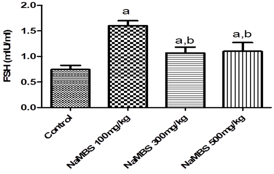
The FSH concentration, as shown in Figure 1, was significantly higher in the groups subjected to SMB treatment at 100mg/kg, 300mg/kg, and 500mg/kg compared to that of the control group (P˂0.05).  Also, there was a significant rise in the FSH levels in the group treated with SMB at 100mg/kg compared to both of the groups treated with SMB at 300mg/kg or 500mg/kg (P˂0.05).

Figure 1. The levels of follicle stimulating hormone (FSH) in Wistar rats.
NaMBS = Sodium metabisulphite; a P<0.05 vs control; b P<0.05 vs NaMBS (100mg/kg); n=6.
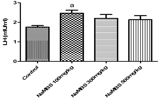
The LH concentration, as depicted in Figure 2, was significantly higher in the groups that received SMB at 100mg/kg and 300mg/kg, compared to that of the control group.  However, there was no significant difference observed in the LH level of the group treated with SMB at 500mg/kg compared to that of the control group (P>0.05).

Figure 2. The levels of luteinizing hormone (LH) in Wistar rats.
NaMBS = Sodium metabisulphite; a P<0.05 vs control; n=6.

Impact of SMB on the Histopathology
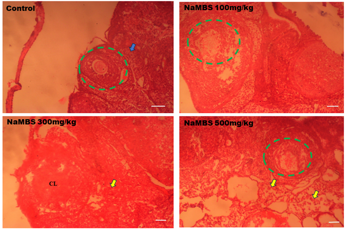
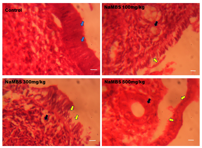
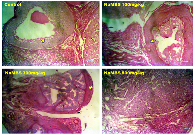
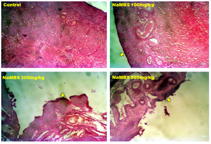
The photomicrograph shown in Figure 3 illustrates the rats’ ovary sections stained with H&E. In the rats that were administered SMB at 300mg/kg or 500mg/kg, the outer layer of the ovaries appeared disorganized and degenerated (yellow arrows). The disruption also affected the follicles within the ovarian cortex. Conversely in the control group, the ovarian cortex appeared well-organized (blue arrows) with many ovarian follicles present. In Figure 4, the image presents the rats uterine sections stained with H&E. In the groups treated with SMB at 300mg/kg or 500mg/kg, both the uterine epithelia and glands were degenerated (yellow & black arrows).
Figure 5 represents sections of the rat ovaries subjected to PAS staining. Both the controls and the group treated with SMB at 100mg/kg exhibited a strong PAS presence in the zona pellucida of the ovarian follicles and the theca interna cells. However, in the groups treated with SMB at 300mg/kg or 500mg/kg, the PAS was distributed throughout the ovarian cortex and the epithelia.  The photomicrographs in Figure 6 illustrate sections of the rat uteri subjected to PAS staining. The PAS staining was detected in the endometrial epithelia in all groups. Nevertheless, in the groups treated with SMB at 300mg/kg or 500mg/kg, the intensity of PAS staining was more intense that that in the control group.

Figure 3. Representative photomicrographs of H&E stain in rat’s ovary.
NaMBS = Sodium metabisulphite; Yellow arrows = Degenerating cortex;
Blue arrow = Properly arranged cortical cells. Green ring = Ovarian follicles. Scale bar =180 µm.


Figure 4. Representative photomicrographs of H & E stain in rats’ uterus.
NaMBS = Sodium metabisulphite; Blue arrow = Normal epithelial lining, No degeneration;
Yellow arrows = Degeneration of endometrial epithelium; Black arrows = Necrosis in glands. Scale bar = 45 µm.

Figure 5. Representative photomicrographs of PAS stain in rat’s ovary.
NaMBS = Sodium metabisulphite; Yellow arrows = Location of PAS expression. Scale bar =180 µm.


Figure 6. Representative photomicrographs of PAS stain in rat’s uterus.
NaMBS = Sodium metabisulphite; Yellow arrows = Location of PAS expression. Scale bar = 180 µm.
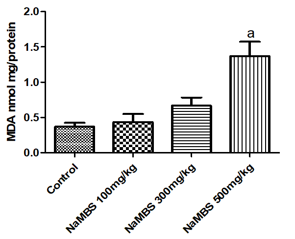
Figure 7. Malondialdehyde (MDA) level in rats’ ovaries.
Data expressed as mean ± SD. NaMBS = Sodium metabisulfite. a P< 0.05 vs Control group.

Effect of SMB on Oxidative Stress
The MDA concentration, as depicted in Figure 7, exhibited a significant increase in the group that received SMB at 500mg/kg as opposed to that of the control group. However, the MDA levels in the groups treated with SMB at 100mg/kg or 300mg/kg did not show a significant difference compared to that of the control group.
Discussion
In this study, the FSH levels exhibited a marked increase in the cohorts exposed to SMB at 100mg/kg, 300mg/kg, or 500mg/kg compared to those of the control group. The observed FSH increase in response to SMB may suggest potential disruptions in the regulatory mechanisms of the hypothalamic-pituitary-gonadal axis. This hormone is a key to the regulation of reproductive processes, such as follicular development in females and spermatogenesis in males [14, 15].
The dose-response relationship between SMB and FSH may suggest that SMB at higher doses could interfere with the feedback loop that controls FSH secretion. Therefore, it is plausible that SMB at higher doses might affect the hypothalamus or the sensitivity of anterior pituitary gland to the negative feedback from the gonads, leading to an elevated release of FSH. Another potential explanation might involve oxidative stress. The SMB compound has been associated with changes in antioxidant enzyme activities and increased lipid peroxidation [8]. Oxidative stress can impact the endocrine system, including the regulation of hormonal synthesis and release [16]. In this context, it is likely that the oxidative stress induced by SMB could influence the production and release of FSH [17].
The LH levels, as illustrated in Figure 2, exhibited a marked increase in the groups treated with SMB at 100mg/kg or 300mg/kg, as compared to that of the control group. However, there was no significant difference in the LH levels between the group treated with SMB at 500mg/kg and the control group. The observed increase in LH levels in response to SMB treatment at 100mg/kg or 300mg/kg could indicate differential interactions between SMB and the hypothalamic-pituitary-gonadal axis. This hormone is critical in triggering ovulation in female and stimulating testosterone production in male rats [18].
Another interpretation of the study’s findings could involve the disruption of the negative feedback that regulates the LH secretion [19]. In this context, SMB might have influenced the hypothalamus or pituitary gland response to the feedback signals from the gonads, leading to a rise in the LH release.  The lack of a significant difference in the LH levels at 500mg/kg SMB might indicate a saturation point beyond which the effects of this compound on LH secretion would be limited. It's conceivable that the dose-response relationship is not linear, and there could be a threshold effect beyond which additional SMB does not further influence the LH levels.
In the rat groups subjected to SMB at 300mg/kg or 500mg/kg, the outer layer of the ovaries exhibited disorganization and signs of cellular degeneration. The disruption also affected numerous follicles on the ovarian cortices.  In contrast, the ovarian cortices in the control group appeared well-structured, with a variety of observable follicles present. Also, in the groups treated with SMB at doses of 300mg/kg or 500mg/kg, both the uterine epithelia and the glands displayed degeneration. These findings might suggest that SMB could be disrupting the normal processes of follicular development and ovulation. This disruption could potentially interfere with the release of mature ova and the ovarian function.
The impact of SMB on the ovarian cortex and follicles may suggest damages due to oxidative stress, since SMB has been linked to alterations in antioxidant activities and increased lipid peroxidation [20]. The degeneration observed in the uterine epithelia and glands in the SMB-treated groups might imply the adverse effect this compound has on the uterine health, which is crucial for successful implantation and pregnancy in animal model and potentially in humans [21]. The degeneration of the uterine lining could hinder the proper attachment of a fertilized ovum and reduce the chance of a successful pregnancy [22].
The primary effect of PAS staining for histological examinations is on glycogen, mucins, and other substances abundant in tissue carbohydrates [23]. Glycogen is an important carbohydrate storage molecule [24], and its presence within ovarian follicles and theca interna cells is indicative of its role in providing energy for various cellular processes [24, 25]. The findings of this study provided evidence that both the controls and the group treated with SMB at 100mg/kg exhibited strong PAS staining in the zona pellucida of the ovarian follicles and theca interna cells. However, in the groups treated with SMB at 300mg/kg or 500mg/kg, the PAS staining was distributed throughout the ovarian cortices and epithelia. Furthermore, in the rat uteri, the PAS staining was observed in the endometrial epithelia in all rat groups. However, in the groups treated with SMB at 300mg/kg or 500mg/kg, the intensity of PAS staining was more intense than that of the control group.
The altered distribution of glycogen in the ovarian tissues of the groups treated with SMB at 300mg/kg or 500mg/kg was suggestive of disruptions in the glycogen regulation and metabolism. The wide distribution of PAS stain in the ovarian cortices and epithelia in these groups might imply that SMB, at high doses affects the processes that control glycogen synthesis, storage and/or utilization. This could be due to the SMB-induced changes in the hormonal levels, metabolic pathways, or cellular signal transduction [26]. The increased intensity of PAS stain in the endometrial epithelia of the groups treated with SMB at doses of 300mg/kg or 500mg/kg may suggest enhancement in the carbohydrate storage or utilization. This event might be linked to the altered metabolic activities in the uterine lining via the SMB effects on the hormonal levels or tissue physiology. The exact mechanisms behind these events would require further investigations into the mechanism by which SMB affects glycogen metabolism in the ovarian and uterine tissues.
Malondialdehyde is a marker of lipid peroxidation, a process that occurs when free radicals react with lipids in cell membranes, leading to cellular damages [27, 28]. The concentration of MDA increased significantly in the rat group treated with SMB at 500mg/kg compared to the controls. However, the MDA levels in the groups treated at 100mg/kg or 300mg/kg SMB were significantly different compared to the controls. The increased MDA level in the group treated at 500mg/kg SMB could be due to the enhanced level of oxidative stress. 
The significant rise in MDA levels suggests that SMB at high concentration (500mg/kg) induces greater oxidative stress, resulting in increased lipid peroxidation [20]. Conversely, the lack of significant differences in the MDA levels between the groups treated with SMB at either 100mg/kg or 300mg/kg, compared to the controls, may imply that these doses do not induce a substantial rise in oxidative stress or lipid peroxidation. It's possible that SMB at lower doses may not cause enough oxidative damage to be detected by the MDA assay. The dose-dependent effect on MDA levels could suggest that SMB might have a threshold, above which its impact on oxidative stress becomes pronounced. Factors such as the duration of exposure, the metabolic capacity of the subjects, and the cumulative effect of the substance could all contribute to the observed differences in MDA levels.
Conclusions
This study explored the effects of SMB on the hormonal levels, ovarian and uterine histological alterations, and oxidative stress markers in rats. The SMB administration resulted in significant alterations in FSH and LH concentrations, and impacted the ovarian and uterine histology. The presence of SMB was associated with disrupted histological architecture and increased oxidative stress, indicating potential adverse effects on the reproductive health in rat model. Further investigations are warranted to fully understand the cytotoxic effects of SMB exposure. For instance, additional research is recommended to uncover how SMB influences glycogen-related processes in ovaries and uterine tissues at varying doses. Furthermore, understanding the precise mechanisms underlying the SMB's impact on FSH levels is also warranted.
Acknowledgements
The authors wish to express their gratitude to Mr. Habeeb Abdul-Rahman, Mr. Dan Ajibade, and Mr. Ezra Lambe for their valuable technical assistance provided toward this study.
Ethical Approval
The study protocol and treatment procedures were approved by the Research Ethics Committee of Adeleke University Ede, Osun State, Nigeria (Approval #: 00670).
Conflicts of Interest
The authors declare no competing interest with any internal or external entities in conducting this study.
Authors' Contributions
NOA: participated in the design and interpretation of the study, data analysis, and the review of the manuscript. NOA and EA: conducted the experiments, collected the tissue samples and was responsible for the data analyses. NOA, and EA: performed the histopathological analyses. All authors contributed fairly equally to the writing of the drafts of the manuscript, reviewed, read, and approved the final article.
Funding
The study did not receive financial support from any external funding agency apart from the personal resources of the authors.
Availability of Datasets
The datasets used in this study are available from the corresponding author on reasonable request.
Type of Study: Research | Subject: General

References
1. Silva M, Lidon F. Food preservatives - An overview on applications and side effects. Emirates Journal of Food and Agriculture. 2016;28(6). [DOI:10.9755/ejfa.2016-04-351]
2. Noorafshan A, Asadi-Golshan R, Monjezi S, Karbalay-Doust S. Sodium metabisulphite, a preservative agent, decreases the heart capillary volume and length, and curcumin, the main component of Curcuma longa, cannot protect it. Folia Biol (Praha). 2014;60(6):275-80.
3. Sadowska B, Sztormowska M, Gawinowska M, Chelminska M. Sodium metabisulfite hypersensitivity in urticaria. Our Dermatology Online. 2021;12(2):106-12. [DOI:10.7241/ourd.20212.2]
4. Lavoie JC, Lachance C, Chessex P. Antiperoxide activity of sodium metabisulfite. A double-edged sword. Biochem Pharmacol. 1994;47(5):871-6. [DOI:10.1016/0006-2952(94)90487-1] [PMID]
5. Mishra L, Yadav B. Assessing the Cytotoxic and Genotoxic Effects of Sodium Metabisulphite: A Food Preservative. Vegetos. 2011;24(1):32-7.
6. Kadi FZE, Bénali AI, Bénali M, Belbraouet S. Effect of Sodium Metabisulphite on Blood Metabolic Status of Wistar Rats. Food and Nutrition Sciences. 2014;05(15):1529-37. [DOI:10.4236/fns.2014.515165]
7. Ercan S, Ozturk N, Celik-Ozenci C, Gungor NE, Yargicoglu P. Sodium metabisulfite induces lipid peroxidation and apoptosis in rat gastric tissue. Toxicol Ind Health. 2010;26(7):425-31. [DOI:10.1177/0748233710369665] [PMID]
8. Shekarforoush S, Ebrahimi P, Fathabad AA, Farzanfar E. Effect of Sodium Metabisulfite on Oxidative Stress and Lipid Peroxidation Biomarkers. Current Nutrition & Food Science. 2020;16(1):114-7. [DOI:10.2174/1573401314666181024130333]
9. Naureen I, Saleem A, Nawaz A, Ghaffar I, Maqbool I. Study The Harmful Effects Of Sodium Metabisulfite On Liver, Testes, And Lipid Peroxidation In Male Mice, (Mus Musculus). International Journal of Engineering Applied Sciences and Technology. 2021;6(5). [DOI:10.33564/IJEAST.2021.v06i05.004]
10. Gibson EK, Mahdy HM. Anatomy, Abdomen and Pelvis, Ovary: StatPearls Publishing; 2023.
11. Zangos S, Marquart F. Female Reproductive System. In: Vogl T, Reith W, Rummeny E, editors. Diagnostic and Interventional Radiology2016. p. 929-55. [DOI:10.1007/978-3-662-44037-7_32]
12. Gasner A, P AA. Physiology, Uterus. StatPearls. Treasure Island (FL) ineligible companies. Disclosure: Aatsha P A declares no relevant financial relationships with ineligible companies.2024.
13. Bancroft JD, Layton C. The hematoxylins and eosin. Bancroft's Theory and Practice of Histological Techniques. 8th ed2019. p. 126-38. [DOI:10.1016/B978-0-7020-6864-5.00010-4]
14. Padmanabhan V, Karsch FJ, Lee JS. Hypothalamic, pituitary and gonadal regulation of FSH. Reprod Suppl. 2002;59:67-82.
15. Das N, Kumar TR. Molecular regulation of follicle-stimulating hormone synthesis, secretion and action. J Mol Endocrinol. 2018;60(3):R131-R55. [DOI:10.1530/JME-17-0308] [PMID] []
16. Chainy GBN, Sahoo DK. Hormones and oxidative stress: an overview. Free Radic Res. 2020;54(1):1-26. [DOI:10.1080/10715762.2019.1702656] [PMID]
17. Shen M, Jiang Y, Guan Z, Cao Y, Sun SC, Liu H. FSH protects mouse granulosa cells from oxidative damage by repressing mitophagy. Sci Rep. 2016;6:38090. [DOI:10.1038/srep38090] [PMID] []
18. Nedresky D, Singh G. Physiology, Luteinizing Hormone. StatPearls. Treasure Island (FL) ineligible companies. Disclosure: Gurdeep Singh declares no relevant financial relationships with ineligible companies.2024.
19. Lincoln GA, Fraser HM. Negative feedback regulation of pulsatile LH secretion during treatment with an LHRH antagonist in rams. J Androl. 1990;11(3):287-92. [DOI:10.1002/j.1939-4640.1990.tb03242.x]
20. Rezaee N, Nematollahi Z, Shekarforous S, Hoseini E. Effect of Sodium Metabisulfite on Rat Ovary and Lipid Peroxidation. Iranian Journal of Toxicology. 2016;10(2):23-8. [DOI:10.32598/IJT.10.2.316.1]
21. Cha J, Sun X, Dey SK. Mechanisms of implantation: strategies for successful pregnancy. Nat Med. 2012;18(12):1754-67. [DOI:10.1038/nm.3012] [PMID] []
22. Vannuccini S, Clifton VL, Fraser IS, Taylor HS, Critchley H, Giudice LC, et al. Infertility and reproductive disorders: impact of hormonal and inflammatory mechanisms on pregnancy outcome. Hum Reprod Update. 2016;22(1):104-15. [DOI:10.1093/humupd/dmv044] [PMID] []
23. Tabatabaei Shafiei M, Carvajal Gonczi CM, Rahman MS, East A, Francois J, Darlington PJ. Detecting glycogen in peripheral blood mononuclear cells with periodic acid schiff staining. J Vis Exp. 2014(94). [DOI:10.3791/52199] [PMID] []
24. Roach PJ, Depaoli-Roach AA, Hurley TD, Tagliabracci VS. Glycogen and its metabolism: some new developments and old themes. Biochem J. 2012;441(3):763-87. [DOI:10.1042/BJ20111416] [PMID] []
25. Daghlas SA, Mohiuddin SS. Biochemistry, Glycogen. StatPearls. Treasure Island (FL) ineligible companies. Disclosure: Shamim Mohiuddin declares no relevant financial relationships with ineligible companies.2024.
26. Zhang Q, Bai Y, Yang Z, Tian J, Meng Z. The molecular mechanisms of sodium metabisulfite on the expression of K ATP and L-Ca2+ channels in rat hearts. Regul Toxicol Pharmacol. 2015;72(3):440-6. [DOI:10.1016/j.yrtph.2015.05.021] [PMID]
27. Ayala A, Munoz MF, Arguelles S. Lipid peroxidation: production, metabolism, and signaling mechanisms of malondialdehyde and 4-hydroxy-2-nonenal. Oxid Med Cell Longev. 2014;2014:360438. [DOI:10.1155/2014/360438] [PMID] []
28. Amedu NO, Omotoso GO. Influence of Vitexin on ataxia-like condition initiated by lead exposure in mice. Toxicology and Environmental Health Sciences. 2020;12(4):305-13. [DOI:10.1007/s13530-020-00041-x]

Add your comments about this article : Your username or Email:
CAPTCHA

Send email to the article author


Rights and permissions
Creative Commons License This work is licensed under a Creative Commons Attribution-NonCommercial 4.0 International License.

© 2025 CC BY-NC 4.0 | Iranian Journal of Toxicology

Designed & Developed by : Yektaweb