Write your message
Volume 18, Issue 2 (May 2024)                   IJT 2024, 18(2): 99-105 | Back to browse issues page

Ethics code: no.8; 30/01/2011


XML Print


Download citation:
BibTeX | RIS | EndNote | Medlars | ProCite | Reference Manager | RefWorks
Send citation to:

saleh M, Al-Masri A, Ezzedin D. Cytotoxic and Genotoxic Effects of Lambda-Cyhalothrin Insecticide on Human Dental Pulp Stem Cells. IJT 2024; 18 (2) :99-105
URL: http://ijt.arakmu.ac.ir/article-1-1335-en.html
1- National Commission for Biotechnology, Department of Biomedical and Animal, Syria , manalcapno@gmail.com
2- National Commission for Biotechnology, Department of Biomedical and Animal, Syria
3- Department of Plant Protection, Faculty of Agriculture, Damascus University, Damascus, Syria
Full-Text [PDF 650 kb]   (149 Downloads)     |   Abstract (HTML)  (414 Views)
Full-Text:   (117 Views)

Introduction

Due to the harmful effects of pesticides on the genetic material of living organisms, it is essential to study and analyze their hazards to human health, including their cytotoxic and genotoxic properties [1]. Such investigations are crucial to preventing the mutagenic and carcinogenic effects of pesticides [2]. Genetic damages can occur in human DNA, such as strand breaks and DNA adducts, or at the level of chromosomes, such as a change in the chromosome number (aneuploidy), deletion, and/or breaks (clastogenicity) [3, 4]. Chromosomal aberrations and DNA damage are considered the primary events that lead to the carcinogenicity and/or mutagenicity of numerous chemicals [5]. The genotoxicity of many pesticides has been studied through in vitro and in vivo experiments. It has been found that most organophosphates, organochlorine, and synthetic pyrethroid pesticides have caused concerns in the development of human cancer. In recent years, 56 pesticides from various groups have been classified as carcinogens [6], with 29 other pesticides being genetically toxic [5].
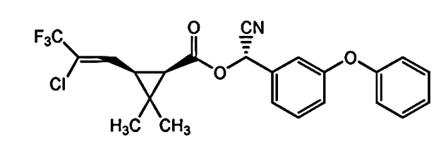
Because of the restrictions and warnings on the use of organophosphate insecticides, pyrethroid pesticides, such as Lambda-cyhalothrin (LCT), have been widely used by farmers for pest control [7]. This compound belongs to synthetic pyrethroid groups, which are analogues to natural pyrethrins and are extracted from dried flowers of Chrysanthemum cinerariaefolium plants [8]. However, the latter agents are more toxic to insects and mammals than the synthetic pyrethrins [9]. Synthetic pyrethroid insecticides, including LCT, are widely used for pest control in agriculture and public health. This compound is a synthetic pyrethroid type-II insecticide (Figure 1) containing an alpha-cyano group [10], which is categorized as a class D carcialbumin contents in the fish kidneys and liver and causes endocrine disruptions [14, 15]. Genotoxic studies on LCT have also shown increases in micronucleus formation in bone marrow cells, the intestinal epithelia of rats [16-18], and the blood cells of Gambusia affinis fish [19]. Moreover, LCT is thought to induce DNA damage in murine macrophage cell lines [7], chromosomal aberrations in rabbits’ lymphocytes [20], and the gill cells of Mystusgulio fish [21]. In an earlier study [22], LCT was also found to be genotoxic to an insect cell line (Sf-9).


Figure 1. Chemical structure of lambda-cyhalothrin.

Most previous studies have focused on the genotoxicity of LCT in animal models. However, few investigations have been conducted in vitro on the cytotoxicity and genotoxicity of LCT in human models. These studies have shown that LCT causes cell death, inhibits cell division, forms micronuclei, and leads to DNA damage in human lymphocytes. This agent also induces oxidative stress in human erythrocytes [23]. In this context, scientific insight is lacking on the effects of LCT on humans, and the available in vitro data are limited to peripheral blood lymphocytes. Therefore, it is necessary to conduct further studies to assess the impact of LCT on cells derived from other human tissues.     
Aim of the Study: The aim of this study was to examine the in vitro cytotoxic and genotoxic effects of LCT insecticide on human cells. To this end, dental pulp stem cells (DPSCs) were chosen as the in vitro model for human cells because they are undifferentiated natural cells that are capable of renewing themselves and can be continuously cultured in their undifferentiated state [24].

Materials and Methods

Chemicals: LCT (RS)-α-cyano-3-phenoxybenzyl (1R)-cis-3-(Z)-(2-chloro-3, 3, 3-trifluorop1-enyl)-2,2-dimethylcyclopropanecarboxylate, with a purity of 98.7%, was obtained from Syngenta©. A stock solution of 10 mM LCT was prepared using dimethyl sulfoxide (DMSO) freshly made before cell treatment.
Cell Culture: In the current study, DPSCs represent healthy and normal cells with a typical fibroblast-like morphology (Figure 2). They express several biomarkers, including the mesenchymal and bone-marrow stem cell markers, STRO-1, CD146, and the embryonic stem cell marker, OCT4 [25]. The DPSCs were isolated from the tooth pulp of a 17-year-old patient [26] and cultured in T25 culture flacks (TPP). These cells were grown in low-glucose Dulbecco’s modified eagle medium (DMEM). The culture medium was supplemented with 10% fetal bovine serum (FBS, 200 mM), L-glutamine (100 U/10 mg), antibiotics (Streptomycin and Penicillin), and 0.25 µg/ml antifungals (Amphotericin B). The culture dishes were kept in a CO2 incubator (Lab Tech-Lco-065 AI) at 37ºC with 5% CO2. The chemicals used for cell culture were purchased from Euroclone©, except for the DMEM medium, which was purchased from Sigma-Aldrich© (St. Gallen, Switzerland). The first and third subculture passages of DPSCs were used for the purpose of cytotoxicity and genotoxicity tests.

Figure 2. (A) Primary culture of DPSCs during the first week, and (B) Culture of DPSCs at 75% confluence.
Cytotoxicity Tests: The cytotoxicity of LCT was assessed by an MTT assay, as described by an earlier study [27]. Cells were seeded at a concentration of 1×10⁵ cells/ml in the DMEM culture medium without phenol red or FBS. A total of 100 μl of the cell suspension was added to each well on 96-well culture plates (TPP, Switzerland). The culture plates were then incubated overnight in a CO2 incubator at 37ºC. The medium was replaced the next day with the test medium at a concentration of 0.5, 1, 2.5,5, 10, 25, or 50 µM of LCT for 24 h. Cell suspensions with 1% DMSO were used as the negative control. Each of the concentrations tested consisted of four replicates, and these tests were repeated in duplicate.
Following the exposure period, the test medium was replaced with 20 µl MTT (2 mg/ml) in PBS and incubated overnight. Next, an aliquot of DMSO (150 µl) was added to each well to solubilize purple formazan crystals. The absorbance of the solution in each well was measured at 540 nm using a microplate reader (SCO, Germany). Regression analyses were also carried out to determine the concentration-response relationships. The inhibition rate (IR) was calculated by the following formula [28]: (1-At/Ac)×100=IR, where At=Absorbance value of the tested wells and Ac=Absorbance value of the control wells.
Alkaline Comet Assay: This assay was performed using the Comet Assay® silver staining kit catalog #4251-050-K (Trevigen, Minneapolis, USA) and Alkaline Comet Assay® following the manufacturer’s instructions with minimal modifications. Briefly, the DPSCs were treated in a 6-well culture plate, then harvested and embedded in low-melting agarose (at 37°C) at a ratio of 1:10. After mixing the sample, a 50 μL aliquot was pipetted onto the Comet Slide™ area immediately. The cells exposed to ultraviolet-C (UVC, 257.3 nm) for 45 min were used as positive controls. The cells treated with DMSO alone were considered the negative controls. To prevent additional damage, all of the steps described above were carried out under dim light. Slides were then transferred to 40 ml pre-chilled lysis solution (cat #: 4250-050-01) containing 10% DMSO and incubated overnight at 4°C. The comet slides™ were immersed in an alkali unwinding solution at pH 13, 300 mM NaOH, and 1 mM EDTA in the dark at room temperature for 30 min.
Electrophoresis was performed at 1 Volt/cm and 300 mA for 30 min. The slides were then immersed twice in distilled water (dH2O) for 5 min and then in 70% ethanol for 5 min. These samples were dried at 37°C for 10-15 min and stained using the silver staining method. The stain intensity was observed under light microscopy at 100× magnification. The reaction was terminated when the comets were easily visible by covering samples with 100 μl 5% acetic acid for 15 min and rinsed in dH2O. The comets were analyzed by visually scoring them, as described in a previous study [29]. The DNA damage was calculated by arbitrary units (AU) according to an earlier method [30], based on the following formula:

AU=0 × N0 + 1 × N1 + 2 × N2 + 3 × N3 + 4 × N4N°  comets analyzed × 100
Where N0, N1, etc. are the numbers of comets in the respective categories. For each treatment, two slides were prepared from two independent experiments.
Statistical Analyses: The statistical analyses were performed using the Mann-Whitney U test in SPSS software (version 17). Error bars represent standard deviations. The results were considered statistically significant at P<0.05.

Results

Cytotoxicity: Figure 3 demonstrates that only the 1 µM concentration of LCT induced a slight inhibition of cell proliferation (about 5%), while at other concentrations (0.5, 2.5, 5, 10, 25, and 50 µM), this compound was non-toxic to cells. These concentrations increased cell proliferation rates by 10, 1, 4, 20, 59, and 76%, respectively. The correlation coefficient was strong at R=0.983. Four concentrations were selected from the cytotoxicity curve of LCT for the genotoxicity study. These were the lowest (0.5 µM) and the two highest concentrations (25 and 50 µM) that increased cell proliferation rates, and the 1 µM concentration that inhibited cell viability.
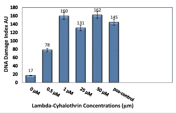
Genotoxicity: Based on the visual scoring method [29], the comets were classified into five different categories, from zero (no tail) to four (almost all DNA in the tail), depending on the tail length and the amount of DNA present in it (Figure 4). Varying degrees of DNA damages were observed by the comet assay (0 to 4) for human stem cells isolated from the tooth pulp. These cells had been exposed to varying concentrations of LCT at 0.5, 1, 25, or 50 µM, in addition to those observed in the negative and positive controls (see Figure 5). As represented by Figure 6, LCT caused DNA damage in DPSCs at all tested concentrations. Based on these tests, DNA damage indices were 78, 160, 131, and 162 AU, corresponding to the LCT concentrations at 0.5, 1, 25, and 50 µM, respectively, as derived from the cytotoxicity curves. The DNA damage indices were significant at P<0.05 for all concentrations compared to those of the negative controls (17 AU). However, they were not significant compared to the positive controls (145 AU). The highest value representing DNA damage was 162 AU at 50 µM LCT.


Figure 4. Silver-stained comet images of human DPSCs classified into five
categories as: Class 0 (undamaged cells) and classes 1, 2, 3, and 4 (damaged cells).

Figure 5. Different degrees of DNA damage from 0 to 4 in human tooth pulp cells treated with different concentrations of lambda-cyhalothrin at 200× magnification
Figure 6. DNA damage induced by Lambda-cyhalothrin in DPSCs expressed in arbitrary units (AU) in the comet assay. Data are means of values for repeated experiments±standard deviations. A statistically significant increase (P<0.05) was determined by comparing the values of DNA damage induced by various concentrations of Lambda-cyhalothrin with those of the negative controls (with 10% DMSO).

Discussion

Pyrethroid insecticides target the central nervous system in insects and other non-target organisms. The principal mechanism of action of these compounds is to interact with voltage-gated sodium channels in the neurons’ cell membrane. They cause neuronal hyper-excitability in the insects’ central nervous system [31]. In addition to their neurotoxicity, these insecticides also affect the endocrine system, exhibiting estrogenic potentials [15], genotoxic effects [31], and oxidative stress [32].
In this study, the cytotoxicity and genotoxicity of LCT insecticides were examined on human DPSCs using MTT and comet assays. The MTT assay results demonstrated that LCT inhibited cell proliferation at 1 µM concentration (~ 5%), while at other concentrations (0.5, 2.5, 5, 10, 25, and 50 µM), this agent increased cell proliferation rate by 10, 1, 4, 20, 59, and 76%, respectively.
Previous studies [13, 33] have found that LCT has been highly toxic to lymphocytes in culture, completely inhibiting cell division and inducing cell death at significantly higher concentrations. However, in vitro results on cells may vary due to differences in the cell types used, the regulation of cell growth, and different cell responses to the toxic effects of various insecticides. The LCT’s stimulating effect on DPSCs’ proliferation may be due to its estrogenic property. Several studies have suggested that the estrogenic potential of LCT [15, 34-37] is due to its stimulating effects on the proliferation of such cells as BG-1 ovarian cancer cells [15] and MCF-7 human breast carcinoma cell line [35]. These studies indicate that LCT possesses estrogenic properties, which may be the reason behind its xenoestrogenic function based on a mechanism similar to that of estradiol [35]. Estrogens are hormones that are important in sexual and reproductive development and bone formation [38, 39]. Estrogens and estrogen-like molecules can modify the biology of several cell types, including MSCs [40]. They regulate the proliferation, differentiation [41], apoptosis, and metabolism of various cell types [42].
In the present study, we used DPSCs as an in vitro human cell model. These cells, which are similar to adult stem cells, have self-renewing potential and multiple differentiation functions and play important roles in the regeneration of dental tissue [38]. Dental cells express estrogen receptors [43], and LCT, by binding to them, increases their proliferation rate, except at a 1 µM concentration. This observation explains the fact that LCT did not affect estrogen receptors at a 1 µM concentration. Furthermore, it is likely that the response of DPSCs to LCT is achieved by cell cycle arrest or cell death via apoptosis, thereby not allowing their continuous proliferation.
In the current study, the genotoxicity of LCT was evident by the alkaline comet assay on DPSCs. The results demonstrated that this insecticide was toxic to the DNA in DPSCs at all concentrations used. This finding was consistent with those reported previously by numerous in vitro and in vivo studies that applied this compound to other organisms [7, 13, 17-19, 22, 33]. The DNA damage indices were evaluated at LCT concentrations of 0.5, 1, 25, and 50 µM using the cytotoxicity curves, which resulted in the AU values of 78, 160, 131, and 162, respectively. Among these concentrations, the 1 µM concentration inhibited cell proliferation, while other concentrations promoted proliferation. In this study, the cells responded to genetic damage by activating specific pathways, resulting in the repair of damaged DNA, activation of checkpoints, and inhibition of the cell cycle. If this repair process does not succeed, the cells are likely to go through programmed cell death, that is, apoptosis, thereby preventing possible mutations [44, 45].
The exact genotoxic mechanism of LCT is currently unknown. However, according to previous studies [7, 16, 23], the possible mechanism could be the induction of oxidative stress by raising the formation of reactive oxygen species (ROS), thereby disrupting the balance between ROS generation and antioxidant defense capability. Further, LCT may also cause damage to cell membrane lipids and proteins [32]. Based on the results of this study, we observed that LCT caused DNA damage in DPSCs while still continuing to proliferate at the tested concentrations. This suggests that LCT may have another operational mechanism that mimics the effects of estrogen.
According to other studies [46, 47], one mechanism by which estrogens promote the proliferation of breast cancer is through inducing DNA damage. Additionally, estrogen alters the DNA damage response (DDR) and repair by regulating proteins that are key effectors. It has been hypothesized that estrogen receptor signaling converges to suppress effective DNA repair and apoptosis in favor of proliferation [47]. Further studies are warranted to discover if the estrogen-mimicking effect of LCT can cause DNA damage and the repair pathways similarly to those induced by estrogen.
Conclusions
Based on the findings of this study, LCT caused DNA damage in human DPSCs but did not induce cytotoxic effects on the cells at the examined concentrations, except for the 1 μM one. Instead, LCT caused increases in cell proliferation. This suggests that LCT may have an additional mechanism of action that mimics the effects of estrogen, and thus it may function as a potential xenoestrogen.
Conflict of Interests
The authors declare no conflict of interest.
Funding
This study was supported by the National Commission for Biotechnology and did not receive any other grants.
Acknowledgement
The authors would like to thank Dr. Ismael Saleh for his help in statistical analysis and Mrs. Banan al-Sheikh and Mrs. Inas Nemer for their assistance provided to this research project.
Compliance with Ethical Guidelines
Human stem cells protocol was reviewed and approved by the Ethics Committee of the Dental Collage of Damascus University (Approval #: 8; 30/01/2011).
Authors' Contributions
Manal Saleh performed the experiments, designed the study, analyzed the data, wrote the manuscript, and reviewed and approved the manuscript.
Aroub Al-Masri supervised and analyzed the data and reviewed and approved the manuscript. Daas Ezzedin supervised, reviewed, and approved the manuscript.
Type of Study: Research | Subject: Special

References
1. Bajpayee M, Pandey AK, Parmar D, Dhawan A. Current status of short-term tests for evaluation of genotoxicity, mutagenicity, and carcinogenicity of environmental chemicals and NCEs. Toxicol Mech Methods. 2005;1;15(3):155-80. [doi:10.1080/15376520590945667]
2. Nagy K, Rácz G, Matsumoto T, Ádány R, Ádám B. Evaluation of the genotoxicity of the pyrethroid insecticide phenothrin. Mutat Res Genet Toxicol Environ Mutagen. 2014 ;1;770:1-5. [doi:10.1016/j.mrgentox.2014.05.001]
3. Mostafalou S, Abdollahi M. Pesticides and human chronic diseases: evidences, mechanisms, and perspectives. Toxicol Appl Pharmacol. 2013;268(2):157-77. [doi:10.1016/j.taap.2013.01.025] [pmid: 23402800]
4. Abdelmigid HM. New trends in genotoxicity testing of herbal medicinal plants. New insights into toxicity and drug testing. InTech. 2013:89-120. [doi.org/10.5772/54858]
5. Mohanty G, Mohanty J, Dipta S, Dutt SK. Genotoxicity Testing in Pesticide Safety Evaluation. Pesticides in the Modern World - Pests Control and Pesticides Exposure and Toxicity Assessment. InTech. 2011. [doi:10.5772/18726]
6. Soloneski S, L. M. Genetic Toxicological Profile of Carbofuran and Pirimicarb Carbamic Insecticides . Insecticides - Pest Engineering. In Tech; 2012. [doi:10.5772/30137]
7. Zhang Q, Wang C, Sun L, Li L, Zhao M. Cytotoxicity of lambda-cyhalothrin on the macrophage cell line RAW 264.7. J Environ Sci (China). 2010; 1;22(3):428-32. [doi:10.1016/S1001-0742(09)60125-X] [pmid: 20614786]
8. Ibrahim HM. Evaluation of the immunotoxic effects of sub-chronic doses of lambda-cyhalothrin in murine model. MOJ. Immunol. 2016;3(6):1-8. [doi:10.15406/moji.2016.03.00108]
9. Aziz KB and Abdel Rahem HM. Lambda, the pyrethroid insecticide as a mutagenic agent in both somatic and germ cells. J Am Sci. 2010;6:317-26. [Link]
10. Chakravarthi K, Naravaneni R, and Philip GH. Study of cypermethrin cytogenesis effects on human lymphocytes using in-vitro techniques. J Appl Sci Environ Manage. 2007;11(2):77-81. [doi:10.4314/jasem.v11i2.54994]
11. U.S. EPA. U.S. Environmental Protection Agency.2012. Office of Pesticide Programs: Chemicals Evaluated for Carcinogenic Potential. Annual Cancer Report: 1-29. [Link]
12. Liu W, Gan J, Schlenk D, Jury WA. Enantioselectivity in environmental safety of current chiral insecticides. The PNAS. 2005;18;102(3):701-06. [doi:10.1073/pnas.0408847102] [pmid: 15632216]
13. Muranli FD. Genotoxic and cytotoxic evaluation of pyrethroid insecticides λ-cyhalothrin and α-cypermethrin on human blood lymphocyte culture. Bull Environ ContamToxicol. 2013;90:357-363. [doi:10.1007/s00128-012-0909-z] [pmid: 23229297]
14. Inyang IR, Obidiozo OZ, Izah SC. Effects of Lambda cyhalothrin in protein and Albumin content in the kidney and liver of Parpohiocephalus obscurus. EC PharmacolToxicol. 2016;2(3):148-53. [Link]
15. Kim CW, Go RE, Choi KC. Treatment of BG-1 ovarian cancer cells expressing estrogen receptors with lambda-cyhalothrin and cypermethrin caused a partial estrogenicity via an estrogen receptor-dependent pathway. Toxicol Res. 2015;31(4):331-7. [doi:10.5487/TR.2015.31.4.331]
16. Fetoui H, Feki A, Salah GB, Kamoun H, Fakhfakh F, Gdoura R. Exposure to lambda-cyhalothrin, a synthetic pyrethroid, increases reactive oxygen species production and induces genotoxicity in rat peripheral blood. Toxicol Ind Health.2015;31(5):433-41. [doi:10.1177/0748233716635003] [pmid: 23406951]
17. Çelik A, Mazmanci B, Çamlica Y, Çömelekoğlu Ü, Aşkin A. Evaluation of cytogenetic effects of lambda-cyhalothrin on Wistar rat bone marrow by gavage administration. Ecotoxicol Environ Sa. 2005a ;1;61(1):128-33. [doi:10.1016/j.ecoenv.2004.07.009]
18. Çelik A, Mazmanci B, Camlica Y, Aşkin A, Çömelekoğlu Ü. Induction of micronuclei by lambda-cyhalothrin in Wistar rat bone marrow and gut epithelial cells. Mutagenesis. 2005b;1;20(2):125-29. [doi:10.1093/mutage/gei020]
19. Muranli FD, Güner U. Induction of micronuclei and nuclear abnormalities in erythrocytes of mosquito fish (Gambusia affinis) following exposure to the pyrethroid insecticide lambda-cyhalothrin. Mutat Res. 2011; 24;726(2):104-8. [doi:10.1016/j.mrgentox.2011.05.004] [pmid: 21620996]
20. Georgieva SV. Investigation of the cytogenetic effect of the insecticide karate on rabbit peripheral blood lymphocytes. Trakia J Sci. 2006;4(2):34-38. [Link]
21. Velmurugan B, Ambrose T, Selvanayagam M. Genotoxic evaluation of lambda-cyhalothrin in Mystusgulio. J Environ Biol. 2006;27(2):247-50. [Link] [Link]
22. Saleh M, Ezz-din D, Al-Masri A. In vitro genotoxicity study of the lambda-cyhalothrin insecticide on Sf9 insect cells line using Comet assay.JJBS. 2021;1;14(2): 213-17. [doi:10.54319/jjbs/140203]
23. Deeba F, Raza I, Muhammad N, Rahman H, ur Rehman Z, Azizullah A, Khattak B, Ullah F, Daud MK. Chlorpyrifos and lambda cyhalothrin-induced oxidative stress in human erythrocytes: in vitro studies. Toxicol Health. 2017; 33(4):297-307. [doi:10.1177/0748233716635003] [pmid: 27102427]
24. Davila JC, Cezar GG, Thiede M, Strom S, Miki T, Trosko J. Use and application of stem cells in toxicology. Toxicol Sci. 2004;1;79(2):214-23. [doi:10.1093/toxsci/kfh100]
25. Chalisserry EP, Nam SY, Park SH, Anil S. Therapeutic potential of dental stem cells. J Tissue Eng. 2017;20;8:1-17. [doi:10.1177/2041731417702531]
26. Hatab TA, Kochaji N, Issa N, Nadra R, Saleh M, Rahmo A, Rekab M. In vivo and immunohisto-chemical study of dentin and pulp tissue regeneration in the root canal. J Chem Pharm Res. 2015;7(5):302-10. [Link]
27. Borenfreund E, Babich H, Martin-Alguacil N. Comparisons of two in vitro cytotoxicity assays - the neutral red (NR) and tetrazolium MTT tests. Toxicol In Vitro.1988;2(1):1-6. [doi:10.1016/0887-2333(88)90030-6] [pmid: 20702351]
28. Liu JW, Cai MX, Xin Y, Wu QS, Ma J, Yang P, Xie HY, Huang DS. Parthenolide induces proliferation inhibition and apoptosis of pancreatic cancer cells in vitro.J Exp Clin Cancer Res. 2010; 29(1):1-7. [doi:10.1186/1756-9966-29-108] [pmid: 20698986]
29. Collins AR. The comet assay for DNA damage and repair: principles, applications, and limitations. Mol Biotechnol.2004;26(3):249-61. [doi:10.1385/MB:26:3:249] [doi:10.1385/MB:26:3:249]
30. García O, Romero I, González JE, Moreno DL, Cuétara E, Rivero Y, Gutiérrez A, Pérez CL, Álvarez A, Carnesolta D, Guevara I. Visual estimation of the percentage of DNA in the tail in the comet assay: Evaluation of different approaches in an intercomparison exercise. Mutation Research/Genetic Toxicology and Environmental Mutagenesis. 2011;720(1-2):14-21. [doi:10.1016/j.mrgentox.2010.11.011]
31. Kakko I, Toimela T, Tähti H. Oestradiol potentiates the effects of certain pyrethroid compounds in the MCF7 human breast carcinoma cell line. ATLA. 2004;32(4):383-90. [doi:10.1289/ehp.99107173] [doi:10.1289/ehp.99107173]
32. Tukhtaev K, Tulemetov S, Zokirova N, Tukhtaev N. Effect of long term exposure of low doses of lambda-cyhalothrin on the level of lipid peroxidation and antioxidant enzymes of the pregnant rats and their offspring. J Res Health Sci. 2012;13:93-9. [doi:10.15208/mhsj.2012.67]
33. Naravaneni R, Jamil K. Evaluation of cytogenetic effects of lambda‐cyhalothrin on human lymphocytes. J Biochem Mol Toxic. 2005;19(5):304-10. [doi:10.1002/jbt.20095] [pmid:16292750]
34. Wang Q, Xia X, Deng X, Li N, Wu D, Zhang L, et al. Lambda-cyhalothrin disrupts the up-regulation effect of 17β-estradiol on post-synaptic density 95 protein expression via estrogen receptor α-dependent Akt pathway. J Environ Sci. 2016; 1;41:252-60. [doi:10.1016/j.jes.2015.04.037] [pmid: 26969072]
35. Zhao M, Zhang Y, Liu W, Xu C, Wang L, Gan J. Estrogenic activity of lambda‐cyhalothrin in the MCF‐7 human breast carcinoma cell line. Environ Toxicol Chem.2008;27(5):1194-200. [doi:10.1897/07-482.1] [pmid: 18419197]
36. Du G, Shen O, Sun H, Fei J, Lu C, Song L, Xia Y, Wang S, Wang X. Assessing hormone receptor activities of pyrethroid insecticides and their metabolites in reporter gene assays. Toxicol Sci. 2010;116(1):58-66. [doi:10.1093/toxsci/kfq120] [pmid: 20410157]
37. Brander SM, Gabler MK, Fowler NL, Connon RE, Schlenk D. Pyrethroid pesticides as endocrine disruptors: molecular mechanisms in vertebrates with a focus on fishes. Environ Sci Technol.2016;50(17):8977-92. [doi:10.1021/acs.est.6b02253]
38. Wang Y, Yan M, Yu Y, Wu J, Yu J, Fan Z. Estrogen deficiency inhibits the odonto/osteogenic differentiation of dental pulp stem cells via activation of the NF-κB pathway. Cell Tissue Res. 2013;352(3):551-59. [doi:10.1091/mbc.e09-01-0085]
39. Ling-Ling E, Xu WH, Liu Y, Cai DQ, Wen Nand Zheng WJ, Estrogen enhances the bone regeneration potential of periodontal ligament stem cells derived from osteoporotic rats and seeded on nano-hydroxyapatite/collagen/poly (L-lactide). Int J Mol Med. 2016;37(6):1475-86. [doi:10.3892/ijmm.2016.2559] [pmid: 27082697]
40. Hong L, Zhang G, Sultana H, Yu Y, Wei Z. The effects of 17-β estradiol on enhancing proliferation of human bone marrow mesenchymal stromal cells in vitro. Stem cells Dev. 2011;20(5):925-31. [doi:10.1089/scd.2010.0125] [pmid: 20735179]
41. Bustamante-Barrientos FA, Méndez-Ruette M, Ortloff A, Luz-Crawford P, Rivera FJ, Figueroa CD, et al. The impact of estrogen and estrogen-like molecules in neurogenesis and neurodegeneration: beneficial or harmful? Front Cell Neurosci.2021;15:636176. [doi:10.3389/fncel.2021.636176] [pmid: 33762910]
42. Froushani SM. The effect of mesenchymal stem cells pulsed with 17 beta-estradiol in an ameliorating rat model of ulcerative colitis. Zahedan J Res Med Sci.2019; 31;21(4). [doi:10.1159/000445583]
43. Kuechler EC, de Lara RM, Omori MA, Schroeder A, Teodoro VB, Baratto-Filho F, et al. Estrogen deficiency affects tooth formation and gene expression in the odontogenic region of female rats. Ann Anat. 2021;236:151702. [doi:10.1016/j.aanat.2021.151702] [pmid:33607226]
44. Pedram A, Razandi M, Evinger AJ, Lee E, Levin ER. Estrogen inhibits ATR signaling to cell cycle checkpoints and DNA repair. Mol Biol cell. 2009;15;20(14):3374-89. [doi:10.1091/mbc.e09-01-0085] [pmid: 19477925]
45. Remington SE. Cellular response to DNA damage after exposure to organophosphates in vitro (Doctoral dissertation, Newcastle University). 2010. [Link]
46. Jiménez-Salazar JE, Damian-Ferrara R, Arteaga M, Batina N, Damián-Matsumura P. Non-Genomic actions of estrogens on the DNA repair pathways are associated with chemotherapy resistance in breast cancer. Front Oncol 2021;11:631007. [doi:10.3389/fonc.2021.631007]
47. Caldon CE. Estrogen signaling and the DNA damage response in hormone dependent breast cancers. Front Oncol. 2014;14;4:106. [doi:10.3389/fonc.2014.00106] [pmid: 24860786]

Add your comments about this article : Your username or Email:
CAPTCHA

Send email to the article author


Rights and permissions
Creative Commons License This work is licensed under a Creative Commons Attribution-NonCommercial 4.0 International License.

© 2025 CC BY-NC 4.0 | Iranian Journal of Toxicology

Designed & Developed by : Yektaweb