Write your message
Volume 17, Issue 4 (October 2023)                   IJT 2023, 17(4): 61-69 | Back to browse issues page


XML Print


Download citation:
BibTeX | RIS | EndNote | Medlars | ProCite | Reference Manager | RefWorks
Send citation to:

John P, Bhatia N, Kale P, Doshi G. Benefit of Betaine in Isoniazid-Rifampicin-Induced Hepatotoxicity in Rats. IJT 2023; 17 (4) :61-69
URL: http://ijt.arakmu.ac.ir/article-1-1241-en.html
1- Department of Pharmacology, SVKM’S Dr. Bhanuben Nanavati College of Pharmacy, Mumbai, 400 056, India
2- Department of Pharmacology, SVKM’S Dr. Bhanuben Nanavati College of Pharmacy, Mumbai, 400 056, India , gaurav.pharmacology@gmail.com
Full-Text [PDF 1422 kb]   (540 Downloads)     |   Abstract (HTML)  (1546 Views)
Full-Text:   (373 Views)
Introduction
 
The toxicity induced by the long-term use of drugs is a serious side effect that significantly contributes to the high drug development cost [1]. Paracelsus stated that all agents are safe at sufficiently low doses and can be toxic at inappropriate high doses [2]. However, we normally consider adverse events when they occur at doses that are relevant to patients’ use of medicines and not accidental drug overdoses. Almost all anti-tuberculosis drugs are associated with hepatotoxicity. Such condition is primarily attributed to the oxidative stress induced by hepatic CYP2E1 during isoniazid (INH) metabolism [3]. Hepatic CYP2E1 is upregulated by the clinical use of INH and rifampicin (RMP), which often leads to increased levels of inflammatory mediators in the liver [4]. This in turn leads to the generation of toxic metabolites and free radicals, which results in ROS generation, mitochondrial injury and glutathione (GSH) down-regulation.
Betaine is a choline metabolite, a naturally-occurring glycine derivative, and an important component of the methionine-homocysteine cycle [5]. Betaine has previously been implicated in GSH upregulation in the liver [6], and has shown many pharmacological properties, including antioxidant, anti-inflammatory, hepatoprotective, anti-thrombotic, anti-depressants, and neuroprotective activities [5]. Betaine ameliorates INH-RMP-induced hepatotoxicity through two pathways. Being a methyl group donor, it is involved in the generation of methionine from homocysteine. In turn, methionine contributes to cysteine synthesis via a trans-sulfuration pathway. It is also involved in glycine synthesis. Both cysteine and glycine are the key components essential for the GSH synthesis [7]. Hence, betaine treatment can promote the upregulation of hepatic GSH process, which leads to the inhibition of ROS formation and alleviation of hepatic injury.
Aim of the Study: Although studies have demonstrated the cytoprotective and hepatoprotective effects of betaine [8-10], the underlying biochemical mechanisms and the full effects of betaine are still unclear. Thus, the current study aimed to assess the hepatoprotective eect of betaine and to explore the related mechanisms in a rat model of drug-induced toxicity. The toxicity was evaluated to determine the protective effects of betaine at three different doses: 125, 250 and 500 mg/kg.
Materials and Methods
Chemicals & Drugs: All chemicals and drugs were obtained from local suppliers in Mumbai, India. Rifampicin was obtained from Lupin Pharmaceuticals, Inc. Isoniazid (pyridine-4-carbohydrazide) and Betaine (2-[trimethylazaniumyl] acetate) was purchased from SRL Chemicals, and TCI Chemicals Pvt. Ltd., respectively. Silymarin (3,5,7-trihydroxy-2-[3-(4-hydroxy-3-methoxyphenyl)-2-(hydroxymethyl)-2,3-dihydro-1,4-benzodioxin-6-yl]-2,3-dihydrochromen-4-one) was purchased from Yucca Enterprises.
Animals: Forty eight healthy Wistar rats (180-200g) were obtained from the National Institute of Biosciences (Mumbai, India). Food and water were provided to the rats ad libitium. Before initiating the experiments, the rats underwent one week of acclimatization. The study protocol was approved by the Institutional Animal Ethics Committee (IAEC-Approval #: CPCSEA/IAEC/P-43/2018) and registered by the “Committee for the Purpose of Control and Supervision of Experiments on Laboratory Animals” (CPCSEA), sponsored by the Government of India.
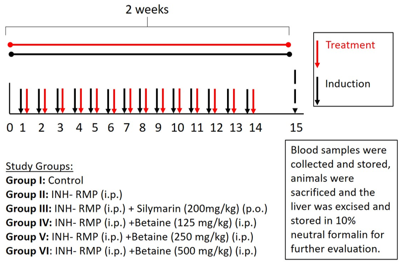
Experimental Design & Protocol: The rats (n=48) were randomly divided into six groups of eight each, as shown in Figure 1. The animals in group I were kept in laboratory environment but did not receive any drugs. Rats in group II received only INH and RMP intraperitoneally, while those in group III were pretreated orally with silymarin (200mg/kg). Rats in groups IV, V, and VI were pretreated with betaine intraperitoneally for 14 days (125 mg/kg, 250 mg/kg, and 500 mg/kg). On day 15, blood samples were collected from all rats and stored for later analyses. At this time, all rats were sacrificed; the liver was removed from each animal, and stored in 10% formalin at pH 7.4 for further evaluations and analyses.
Betaine Administration Schedule: All of the rats were subjected to a standardized 14-day treatment protocol per group. Betaine was administered to rats in groups IV to VI, 4-5 hours before injecting combined INH-RMP to determine the hepatoprotective activity of betaine for 14 consecutive days. The rats were treated with three doses of betaine at 125, 250, or 500mg/kg, to determine its effective dosage [6]. The INH and RMP were administered through the intraperitoneal route for 14 days at a therapeutic dose of 50 or 100mg/kg, respectively [11, 12].
Estimation of Liver Indices and Serum Analyses: About 18-20 hours post-treatment, blood samples were collected from the rats in all groups. The sera were prepared from the blood samples after centrifugation at 3,000 rpm for 15 min. On the 15th day, the rats’ livers were weighted and their samples examined histologically. In addition, the liver indices were determined, using the following formula: liver weight (g)/body weight (g) x100%. The liver samples were further used for the determination of serum biochemical parameters through liver function tests. These tests quantified such parameters as serum glutamic oxaloacetic transaminase (SGOT), lactic acid dehydrogenase (LDH), alkaline phosphatase (ALP), serum glutamic pyruvic transaminase (SGPT), and the total protein levels, using commercial kits; all assays were performed on a biochemical analyzer (ERBA Chem, India) [3, 7].
Determination of TNF-α & IL-1β Levels: The liver samples were rinsed in phosphate-buffered saline (PBS) at pH 7.4, and stored at -80°C before homogenization. The tissues were minced and homogenized in PBS at pH7.4 in a tissue homogenizer (Polytron, India) under ice-cold conditions. The homogenate samples were centrifuged at 2000-3000 rpm for approximately 20 min (Eltrec, India) and the supernatants were used for the various assays. The subsequent steps were conducted as described in the manual of the GENLISA™ ELISA kits by Krishgen Biosystems (Mumbai, India), using sandwich ELISA technique. Monoclonal antibodies were pre-coated onto microwells. 100µl of test samples and standards were pipetted into each microwell. Further, we covered the plates with a sealer and incubated them at 37°C for 90 minutes. This step was followed by aspiration and washing the plate four times with diluted Buffer and blotting the residual buffer by firmly tapping the plates upside down on absorbent papers.
We then pipetted 100µl biotinylated IL-1β antibody working solution into all wells, covered each plate with a sealer, and incubated them at 37°C for 60 minutes. The plates were aspirated and washed again as discussed above. We then pipetted the working solution of  streptavidin and conjugate (100µl, v/v) into all wells and mixed them well, and covered each plate with a sealer and incubated them at 37°C for 30 minutes. Finally, aspiration and washing the plates were done as described earlier. Next, we pipetted 90µl TMB (3,3′,5,5′-Tetramethylbenzidine) substrate into all wells and checked the color development. Lastly, the plates were incubated at 37°C for 10 minutes. Then, we pipetted 50µl of stop solution into all wells. The solutions turned from blue to yellow, and the absorbencies were read at 450nm on a microplate reader within 10-15 minutes after adding the stop solution to each well [11].
Enzymatic & Nonenzymatic Estimations: In this part of the study, we estimated the levels of glutathione, catalase, and superoxide dismutase activities, and the extent of lipid peroxidation. For this purpose, liver samples were homogenized in a solution containing 1.15% potassium chloride and 50mM Tris-HCL buffer at pH 7.4. The homogenates were centrifuged at 10,000 rpm at minus 4°C for 20 min. The supernatants were used to assess the levels of reduced glutathione (GSH), the catalase (CAT) activities, and superoxide dismutase (SOD), and the extent of lipid peroxidation (MDA) [13].
Assessment of Reduced Glutathione: The GSH level was assessed as described previously by Beutler [14] [14]. One ml of the supernatant was mixed with 2 ml phosphate buffer and 0.5 ml Ellman’s reagent (10 mM). The resultant solution developed a yellow color, the absorbance of which was read at 412 nm (Shimadzu UV-1800). The GSH level of the test samples was evaluated using a series of standards, and expressed in μmol/mg protein [13].
Assessment of Catalase Activity: The CAT activity was assessed as described previously by Chance and Oshino [15]. Two μl of the supernatant were mixed with 191μl phosphate buffer, the absorbance was read, and then 7 μl hydrogen peroxide was added to the mixture. The absorbance of the mixture was read for a second time at 240 nm on a Shimadzu UV-1800 unit. The results were expressed as CAT activity per mg protein [13, 15].
Assessment of Lipid Peroxidation: The extent of lipid peroxidation is measureable based on the malondialdehyde (MDA) levels as described previously by Ohkawa, et al. [16]. The mixture contained 0.5 ml 24% TCA, phosphate buffer (0.1 M, pH 8.0), and the supernatant. It was incubated at room temperature for 10 min and then centrifuged for 20 min at 2000 rpm. One ml of the supernatant was mixed with 0.25 ml 0.33% TBA in 20% acetic acid and was incubated at 95°C for 1hr, and then the absorbance was read at 532 nm [12, 16].
Assessment of Superoxide Dismutase: The activity of superoxide dismutase (SOD) was assessed as described previously by Misra and Fridovich [17]. A 200µl aliquot of the homogenate was mixed with 30 mM EDTA, 300 μl 2 mM pyrogallol, and 2.5 ml phosphate buffer. The change in the absorbance of the mixture was read on a spectrophotometer at 420 nm.  One unit of SOD activity is known to inhibit pyrogallol auto-oxidation rate by 50%, and the activity was expressed as μg/mg protein [13, 17].
Histopathological Examinations: The rat liver samples were extracted, washed, rinsed with 0.9% saline, and stored in 10% formalin in the fridge. Then, the sectioned slides were stained with hematoxylin and eosin (H&E) for further histopathological examinations [11, 12].
Statistical Analyses: The statistical evaluations of the data were performed, using GraphPad Prism software designed for 32bit MS Windows. All statistical comparisons were made using one-way analysis of variance (ANOVA) and Tukey’s post-hoc tests. The data were represented as the means ± the standard deviations. The statistical significance was set at P<0.05, P<0.01, and P<0.001, based on the various comparisons between the pairs and among the entire groups.


Graphical Abstract. Benefit of Betaine in Isoniazid-Rifampicin (INH-RMP)-induced hepatotoxicity in rats


Figure 1. Experimental protocol for Isoniazid-Rifampicin (INH-RMP)-induced hepatotoxicity.


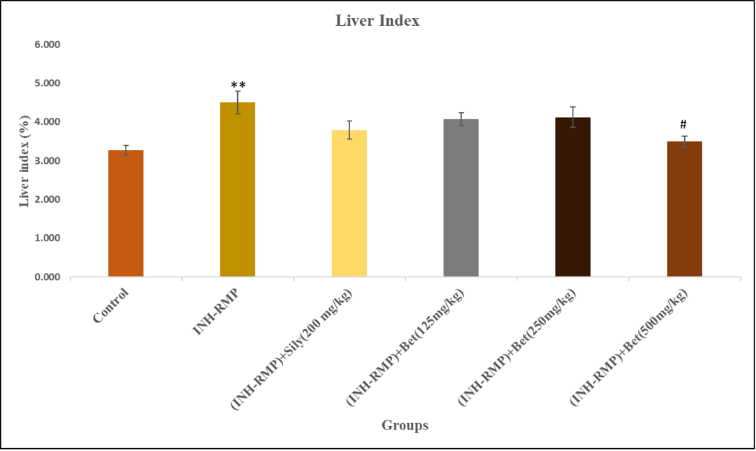
Figure 2. Betaine treatment on the liver index of INH-RMP-intoxicated animals after 14 days of treatment.


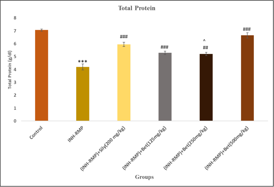
Figure 3. Betaine treatment on the Protein levels of INH-RMP-intoxicated animals after 14 days of treatment.


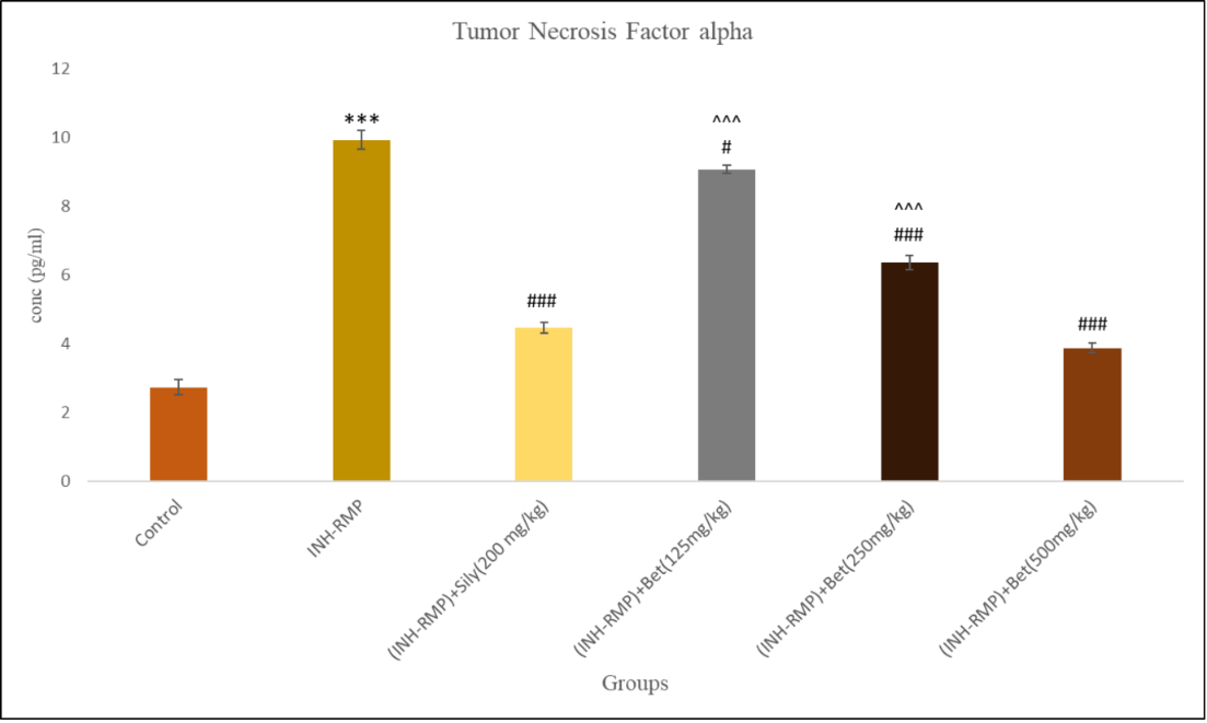
Figure 4. Betaine treatment on the tumor necrosis factor-α (TNF-α) levels of INH-RMP-intoxicated animals after 14 days of treatment.


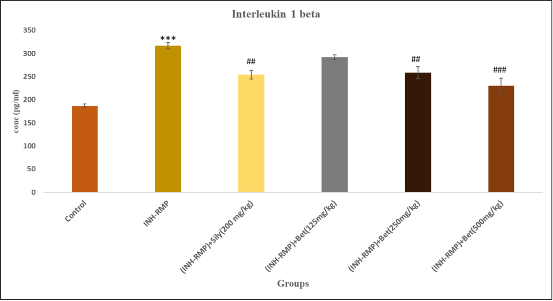
Figure 5. Betaine treatment on the interleukin-1β (IL-1β) levels of INH-RMP-intoxicated animals after 14 days of treatment.


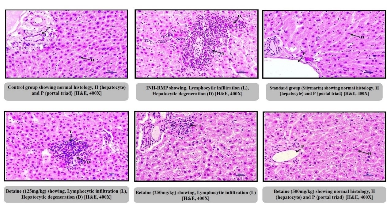
Figure 6. Histopathological studies in Isoniazid-Rifampicin (INH-RMP)-induced toxicity.

Table 1. Estimation of SGPT, SGOT, ALP, and LDH levels of INH-RMP-intoxicated animals after 14 days of treatment.
Serial No. Group (U/L)
SGPT SGOT ALP LDH
1. Control 44.38 ±1.793 116.9±4.989 111.3±5.009 301.6±16.45
2. INH-RMP 105.1± 6.263*** 202.9±8.356*** 401.7±19.28*** 1224±156.2***
3. INH-RMP +Silymarin (200mg/kg) 65.18± 1.892### 160.4±4.029### 148.8±7.558### 570.6±38.36###
4. INH-RMP +Betaine (125 mg/kg) 97.70 ± 2.893^^^ 193.5±5.188^^ 263.9±10.88^^^ ## 921.6±21.30^^#
5. INH-RMP +Betaine (250 mg/kg) 72.52± 1.215## 159.9±6.088### 202.2±5.406^^ 769.9±28.25###
6. INH-RMP +Betaine (500 mg/kg) 56.90± 3.275### 149.2±5.962### 151.1±6.010### 518.9±26.25###
Data are presented as Means ± SEM (n=8).  ***Indicates significantly different when compared with the control group (P<0.001). ### Indicates significantly different when compared with INH-RMP group (P<0.001).
^^^ Indicates significantly different as compared with Silymarin group (P<0.001).

Table 2. Estimation of GSH, MDA, SOD, and CAT levels of INH-RMP-intoxicated animals after 14 days of treatment.
Serial No. Group GSH (umol/mg protein) MDA (nmol/mg protein) SOD (U/mg protein) CAT (U/mg protein)
1 Control 26.90±1.424 2.895±0.08136 206.3±5.175 43.40±1.543
2 INH-RMP 14.34± 0.1939*** 5.451±0.169*** 147±5.471*** 21.32±1.059***
3 INH-RMP +Silymarin (200mg/kg) 22.04± 0.4757### 3.912±0.2765### 169.7±7.297# 28.17±1.352#
4 INH-RMP +Betaine (125mg/kg) 17.52± 0.3149## 4.730±0.1548^ 151.3±4.566 18.80±1.020^^
5 INH-RMP +Betaine (250mg/kg) 18.94± 0.6851### 4.238±0.1586### 170±2.978# 25.93±1.485
6 INH-RMP +Betaine (500mg/kg) 22.54± 0.6354### 3.662±0.1927 ### 174.5±3.801## 35.91±2.310^###
Data are presented as Means ± SEM (n=8).  ***Indicates significantly different when compared with the control group (P<0.001). ### Indicates significantly different when compared with INH-RMP group (P<0.001).
^^^ Indicates significantly different as compared to the Silymarin group (P<0.001).
Results
Liver Indices and Serum Biochemical Analyses: Treatment with INH-RMP enhanced the liver indices of the rats in a dose-dependent manner, showing that INH-RMP led to hepatic hypertrophy. Conversely, the liver indices of the rats treated with betaine (500 mg/kg) were significantly reduced compared to the INH-RMP group. The animals treated with silymarin (200 mg/kg), betaine at 125 or 250 mg/kg showed a minor reduction in their liver indices compared to the group treated with INH-RMP only (Figure 2).
The effects of betaine treatment on the SGOT, SGPT, LDH, and ALP levels in the rats treated with INH-RMP were assessed after 14 days of treatment. Compared to the control group, the liver function in the INH-RMP-treated group was markedly elevated. Animals treated with silymarin exhibited lower liver function as compared to those treated with INH-RMP only. The rat groups treated with either 125 or 250 mg/kg betaine showed a minor reduction in their liver function test compared to the INH-RMP group. Animals that received betaine at 500 mg/kg exhibited significantly lower liver function compared to those treated with INH-RMP only. The control group exhibited normal liver function. The results are presented in Table 1.
The protein levels significantly declined in the rat group treated with INH-RMP only compared to those in the control group. The animals treated with silymarin showed increased levels of protein compared to those that received INH-RMP only. The animals that received betaine at 500 mg/kg showed significantly increased levels of protein compared to that of the INH-RMP group. The groups that were treated with betaine at 125 or 250 mg/kg exhibited marked upregulation of the protein levels compared to those treated with INH-RMP only. The control group exhibited normal levels of total protein (Figure 3).
TNF-α and IL-1β Levels: The levels of pro-inflammatory mediators were elevated in the INH-RMP group compared to the controls. The silymarin-treated group exhibited a marked reduction in the levels of TNF- α and IL-1β. The group treated with 500 mg/kg betaine exhibited a marked down-regulation of TNF- α and IL-1β levels. The group treated with 250 mg/kg betaine exhibited a marked down-regulation in the levels of these mediators compared to those treated with INH-RMP only. Group treated with 125 mg/kg betaine showed minimal reductions in the TNF-α and IL-1β levels compared to those that received INH-RMP only (Figures 4 & 5).
Estimation of Biochemical Parameters: The rats treated with INH-RMP exhibited marked reductions in the levels of hepatic GSH, MDA, SOD, and CAT compared to those in the control group (Table 2).
Histopathological Examinations: There were no marked hepatic abnormalities in the controls, whereas in the INH-RMP treated groups, lymphocytic infiltration and hepatocytic degeneration were observed. Also as observed during the microscopic examinations, the betaine pretreatment in the INH-RMP group led to a significant attenuation of hepatic lesions and normalization of the histopathological findings. Further, the results indicated that betaine at 125, 250 or 500 mg/kg protected the liver tissue dose-dependently (Figure 6).
Discussion
Despite the high risk of hepatic injury and toxicity, INH and RMP remain the first-line of treatment in patients with tuberculosis (TB) [18]. In the current study, we assessed the protective effects of betaine against the hepatotoxic effects of INH-RMP in rats. The upregulation of ALT, AST, LDH, and ALP were associated with signs of liver injury. Hence, these enzymes are utilized as the biomarkers of hepatotoxicity [18]. We observed a two-fold increase in the serum levels of the above-mentioned enzymes after treatment with INH-RMP. The findings suggest that INH and RMP adversely affected the liver, which corroborated the findings reported by previous studies [19-21]. The upregulation in the serum levels of the above enzymes is primarily attributed to their leakage from damaged hepatic parenchymal cells [22, 23]. Previous studies have relied on the assessment of the serum levels of these biomarkers to evaluate the hepatotoxic effects of anti-TB drugs and their amelioration [12, 18]. 
Our findings from the current study indicated that pretreatment with betaine at 250 or 500 mg/kg led to a significant amelioration of the hepatotoxicity due to INH-RMP treatment. Further, the hepatoprotective effect of the low dose of betaine (125 mg/kg) was not significant. In comparison to silymarin at 200mg/kg, betaine showed an equivalent effect at 500 mg/kg for its hepatoprotective effect. Our findings were consistent with those of previous studies that assessed the protective effects of betaine against the hepatotoxic effects of chloroform and lipopolysaccharides [17, 20]. The total protein levels decreased in INH-RMP treated group, proving that the administration of the drugs caused impairment in the liver function. However, the betaine pretreatment at 125, 250, or 500 mg/kg increased the protein levels, consistent with the findings reported by previous studies [18, 21, 24].
The hepatotoxicity induced by anti-TB drugs has also been associated with a rise in the oxidative stress [18]. Similarly, INH-RMP treatment also led to ROS generation and oxidative stress, indicating deterioration of the antioxidant defense system. Another important biomarker of hepatotoxicity due to the INH-RMP side effect is MDA, which is an end-product of lipid peroxidation [6, 7, 16]. It is noteworthy that the agents that protect against the hepatotoxicity act via inhibition of ROS and MDA. Corroborating the previous studies, we observed a marked elevation in the lipid peroxidation in rats treated with INH-RMP [24-27].  However, these effects were ameliorated after the rats received betaine, likely due to its ROS scavenging capacity. Therefore, the findings suggest that treatment with betaine effectively restores the mitochondrial GSH at normal level.
Rats treated with INH-RMP exhibited a marked down-regulation of hepatic GSH, CAT and SOD activities. The SOD is important for the protection against the superoxide radicals generated as INH is metabolized. Hence, we assessed the mitochondrial SOD (mSOD) activity in the livers of rats treated with both betaine and INH-RMP combined. Compared to the control group, the INH-RMP-treated rats that were pretreated with 250 or 500 mg/kg betaine exhibited significantly higher mSOD activity. However, the INH-RMP-treated group that was pretreated with 125 mg/kg betaine did not exhibit marked amelioration or reduction in the SOD activity. Overall, our findings suggest that betaine treatment attenuates mitochondrial toxicity via restoration of the SOD activity.
Based on the published literature, GSH helps protect against oxidative stress-induced toxicity by attenuating the levels of oxidants. Similarly, GPx, SOD, and CAT also protect against tissue damages from ROS [28]. The elevation in the levels and activities of the elements of the antioxidant system in the INH-RMP-treated rats could be attributed to the involvement of the elements in the scavenging of free radicals. However, pretreatment with betaine at 125, 250, or 500 mg/kg ameliorated the hepatic GSH levels and the high SOD and CAT activities. These observations were consistent with a previously published report, suggesting that betaine relieved the hepatotoxicity due to the high dose of acetaminophen [6, 29, 30]. It is likely that betaine exerted its protective effect via potentiating the antioxidant defense enzymes. However, the protective effects of betaine were milder at a dose of 125 mg/kg than the higher doses. These findings suggest that betaine might be involved in the attenuation of NH-RMP-induced toxicity, particularly via inhibition of oxidative stress.
The elevated release of several inflammatory cytokines during drug-induced hepatotoxicity is associated with elevated tissue damages [31]. In the current study, we observed that the INH-RMP-treated rats exhibited elevated levels of TNF-α and IL-1β. Previous studies have also shown that the cytokines are associated with the promotion of liver necrosis, inflammatory cell activation, hepatocyte apoptosis, and increased vascular permeability [32]. In this context, IL-1β mediates inflammatory effects via binding to its receptors and subsequently activates transcription factors that belong to the NF-κB family [33]. On the other hand, TNF-α binds to its receptor and activates the proapoptotic caspase cascade [34]. In the current study, the elevated TNF-α and IL-1β levels observed in the INH-RMP-treated rats might be attributed to ROS-mediated upregulation of the hepatic NF-κB. Conversely, the rats pretreated with betaine exhibited significantly lower levels of TNF-α and IL-1β levels, which are corroborated by the findings reported by previous studies [35-37].
The histopathological analyses conducted in this study revealed a rise in lymphocytic infiltration and hepatocytic degeneration in INH-RMP-treated rats. Conversely, pretreatment with betaine significantly reduced the hepatic lesions in rats pretreated with INH-RMP and restored the liver’s normal histological features. In addition, we also assessed the effects of silymarin (milk thistle) in the current study and our findings were consistent with those reported by a previous study [8]. Our data also indicated that at 125 mg/kg, the effects of betaine were relatively mild. The hepatoprotective effects of betaine were more prominent at high doses of 250 or 500 mg/kg. The most effective betaine dosage was 500 mg/kg, at which the effects of betaine were quite comparable to those of silymarin.
Conclusions
Based on the findings of the current study, we conclude that treatment with betaine in animal model helps in the alleviation of INH-RMP-induced liver damages. Specifically, betaine reduced the inflammation and oxidative stress in the liver via regeneration of the mitochondrial GSH, reduction of ROS, and protection against mitochondria complex II. Future investigations are warranted to explore the clinical outcomes after treatment of animals and humans with betaine.
Conflicts of Interest
The authors declare no conflicts of interest.
Funding
The authors are thankful for computational and infrastructural facilities provided by the Department of Science and Technology, New Delhi through their FIST program (SR/FST/College-054/2017).
Acknowledgement
The authors are thankful to the management of Shri Vile Parle Kelavani Mandal in Mumbai, India, for their financial support.
Compliance with Ethical Guidelines
The study protocol was approved by the Ethics Committee (Approval #: CPCSEA/IAEC/P-43/2018) and registered under the “Committee for the Purpose of Control and Supervision of Experiments on Laboratory Animals” (CPCSEA), Government of India
Authors' Contributions
Prerna John: Investigation, Literature review, Data collection, Data
Analysis, Writing Draft; Nirav Bhatia: Investigation, Data collection, Data Analysis;
Pravin Kale: Investigation, Methodology & Research Designing, Data Analysis;
Gaurav Doshi: Materials, Data analysis, Critical review;
Gaurav Doshi and Pravin Kale: Conception, Methodology & Research Designing, Supervision, Critical review.
Type of Study: Research | Subject: General

References
1. Guengerich FP. Mechanisms of drug toxicity and relevance to pharmaceutical development. Drug metabolism and pharmacokinetics. 2011;26(1):3-14. [DOI:10.2133/dmpk.DMPK-10-RV-062] [PMID] [PMCID]
2. Borzelleca JF. Paracelsus: herald of modern toxicology. Toxicological sciences : an official journal of the Society of Toxicology. 2000;53(1):2-4. [DOI:10.1093/toxsci/53.1.2] [PMID]
3. Lian Y, Zhao J, Wang YM, Zhao J, Peng SQ. Metallothionein protects against isoniazid-induced liver injury through the inhibition of CYP2E1-dependent oxidative and nitrosative impairment in mice. Food and chemical toxicology : an international journal published for the British Industrial Biological Research Association. 2017;102:32-8. [DOI:10.1016/j.fct.2017.01.016] [PMID]
4. Hassan HM, Guo H, Yousef BA, Ping-Ping D, Zhang L, Jiang Z. Dexamethasone Pretreatment Alleviates Isoniazid/Lipopolysaccharide Hepatotoxicity: Inhibition of Inflammatory and Oxidative Stress. Frontiers in pharmacology. 2017;8:133. [DOI:10.3389/fphar.2017.00133] [PMID] [PMCID]
5. Wang Z, Yao T, Pini M, Zhou Z, Fantuzzi G, Song Z. Betaine improved adipose tissue function in mice fed a high-fat diet: a mechanism for hepatoprotective effect of betaine in nonalcoholic fatty liver disease. American journal of physiology Gastrointestinal and liver physiology. 2010;298(5):G634-42. [DOI:10.1152/ajpgi.00249.2009] [PMID] [PMCID]
6. Khodayar MJ, Kalantari H, Khorsandi L, Rashno M, Zeidooni L. Betaine protects mice against acetaminophen hepatotoxicity possibly via mitochondrial complex II and glutathione availability. Biomedicine & pharmacotherapy = Biomedecine & pharmacotherapie. 2018;103:1436-45. [DOI:10.1016/j.biopha.2018.04.154] [PMID]
7. Heidari R, Niknahad H, Sadeghi A, Mohammadi H, Ghanbarinejad V, Ommati MM, et al. Betaine treatment protects liver through regulating mitochondrial function and counteracting oxidative stress in acute and chronic animal models of hepatic injury. Biomedicine & pharmacotherapy = Biomedecine & pharmacotherapie. 2018;103:75-86. [DOI:10.1016/j.biopha.2018.04.010] [PMID]
8. Hasanzadeh Moghadam M, Khadem Ansari MH, Farjah GH, Rasmi Y. Hepatoprotective effects of betaine on liver damages followed by myocardial infarction. Vet Res Forum. 2018;9(2):129-35.
9. Jung YS, Kim SJ, Kwon DY, Ahn CW, Kim YS, Choi DW, et al. Alleviation of alcoholic liver injury by betaine involves an enhancement of antioxidant defense via regulation of sulfur amino acid metabolism. Food and chemical toxicology : an international journal published for the British Industrial Biological Research Association. 2013;62:292-8. [DOI:10.1016/j.fct.2013.08.049] [PMID]
10. Ghartavol MM, Gholizadeh-Ghaleh Aziz S, Babaei G, Hossein Farjah G, Hassan Khadem Ansari M. The protective impact of betaine on the tissue structure and renal function in isoproterenol-induced myocardial infarction in rat. Molecular genetics & genomic medicine. 2019;7(4):e00579. [DOI:10.1002/mgg3.579] [PMID] [PMCID]
11. Eminzade S, Uraz F, Izzettin FV. Silymarin protects liver against toxic effects of anti-tuberculosis drugs in experimental animals. Nutrition & metabolism. 2008;5:18. [DOI:10.1186/1743-7075-5-18] [PMID] [PMCID]
12. Basheer AS, Siddiqui A, Paudel YN, Hassan MQ, Imran M, Najmi AK. Hepatoprotective and antioxidant effects of fish oil on isoniazid-rifampin induced hepatotoxicity in rats. Pharma Nutrit. 2017;5:29-33. [DOI:10.1016/j.phanu.2017.01.002]
13. Sedlak J, Lindsay RH. Estimation of total, protein-bound, and nonprotein sulfhydryl groups in tissue with Ellman's reagent. Analytical biochemistry. 1968;25(1):192-205. [DOI:10.1016/0003-2697(68)90092-4] [PMID]
14. Beutler E, Duron O, Kelly Bm. An improved method for the determination of blood glutathione. J Lab Clin Med. 1963;61:882-8.
15. Chance B, Oshino N. Kinetics and mechanisms of catalase in peroxisomes of the mitochondrial fraction. The Biochemical journal. 1971;122(2):225-33. [DOI:10.1042/bj1220225] [PMID] [PMCID]
16. Ohkawa H, Ohishi N, Yagi K. Assay for lipid peroxides in animal tissues by thiobarbituric acid reaction. Analytical biochemistry. 1979;95(2):351-8. [DOI:10.1016/0003-2697(79)90738-3] [PMID]
17. Misra HP, Fridovich I. The role of superoxide anion in the autoxidation of epinephrine and a simple assay for superoxide dismutase. The Journal of biological chemistry. 1972;247(10):3170-5. [DOI:10.1016/S0021-9258(19)45228-9]
18. Metushi I, Uetrecht J, Phillips E. Mechanism of isoniazid-induced hepatotoxicity: then and now. British journal of clinical pharmacology. 2016;81(6):1030-6. [DOI:10.1111/bcp.12885] [PMID] [PMCID]
19. Hassan HM, Guo H, Yousef B, Guerram M, Hamdi AM, Zhang L. Role of Inflammatory and Oxidative Stress, Cytochrome P450 2E1, and Bile Acid Disturbance in Rat Liver Injury Induced by Isoniazid and Lipopolysaccharide Cotreatment. Antimicrob Agent Chemother. 2016;60(9):5285-93. [DOI:10.1128/AAC.00854-16] [PMID] [PMCID]
20. Metushi IG, Sanders C, Acute Liver Study G, Lee WM, Uetrecht J. Detection of anti-isoniazid and anti-cytochrome P450 antibodies in patients with isoniazid-induced liver failure. Hepatology. 2014;59(3):1084-93. [DOI:10.1002/hep.26564] [PMID] [PMCID]
21. Metushi IG, Cai P, Zhu X, Nakagawa T, Uetrecht JP. A fresh look at the mechanism of isoniazid-induced hepatotoxicity. Clinical pharmacology and therapeutics. 2011;89(6):911-4. [DOI:10.1038/clpt.2010.355] [PMID]
22. Okwa IB, Akindele AJ, Agbaje EO, Oshinuga OT, Anunobi CC, Adeyemi OO. Effect of subclinical, clinical, and supraclinical doses of calcium channel blockers on models of drug-induced hepatotoxicity in rats. EXCLI J. 2013;12:231-50.
23. Sallie R, Tredger JM, Williams R. Drugs and the liver. Part 1: Testing liver function. Biopharmaceutics & drug disposition. 1991;12(4):251-9. [DOI:10.1002/bdd.2510120403] [PMID]
24. Hassan HM, Guo HL, Yousef BA, Luyong Z, Zhenzhou J. Hepatotoxicity mechanisms of isoniazid: A mini-review. Journal of applied toxicology : JAT. 2015;35(12):1427-32. [DOI:10.1002/jat.3175] [PMID]
25. Evan Prince S, Udhaya LB, Sunitha PS, Arumugam G. Reparation of Isoniazid and Rifampicin Combinatorial Therapy-Induced Hepatotoxic Effects by Bacopa monnieri. Pharmacology. 2016;98(1-2):29-34. [DOI:10.1159/000444856] [PMID]
26. Kim SK, Kim YC, Kim YC. Effects of singly administered betaine on hepatotoxicity of chloroform in mice. Food and chemical toxicology : an international journal published for the British Industrial Biological Research Association. 1998;36(8):655-61. [DOI:10.1016/S0278-6915(98)00024-6] [PMID]
27. Balkan J, Parldar FH, Dogru-Abbasoglu S, Aykac-Toker G, Uysal M. The effect of taurine or betaine pretreatment on hepatotoxicity and prooxidant status induced by lipopolysaccharide treatment in the liver of rats. European journal of gastroenterology & hepatology. 2005;17(9):917-21. [DOI:10.1097/00042737-200509000-00006] [PMID]
28. Sodhi CP, Rana SV, Mehta SK, Vaiphei K, Attari S, Mehta S. Study of oxidative-stress in isoniazid-rifampicin induced hepatic injury in young rats. Drug and chemical toxicology. 1997;20(3):255-69. [DOI:10.3109/01480549709003881] [PMID]
29. Palanisamy N, Manian S. Protective effects of Asparagus racemosus on oxidative damage in isoniazid-induced hepatotoxic rats: an in vivo study. Toxicology and industrial health. 2012;28(3):238-44. [DOI:10.1177/0748233711410911] [PMID]
30. Franco R, Schoneveld OJ, Pappa A, Panayiotidis MI. The central role of glutathione in the pathophysiology of human diseases. Archives of physiology and biochemistry. 2007;113(4-5):234-58. [DOI:10.1080/13813450701661198] [PMID]
31. Ramappa V, Aithal GP. Hepatotoxicity Related to Anti-tuberculosis Drugs: Mechanisms and Management. Journal of clinical and experimental hepatology. 2013;3(1):37-49. [DOI:10.1016/j.jceh.2012.12.001] [PMID] [PMCID]
32. Viswanatha Swamy AH, Kulkarni RV, Thippeswamy AH, Koti BC, Gore A. Evaluation of hepatoprotective activity of Cissus quadrangularis stem extract against isoniazid-induced liver damage in rats. Indian journal of pharmacology. 2010;42(6):397-400. [DOI:10.4103/0253-7613.71920] [PMID] [PMCID]
33. Ishida Y, Kondo T, Ohshima T, Fujiwara H, Iwakura Y, Mukaida N. A pivotal involvement of IFN-gamma in the pathogenesis of acetaminophen-induced acute liver injury. FASEB journal : official publication of the Federation of American Societies for Experimental Biology. 2002;16(10):1227-36. [DOI:10.1096/fj.02-0046com] [PMID]
34. Tarantino G, Savastano S, Colao A. Hepatic steatosis, low-grade chronic inflammation and hormone/growth factor/adipokine imbalance. World journal of gastroenterology. 2010;16(38):4773-83. [DOI:10.3748/wjg.v16.i38.4773] [PMID] [PMCID]
35. Dinarello CA. Interleukin-1 in the pathogenesis and treatment of inflammatory diseases. Blood. 2011;117(14):3720-32. [DOI:10.1182/blood-2010-07-273417] [PMID] [PMCID]
36. Dinarello CA. An expanding role for interleukin-1 blockade from gout to cancer. Molecular medicine. 2014;20 Suppl 1(Suppl 1):S43-58. [DOI:10.2119/molmed.2014.00232] [PMID] [PMCID]
37. Tacke F, Luedde T, Trautwein C. Inflammatory pathways in liver homeostasis and liver injury. Clinical reviews in allergy & immunology. 2009;36(1):4-12. [DOI:10.1007/s12016-008-8091-0] [PMID]

Add your comments about this article : Your username or Email:
CAPTCHA

Send email to the article author


Rights and permissions
Creative Commons License This work is licensed under a Creative Commons Attribution-NonCommercial 4.0 International License.

© 2025 CC BY-NC 4.0 | Iranian Journal of Toxicology

Designed & Developed by : Yektaweb