Write your message
Volume 17, Issue 4 (October 2023)                   IJT 2023, 17(4): 50-60 | Back to browse issues page


XML Print


Download citation:
BibTeX | RIS | EndNote | Medlars | ProCite | Reference Manager | RefWorks
Send citation to:

Akinrinde A, Omotosho O, Adetiba R, Oyagbemi A, Omobowale T. Rutin and Melatonin Ameliorate the Gastrointestinal and Hepatic Injuries Induced by Oral Lead Acetate in Rats. IJT 2023; 17 (4) :50-60
URL: http://ijt.arakmu.ac.ir/article-1-1242-en.html
1- Gastrointestinal and Environmental Toxicology Laboratory, Department of Veterinary Physiology and Biochemistry, Faculty of Veterinary Medicine, University of Ibadan, Nigeria
2- Department of Veterinary Medicine, Faculty of Veterinary Medicine, University of Ibadan, Nigeria , oo.omotosho@gmail.com
3- Department of Veterinary Medicine, Faculty of Veterinary Medicine, University of Ibadan, Nigeria
Full-Text [PDF 1462 kb]   (507 Downloads)     |   Abstract (HTML)  (1270 Views)
Full-Text:   (330 Views)
Introduction
 
Environmental contamination of foods and water with heavy metals is inevitable [1]. Chronic ingestion of environmentally-derived heavy metals, such as lead (Pb), has been linked to gastrointestinal (GI) diseases through mechanisms, i.e., excessive production of reactive oxygen species (ROS) and/or dysregulation of immune responses [2]. Incomplete absorption of metal ions in the GI tract raises the likelihood of ingested metal to stay in and interact with the GI lumen [3]. These interactions may involve selective disruption of the metabolism of specific macromolecules, such as lipids, which could aid in the diagnosis of heavy metal toxicities.
Fatty acids are essentially carbon chains that are either saturated or unsaturated molecules, i.e., contain only single carbon-hydrogen (C-H) bonds or unsaturated molecules containing one or more double C=H bonds [4]. Free fatty acids in the GI lumen predominantly originate from diet and endogenous components derived from bile, bacteria, and exudation, or due to cell losses in the mucosa [4]. Fatty acids play important roles as energy sources and components of the enterocytes plasma membranes. In addition, they participate in biochemical pathways, e.g., cell-signaling molecules and as substrates, while some fatty acids serve in the immune-modulatory functions. Further, experimental animal models and clinical trials have shown that n-3 polyunsaturated fatty acids (PUFAs) exert protective effects on the intestines against inflammatory bowel diseases [5-7].
Studies have advanced the benefits of short-chain fatty acids, with less than six carbon atoms, in the maintenance of the host health [8-10], and their utility as biomarkers in bowel disorders, such as inflammatory bowel disease (IBD) [11-13]. In contrast, the role and utility of medium- and long-chain fatty acids (MLCFAs) as measures of intestinal health have not received ample attention. Few studies have indicated that MLCFAs present in the faeces may be the diagnostic indicators of chronic IBD. For instance, the faecal level of hexanoates has been found to be a good indicator of predicting patients with IBD [14]. Studies on the association of colorectal cancer with faecal contents of monounsaturated or polyunsaturated fatty acids have been carried out although with contradictory results [15, 16]. It is clear that the faecal contents of long-chain fatty acids can be affected by a number of factors, including the amount of dietary fat intake, impaired rate of fat absorption from the small intestine, utilization and modification of the gut microbiota and/or inflammation within the intestinal mucosa [17].
There is evidence to suggest that supplementation of animal or human diet with phytochemicals, such as rutin and/or melatonin help improve the gut barrier structure, function as antioxidants, and reduce the oxidative stress [18, 19]. Rutin is a flavonoid glycoside (3,3′,4′,5,7-pentahydroxyflavone-3-rhamnoglucoside), which is found abundantly in a variety of fruits and vegetables, such as apples, citrus fruits, figs and tea leaves. In contemporary scientific literature, rutin is recognized as a bioactive agent with a number of pharmacological effects, such as analgesic, antidiabetic, anticonvulsant, and antihypertensive activities [20, 21]. A few reports have also pointed out to the protective effects of rutin in the GI tract, including its anti-ulcer activity against ethanol-induced ulcers via suppression of the oxidative stress [22, 23].
Melatonin (or N-acetyl-5-methoxytryptamine) is a ubiquitous natural hormone found in humans, animals, plants and even microbes [24, 25]. At any time of the day, the gut contains over 400 times the amount of melatonin found in the pineal gland, where its synthesis was first discovered [26]. In the gut, melatonin is known to exert antioxidant effect and thereby prevents the development of mucosal ulcerations. It is also involved in the stimulation of the immune system, epithelial regeneration and improvement of microcirculation in the gut [27]. Previous studies in rats suggest that melatonin supplementation increases its secretion from pineal gland via direct action on the circardian rhythm [27, 28]. Reports in humans have also suggested that melatonin supplementation does not suppress its endogenous production even after long-term use [28].
To our knowledge, no published literature has investigated the effect of chronic lead administration on the composition of medium- and long-chain fatty acids in the rats’ faeces. Reports on the protective effects of rutin or melatonin on the chronic, lead-induced oxidative damage in various segments of the GI tract and liver are also very limited.
Aim of the Study: This study aimed to investigate the following questions: a) investigate whether chronic lead acetate administration induces significant alterations in the composition of faecal medium and long chain fatty acids (MLCFAs); b) compare the composition of faecal MLCFAs in rats subjected to chronic exposure to lead acetate with the healthy controls;
c) develop a basis for the potential use of MLCFAs as biomarkers of heavy metal induced disruption of the intestinal barrier and integrity; d) investigate the protective roles of rutin and melatonin against lead-induced oxidative damage to the stomach, liver and intestinal tissues; and e) whether rutin and melatonin can reverse the alterations in the faecal fatty acids caused by the chronic ingestion of lead in rats.
Materials and Methods
Chemicals: Lead acetate (PbA), rutin and melatonin were purchased from Sigma-Aldrich (St. Louis, MO, USA). Thiobarbituric acid, trichloroacetic acid (TCA), Tris buffer, Xylenol orange, sodium hydroxide, 1,2-dichloro-4-nitrobenzene, reduced glutathione (GSH), 5,5’-dithio-bis-2-nitrobenzoic acid, and other major reagents were obtained from Sigma-Aldrich. The dose of PbA was decided based on previous studies where 1% lead acetate administered for 12 weeks was reported to induce stress reactions and hematologic changes in rats [29-31]. The dosage of rutin (50 mg/kg) and melatonin (25 mg/kg) used in this study were selected based on previous studies, which showed that the doses were effective against the toxicity of heavy metals and chemicals, such as mercuric chloride, carbon tetrachloride [32-35].
Animals and Treatments: Fifty male Wistar rats, weighing 100-120g were used in the present study. All of the animals were housed in plastic cages in a well-ventilated environment, and were allowed an acclimatization period of one week before starting the experiments. The study protocols were based on the guidelines of animal care and use published by the National Institutes of Health (NIH), and those approved by the local Animal Ethics Committee of the University of Ibadan, Nigeria. The rats were randomly divided into five groups of ten each and were treated for the duration of the study (90 days) as follows:
Group A - Control Group: Rats received the vehicle, i.e., corn oil.
Group B - PbA Group: Rats were treated with PbA (1% w/v) in drinking water.
Group C - PbA + Rutin Group: Rats were treated with PbA as in group B and were concurrently given rutin (50 mg/kg) by oral gavage.
Group D - PbA + Melatonin Group: Rats were treated with PbA as in group B along with melatonin (25 mg/kg) by oral gavage.
Group E - PbA + Rutin + Melatonin Group: Rats were treated with PbA as in group B and were given combined rutin (50 mg/kg) and melatonin (25 mg/kg) by oral gavage.
Sample Collection and Tissue Preparation: At the end of the experimental period, freshly excreted faeces were collected from each rat into plain sample bottles and were immediately frozen at -20ºC until further analyses. Blood samples were collected from the retro-orbital venous plexus into non-heparinized test tubes. The animals were then sacrificed by cervical dislocation, and the liver, stomach, small intestines and colon were immediately removed, and prepared for biochemical assays after homogenization using a Potter-Elvehjem homogenizer (Bellcoglass Co., Vineland, NJ, USA) and centrifugation at 4ºC for 10 min at 10,000 g. Smaller portions of each tissue samples were also processed for histopathological analysis. The sera were obtained by centrifugation of the coagulated blood samples in non-heparinized tubes at 3000 rpm for 10 minutes.
Serum Biochemical Parameters: The serum samples were used for the determination of biochemical parameters including erythrocyte sedimentation rate (ESR), Total protein (TP), albumin, globulin, alanine aminotransferase (ALT), aspartate aminotransferase (AST), alkaline phosphatase (Alk Phos), blood urea nitrogen (BUN) and creatinine.
Oxidative Stress & Antioxidant Assays: Hydrogen peroxide (H2O2) concentrations in the liver, stomach, small intestine and colon were determined spectrophotometrically based on the Wolff’s method [36]. The concentrations of malondialdehyde (MDA) in the tissue samples were used as the index of lipid peroxidation using the methods described by Varshney and Kale [37].
The total sufhydryl (thiol) groups and the content of reduced glutathione (GSH) in the samples were quantified using Ellman’s method [38] and Jollow, et al. [39], respectively.  The activity of Glutathione S-transferase (GST) was assayed by its ability to catalyze the conjugation of GSH with 1-chloro-2, 4-dinitrobenzene, monitored over a period of 3 min, and the absorbance was read at 340nm at 30-second intervals. The assay was performed according to the method of Habig, et al. [40]. The superoxide dismutase (SOD) activity was measured according to the method of Misra and Fridovich [41], and with slight modification recommended by Oyagbemi, et al. [42].
Faecal Fatty Acid Profiles: Prior to analysis, faecal samples were processed for extraction of fatty acids according to the method described by Scortichini, et al. [43], with slight modifications. Briefly, pulverized faecal samples were acidified with sulphuric acid followed by extraction with diethyl ether. Following centrifugation, the collected organic phase was then injected into the GC column calibrated with fatty acid methyl ester mix standards for the analysis of the medium- and long-chain fatty acids. The chromatography conditions were set according to previous methods described by Akinrinde, et al. [44]. The eluting components were detected by flame ionization detector and the signal output was captured and recorded on a computer, using Total Chrome software.
Histopathology: Small portions of the liver, stomach, jejunum and colon were collected and fixed in 10% phosphate-buffered formalin. The tissues were processed routinely in graded alcohol concentrations and then embedded in paraffin wax. Five µm-thick sections were then made on a microtome and mounted on slides followed by staining with hematoxylin and eosin (H&E). The slides were examined on an Olympus light microscope.
Statistical Analyses: The data were expressed as the means ± standard deviations and differences in means across the groups were statistically analyzed by one-way analysis of variance (ANOVA) followed by Tukey’s post hoc test for multiple comparisons. P-values less than 0.05 were considered statistically significant. The statistical analysis was performed using GraphPad Prism software (version 7).
Results
Effects of Rutin and Melatonin Vs Faecal Fatty Acid Profiles: The fatty acid profiles of the faecal samples from the rat groups are shown in Table 1. The analysis of fatty acid compositions in the faecal materials exposed to PbA showed significant increases in the levels of saturated long-chain fatty acids compared to those of the controls. The lengths of fatty acid chains in the PbA group ranged from C10 to C17 and were made up predominantly of undecylic acid (C11:0), tridecylic acid (C13:0) and palmitic acid (C16:0). On the other hand, the predominant fatty acids detected in the faecal materials of the control rats consisted of unsaturated fatty acids including eicosatrienoic acid (C20:3n6) and palmitoleic acid (C16:1). Tridecylic acid (C13:0) and heptadecanoic acid (C17:0) were the predominant saturated fatty acid species in the faeces from the control rats.
Unlike the PbA group, faeces from rats treated with rutin produced a much greater diversity of fatty acids (range: C11 to C20) and a higher content of unsaturated fatty acids mainly due to an increase in eicosadienoic acid (C20:2). Other unsaturated fatty acids such as pentadecenoic acid (C15:1), heptadecenoic acid (C17:1), oleic acid (C18:1n9c), linoleic acid (C18:2n6c) and eicosenoic acid (C20:1) were also detected in the faeces from the rutin-treated rats. However, other fatty acids, such as caprylic acid (C8:0), capric acid (C10:0), Myristic acid (C14:0) and Myristoleic acid (C14:1), which were found in the analysis of faeces from other groups were not detected in the faeces from the PbA+rutin group. Further, the analysis of fatty acids in the faeces from the rats treated with PbA+melatonin or PbA+rutin+melatonin showed a much similar profile as that obtained from those treated with PbA alone. The feaces from the latter group were also dominated by saturated fatty acids including tridecylic and palmitic acids. However, the PbA+rutin+melatonin group showed high levels of tricosylic acid (C23:0) in their faecal samples.
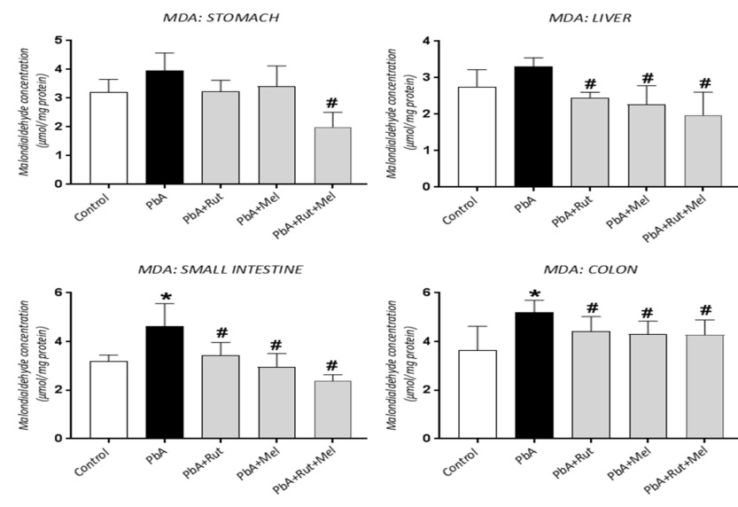
Effects of Rutin and Melatonin Vs Oxidative Stress: The concentrations of hydrogen peroxide (H2O2) and MDA, which is a product of lipid peroxidation and thiol groups in the stomach, liver, small intestine and colon, are shown in Figures 1-3. The results further showed that treatment of rats with PbA caused a significant increase in H2O2 concentration in the liver, small intestines and colon compared to those of the controls (P<0.05; Figure 1). Similarly, the MDA level was significantly high in the small intestine and colon of the rats exposed to PbA compared to that of the controls (Figure 2). Conversely, PbA treatment caused a significant reduction in the concentration of thiol groups in the stomach, liver, small intestines and colon as compared to those of the controls (Figure 3).
The elevation in the levels of H2O2 and MDA induced by PbA was decreased in the presence of rutin, melatonin or their combination. Specifically, rutin (liver, small intestine and colon), melatonin (liver and colon) and their combination (liver, small intestine and colon) significantly reduced the levels of H2O2 compared to that found in the rats treated with PbA (Figure 1). In a similar fashion, treatment with rutin, melatonin singly or their combination resulted in a significant reduction in the MDA levels of the liver, small intestine and colon compared to those of rats exposed to PbA alone (Figure 2). However, treatment of rats with PbA combined with rutin+melatonin produced a more significant reduction in the levels of H2O2 (liver, small intestine and colon) and MDA (stomach, liver and small intestine) compared to rats treated with either rutin or melatonin alone. In most of the stomach, liver and colon tissue samples examined, there was a significant improvement in the total thiol concentrations in rats treated with either melatonin alone or with rutin and melatonin combined. Treatment with rutin alone produced a significant rise in the thiol concentration of the stomach only. Again, the improvement in thiol levels was greater when rats were pretreated with both rutin and melatonin, rather than with either compound alone.
Exposure of rats to PbA led to a significant reduction in the hepatic levels of GSH, whereas the GSH levels in the stomach, small intestine and colon were not significantly changed in all groups at the end of the study (Table 2). Interestingly, treatment with rutin, melatonin or the combination led to significantly improved GSH levels in the liver. Further, the results demonstrated that exposure of rats to PbA caused a significant decline in the GST activities (liver and colon) and SOD (stomach, liver and colon) compared to those of the controls (Table 2). However, in rats treated with rutin (liver), melatonin (liver and colon) or rutin+melatonin (liver and colon), the decline in GST activity was significantly inhibited, compared to the rats treated with PbA alone. The SOD levels significantly improved in the stomach, liver and colon of the rats that had been treated with rutin alone or with rutin and melatonin combined. No significant changes were noticed in the SOD activity in the small intestines in all groups.
Effects of Rutin and Melatonin Vs Serum Parameters: Serum biochemical parameters in rats from various groups are presented in Table 3. The results showed that chronic administration of PbA produced significant reductions in their serum albumin levels compared to that of the controls, although the total protein levels were not affected. Other parameters including ESR, AST, ALT and Alk Phos activities, and the serum concentrations of BUN and creatinine were not significantly altered in the PbA-treated rats. However, the protective effects of rutin and melatonin were clearly demonstrated by the significant reductions in the activities of AST, ALT, Alk Phos, along with the significant restoration of the albumin, BUN and creatinine levels.
Effects of Rutin and Melatonin on Histopathological Features: The histopathological alterations in the liver, stomach, jejunum and colon are presented in Figures 4-7. The images from the control rats (Figure 4A) indicated that the hepatocytes had normal morphology with polyhedral shapes, distinct nuclei and eosinophilic cytoplasm, while the sinusoids were mostly clear. Major histopathological changes were observed in the PbA group (Figure 4B), demonstrating abnormal congestion, severe inflammatory cell infiltrations around the central venules, and the portal vein. However, there was much less portal congestion around central venules in the groups pretreated with rutin (Figure 4C), melatonin (Figure 4D) or their combination (Figure 4E).
The histological analyses of the gastric tissue samples showed impaired mucosal and sub-mucosal structures in rats treated with PbA compared to that of the controls (Figure 5). The gastric mucosal layer in the control rats showed well-arranged epithelial cells and submucosal glands with distinct muscular fibers (Figure 5A). However, the stomach tissue samples from the PbA group showed major loss of the epithelial cells and erosion of the submucosa with marked degeneration of the muscular layer (Figure 5B). Also, there was moderate infiltration of inflammatory cells in the gastric glands and lamina propria in this group. Treatment with rutin (Figure 5C) or melatonin (Figure 5D) or the combination (Figure 5E) resulted in significant improvement of the histopathological alterations caused by PbA, with moderate restoration of the epithelial integrity. However, mild degrees of inflammatory cell infiltration of the submucosa were observed.
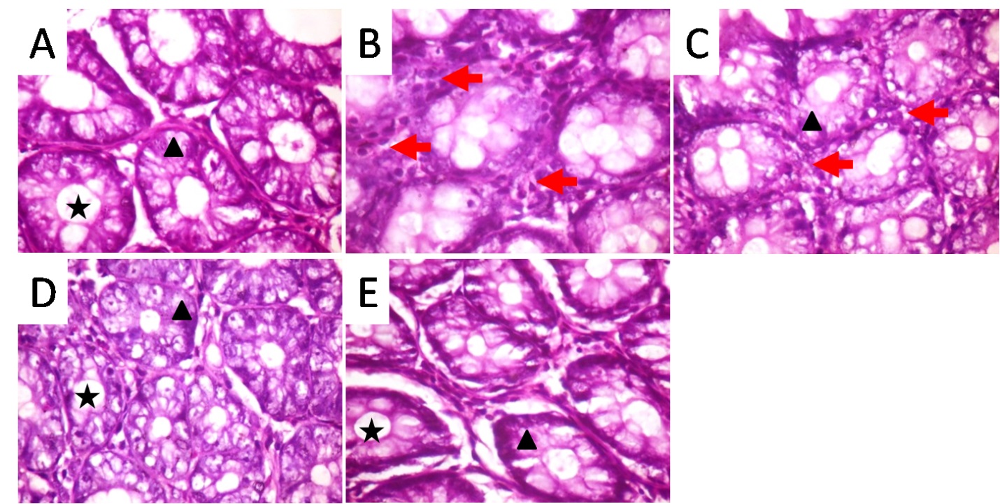
The histological evaluations of the jejunal tissue samples and a representative segment of the small intestine from rats in various groups are illustrated in Figure 6. Compared to the control rats, the various treatments administered caused no significant alterations in the jejunal structures. Normal arrangement of villi without erosions, and vacuolations and/or inflammatory cell infiltrations were observed in all groups. Figure 7 illustrates sections of the colonic histology examined under light microscopy. The colonic sections from the control rats showed normal mucosa with clearly visible crypt, lumen, well arranged epithelia and stroma, and absence of inflammatory cell infiltrations. Administration of PbA altered the morphology of the colonic histology, showing indistinct epithelia with inflammatory cells infiltration in the stroma (Figure 7B). However, treatment with rutin, melatonin or the combination reduced the severity of the PbA-induced lesions with the protection being more evident in the groups treated with melatonin alone or combined with rutin (Figures 7D & 7E), respectively.


Figure 1. Hydrogen peroxide (H2O2) concentration in the stomach, liver, small intestines and colon of male rats as affected by treatment with lead acetate (PbA), Rutin (Rut) and/or melatonin (Mel). Bars represent values expressed as means ±SDs; n = 10 for each group. *Significant (P<0.05) compared to the controls.  #Significant (P<0.05) compared to the PbA group.


Figure 2. Malondialdehyde (MDA) concentration in the stomach, liver, small intestines and colon of male rats as affected by treatment with lead acetate (PbA), Rutin (Rut) and/or melatonin (Mel). Bars represent values expressed as means ±SDs; n = 10 for each group. *Significant (P<0.05) compared to the controls. #Significant (P<0.05) compared to the PbA group.


Figure 3. Total thiol concentration in the stomach, liver, small intestines and colon of male rats as affected by treatment with lead acetate (PbA), Rutin (Rut) and/or melatonin (Mel). Bars represent values expressed as means ±SDs; n = 10 for each group. *Significant (P<0.05) compared to the controls.  #Significant (P<0.05) compared to the PbA group.


Figure 4. Representative images of liver sections stained with H&E observed with light microscopy (Mag. x400).Plate A (control) shows normal central venules and normal morphology of hepatocytes (white arrow) and sinusoids.
Plate B (PbA) shows moderately congested portal vein with inflammatory cell infiltration around the portal veins (portal triaditis) (black arrow) while the morphology of the hepatocytes (white arrow) and sinusoids appear normal. Plates C (PbA+Rut) and D (PbA+Mel) show moderately congested central venules and portal vein with normal morphology of the hepatocytes (white arrow) and sinusoids. Plate E (PbA+Rut+Mel) shows central venules CV with mild perivascular infiltration of inflammatory cells but normal morphology of the hepatocytes (white arrow) and sinusoids. CV: Central veins; PV: Portal vein.


Figure 5. Representative images of stomach sections stained with H&E observed with light microscopy (Mag. Upper plates x100 and lower plates x400). Plate A (Control) shows normal and well preserved mucosal epithelial layer, submucosa and circular muscle layer. Plate B (PbA) shows poorly preserved mucosal epithelial cells and sub-mucosal layer which are sloughed in many areas (white arrows) and mild inflammatory cell infiltration of gastric glands and lamina propria. The circular muscle layer is indistinct and appears degenerated. Plate C (PbA+Rut) also shows poorly preserved mucosal epithelial layer (white arrow) with mild infiltration of gastric glands by inflammatory cells, although the sub-mucosal and circular muscle layers appear normal. Plate D (PbA+Mel) and E (PbA+Rut+Mel) show well preserved mucosal, sub-mucosal and circular muscle layers with mild inflammatory cell infiltration of the gastric glands.
ML: mucosal layer; SML: sub-mucosal layer; CML: circular muscle layer.


Figure 6. Representative images of jejunum sections stained with H&E observed with light microscopy (Mag. Upper plates x40 and lower plates x100). All plates show no significant lesions. A: Controls; B: PbA; C: PbA+Rut; D: PbA+Mel; E: PbA+Rut+Mel.


Figure 7. Representative images of transverse sections of colonic mucosa stained with H&E observed with light microscopy (Mag. x400).
Plate A (Control): Normal colonic mucosa showing clearly visible crypt lumen; well arranged/intact epithelial cells/epithelium and stroma without inflammatory cell infiltration; Plate B (PbA): Damaged/indistinct epithelium with infiltration of stroma by inflammatory cells (red arrows); Plate C (PbA+Rut): Crypts are identifiable, but there is mild inflammatory cell infiltration in the stroma. Plates D (PbA+Rut) and E (PbA+Rut+Mel) show clearly visible crypts and well-arranged epithelial cells without inflammatory cell infiltration. Crypt lumen,  Epithelial cells.

Table 1. Faecal Fatty acid profiles of rats chronically exposed to Lead acetate and treated with Rutin (Rut) and/or melatonin (Mel).
S/N Fatty acid nomenclature Name Control PbA PbA+Rut PbA+Mel PbA+Rut+Mel
1 C8:0 Caprylic - - - 3.11±0.02 2.33±0.04
2 C10:0 Capric 0.84±0.03b 2.91±0.73a - 2.61±0.12a 2.46±0.02a
3 C11:0 Undecylic 3.04±0.21a 11.32±1.21b 3.41±0.14a 4.10±0.25a 1.17±0.02a
4 C12:0 Lauric - 5.74±0.23b 0.94±0.01a 1.50±0.01a 1.37±0.14a
5 C13:0 Tridecylic 11.26±0.19a 38.19±0.45b 18.53±1.93c 23.70±0.03d 30.80±0.54e
6 C14:0 Myristic 0.31±0.37b 1.24±0.62a - 5.24±0.51c 2.10±0.02a
7 C14:1 Myristoleic 3.30±0.12a 5.79±0.34b - 8.44±0.29c 0.84±0.02d
8 C15:0 Pentadecanoic 4.62±0.14a 6.20±0.26b 1.52±0.05a 3.26±0.97a 0.37±0.01c
9 C15:1 Pentadecenoic - - 2.95±0.32 - -
10 C16:0 Palmitic 5.94±0.17c 25.11±0.35a 12.41±1.23b 23.19±0.68a 16.48±0.69b
11 C16:1 Palmitoleic 10.44±0.21b 2.91±0.19a 3.05±0.24a 3.04±0.12a 2.53±0.41a
12 C17:0 Heptadecanoic 12.17±0.69a 0.59±0.59b 11.43±0.92a 5.88±0.45c -
13 C17:1 Heptadecenoic - - 3.91±0.004a 3.36±0.02a -
14 C18:0 Stearic - - 1.11±0.001a 4.49±0.27b -
15 C18:1n9c Oleic - - 0.98±0.003 - -
16 C18:2n6c Linoleic - - 1.83±0.26 - -
17 C20:1 Eicosenoic - - 1.74±0.002 - -
18 C20:2 Eicosadienoic - - 36.17±0.29 - -
19 C20:3n6 Eicosatrienoic 48.08±0.21 - - - -
20 C22:0 Behenic - - - 2.41±0.04 -
21 C22:1n9 Erucic - - - 5.65±0.51 -
22 C23:0 Tricosylic - - - - 39.54±0.59
Data are expressed as relative values (%) i.e. mean % of total fatty acids ± SEM
abcdeValues with different superscripts within a row differ significantly at P<0.05.
Table 2. Changes in Reduced glutathione (GSH) concentration, Glutathione S-transferase (GST) and Superoxide dismutase (SOD) activities in stomach, liver, small intestines and colon of male rats as affected by treatment with lead acetate (PbA), Rutin (Rut) and/or melatonin (Mel).
Parameter Groups
Control PbA PbA + Rut PbA + Mel PbA + Rut + Mel
GSH
Stomach 88.57±8.82 82.09±2.96 81.96±4.86 86.59±3.33 85.28±3.62
Liver 234.16±51.86 149.53±28.15* 213.81±49.25# 240.17±27.09# 276.08±28.20#
Small intestine 46.21±5.67 39.49±3.71 41.28±1.86 39.79±2.36 40.72±4.30
Colon 47.35±6.84 40.26±1.15 44.04±6.41 40.81±1.64 40.35±2.38
GST
Stomach 0.04±0.02 0.03±0.01 0.03±0.01 0.02±0.01 0.03±0.01
Liver 0.40±0.04 0.24±0.06* 0.43±0.09# 0.46±0.15# 0.50±0.13#
Small intestine 0.10±0.01 0.11±0.03 0.11±0.02 0.12±0.04 0.11±0.03
Colon 0.09±0.03 0.03±0.01* 0.06±0.01 0.09±0.03# 0.09±0.03#
SOD
Stomach 3.34±0.65 2.59±0.71* 3.68±0.40# 2.90±0.71 3.65±0.58#
Liver 12.59±1.64 6.05±0.61* 11.43±2.91# 9.60±2.48 12.88±2.56#
Small intestine 1.91±0.28 2.06±0.23 2.42±0.52 1.91±0.32 2.08±0.71
Colon 4.26±0.17 2.93±0.38* 4.02±0.77# 2.95±0.43 4.05±0.56#
Values are expressed as means ±SD; n = 10 for each group. *Significant (P<0.05) when compared with the control #Significant (P<0.05) when compared with the PbA group. GSH concentration (mol/g tissue). GST specific activity (mmol CDNB-GSH complex formed/min per mg protein). SOD activity (units/mg protein)

Table 3. Serum chemistry profiles of rats chronically exposed to Lead acetate and treated with rutin (Rut) and/or melatonin (Mel).
Parameter Groups
Control PbA PbA + Rut PbA + Mel PbA + Rut + Mel
ESR (mm/hr) 0.96±0.09 0.96±0.15 0.84±0.06 0.88±0.08 0.76±0.06#
TP (mg/dL) 8.60±0.46 8.32±0.36 8.10±0.40 8.08±0.47 8.14±0.30
Albumin (g/dL) 3.64±0.17 3.28±0.19* 2.90±0.34# 2.88±0.36# 2.84±0.23#
Globulin (g/dL) 4.96±0.37 4.82±0.50 5.20±0.10 5.20±0.14 5.30±0.29
A:G 0.74±0.05 0.66±0.05* 0.57±0.06# 0.55±0.06# 0.54±0.06#
AST (U/L) 50.75±2.22 51.75±0.96 48.25±0.96# 46.60±0.89# 47.00±1.41#
ALT (U/L) 31.00±2.16 31.67±1.53 27.25±1.71# 26.75±1.26# 26.80±2.49#
ALP (U/L) 132.00±4.32 131.00±1.73 123.50±3.70# 123.80±6.98# 122.50±3.42#
BUN (mg/dL) 18.6±1.14 18.08±0.64 17.66±0.57 18.18±0.84 16.96±0.48#
Creatinine 0.78±0.05 0.80±0.07 0.74±0.06 0.78±0.08 0.66±0.06#
Data are expressed as means ±standard deviations (n = 5). *Significant (P<0.05) compared to the controls. #Significant (P<0.05) when compared with the PbA group. ESR, erythrocyte sedimentation rate; TP, total protein; A:G, albumin-globulin ratio; AST, aspartate transaminase; ALT, alanine transaminase; ALP, alkaline phosphatase; BUN, blood urea nitrogen.

Discussion
Oral exposure to lead is capable of stimulating local oxidative stress and inflammation in the GI tract leading to profound alterations in the tissue morphology and function. These include impairments in the intestinal permeability, nutrient absorption and metabolism of essential nutrients such as lipids [45]. The current study provides ample evidence that chronic exposure to lead disrupts intestinal fatty acid profiles, causing alterations in the oxidant-antioxidant balance and the morphology of the GI and hepatic tissues in rats. Recent studies have shown that dietary lead exposure also alters the homeostasis of gut microbiota accompanied by changes in the profiles of microbiota-derived metabolites, such as short-chain fatty acids [46, 47]. However, little information is available on alterations in the metabolism and profiles of medium- and long-chain fatty acids following chronic exposure to lead in animal models.
Our findings showed a preponderance of saturated fatty acids, mainly tridecylic and palmitic acids in the faeces of PbA-treated rats together with deficiencies in mono or polyunsaturated fatty acids. This observation was in contrast to the results of the faecal analyses from both the control rats and those treated with rutin, in which high levels of unsaturated fatty acids were found. Ample epidemiological evidence has linked a high incidence of inflammatory bowel diseases (IBD) with an abundance of faecal saturated long-chain fatty acids [48]. Also, an increase in the faecal level of n-3 PUFAs has been shown to mitigate intestinal histological alterations, such as ulcers, necrosis and inflammation. Specifically, these conditions have been associated with such enteritis models induced by trinitrobenzene sulfonic acids, dextran sulfate sodium, and lipopolysaccharides [49, 50]. Consistent with the findings from the current study, these reports suggest complex relationships involving medium- and long-chain fatty acids, which may serve as important metabolic biomarkers of toxic and/or disease-related changes in the GI tract. Hence, additional studies are warranted to unravel the underlying mechanisms and to explain the role of medium- and long-chain fatty acids in the development of intestinal diseases.
Earlier studies have suggested that susceptibility to intestinal oxidative stress could be influenced by the level of unsaturated fatty acids in the cell membranes [51, 52]. Our findings suggest that a rise in saturated fatty acids in the faeces of rats treated with lead acetate may accompany an increase in the hepatic and GI tract oxidative stress. Also, high levels of H2O2 and MDA in the liver and intestines are indicative of oxidative stress due to ROS generation. This finding was corroborated by profound reductions in thiol groups found in all of the animal tissue samples examined, particularly the reduced GSH levels in the liver and colon, and inhibition of GST and SOD activities in the liver and most of the intestinal tissue samples.
It has been previously established that increased lipid peroxidation in tissues is usually reflected by higher MDA levels [53]. The antioxidant enzyme, SOD, plays important roles in protecting cells against damage while GST is involved in inhibiting toxic metabolites generated in tissues following exposure to toxins [54]. Based on the extent of oxidative stress observed in this study, it was not surprising that our histopathological examinations revealed alterations in the hepatic tissue morphology. We found congested portal vein and central venules with inflammatory cell infiltration, although the hepatocytes morphology was not considerably affected. Also, the gastric and colonic tissue samples showed poorly preserved epithelial and sub-mucosal layers, while inflammatory cell infiltrations were widely evident in the GI tissue samples of the rats treated with lead acetate.
Current research trends have focused on search for agents that can reduce oxidative stress and inflammation in the gut and thus protect the organ’s barrier against lead toxicity [55]. In the current study, we found that supplementation of rat foods with rutin at 50 mg/kg and/or with melatonin at 25 mg/kg significantly protected against chronic lead toxicity in the rats’ GI and liver tissues. Our results demonstrated the profound antioxidant potentials of rutin and melatonin as evident by their significant capacity to reduce H2O2 and MDA levels. These events corresponded to increases in the thiol groups, and SOD/GST activities, compared to those in rats treated with lead acetate alone. Rutin and melatonin, as natural compounds, resulted in much alleviation of tissue lesions induced by lead acetate, particularly in the stomach, liver and colon. With respect to variations in the extent of lesions along the GI tract, the results revealed that orally administered lead acetate impacted the morphology of GI structures to greater extents at sites where the heavy metals probably had longer transit times, i.e. stomach and colon versus jejunum. This finding consistently reflected in the status of antioxidant systems in the small intestine where the concentration of GSH and the activities of GST and SOD remained unaltered in all of the experimental groups.
Fatty acids detected in the faeces usually originate from the diet, although some studies indicate that faecal fatty acid composition may substantially reflect the metabolic activity of the bacteria in the GI tract. For instance, there is evidence that stearic, oleic, and linoleic acids can be converted to hydroxystearic acid by the intestinal bacteria and the extent of oxidation is linked to the overgrowth of certain bacterial species [56]. Notably, our results showed a greater diversity of medium- and long-chain fatty acids in rats treated with rutin, compared to those found in other groups. It appears that rutin had shifted fatty acid metabolism by intestinal bacteria in favor of unsaturated fatty acids. This may be responsible for the greater diversity of fatty acids found in the rat group treated with rutin. Future studies are required to elucidate the specific bacterial populations that are favored or inhibited during exposure to lead acetate and how this is correlated with fatty acid profile in the intestines.
The results of the current study are consistent with previous reports indicating that rutin or melatonin supplementation reduces oxidative stress via increases in the activities of antioxidant enzymes, SOD, GST, catalase, glutathione reductase and/or peroxidase, while also decreasing the MDA levels [57, 58]. A number of studies have also shown that melatonin protects against lead toxicity, although they used shorter periods of lead exposure [59, 60]. In the current study, the protective roles of rutin and melatonin were clearly demonstrated by a significant reduction in the lead acetate ability to increase such liver enzymes’ levels, as AST, ALT and Alk Phos in the serum, and reduced concentrations of BUN and creatinine, indicative of renal injury [61].
Conclusions
The present study investigated the protective roles of rutin and melatonin in a chronic lead acetate-induced model of liver and GI tract injury along with assessing the potential alterations in the metabolism of intestinal fatty acids. Our results provided evidence that lead acetate induced disruptions in fatty acid composition in favor of saturated fatty acids, and showed alterations in the tissue morphology and oxidant-antioxidant balance. In addition, rutin, melatonin and the combination showed protective effects toward the liver, stomach and intestines by reducing oxidative damages and preventing against histopathological alterations due to lead acetate toxicity. Our results provide a basis for the clinical application of rutin and/or melatonin in support of treating liver and gastrointestinal injuries in humans. The results also suggest that a complete faecal and plasma fatty acid profile deserve greater attention as non-invasive diagnostic indicators of chronic GI injury in response to lead acetate toxicity.
Competing Interests
The authors have no relevant financial or non-financial interests to disclose.
Ethical Approval
All the animal studies were based on international guidelines published by the National Institutes of Health for the care and use of laboratory animals (NIH Publications No. 8023, revised 1978). The design of the experiments and experimental animal handling followed guidelines approved by the local ethics committee of the University of Ibadan.
Funding
The authors declare that no funds, grants, or other support were received from other agencies.
Author’s Contributions
Conceptualization: Akinrinde AS, Omotosho OO, Omobowale TO; Methodology: Akinrinde AS, Adetiba R, Omotosho OO, Oyagbemi AA; Formal analysis and investigation: Akinrinde AS, Adetiba R, Omotosho OO; Writing – original draft preparation: Akinrinde AS; Writing – review and editing: Omotosho OO, Omobowale TO; Funding acquisition: Akinrinde AS, Omotosho OO; Resources: Akinrinde AS, Oyagbemi AA, Omotosho OO, Supervision: Akinrinde AS, OyagbemiAA, Omobowale TO.
Type of Study: Research | Subject: General

References
1. Luo L, Wang B, Jiang J, Fitzgerald M, Huang Q, Yu Z, et al. Heavy Metal Contaminations in Herbal Medicines: Determination, Comprehensive Risk Assessments, and Solutions. Frontiers in pharmacology. 2020;11:595335. [DOI:10.3389/fphar.2020.595335] [PMID] [PMCID]
2. Omobowale TO, Oyagbemi AA, Akinrinde AS, Saba AB, Daramola OT, Ogunpolu BS, et al. Failure of recovery from lead induced hepatoxicity and disruption of erythrocyte antioxidant defence system in Wistar rats. Environmental toxicology and pharmacology. 2014;37(3):1202-11. [DOI:10.1016/j.etap.2014.03.002] [PMID]
3. Nkwunonwo UC, Odika PO, Onyia NI. A Review of the Health Implications of Heavy Metals in Food Chain in Nigeria. TheScientificWorldJournal. 2020;2020:6594109. [DOI:10.1155/2020/6594109] [PMID] [PMCID]
4. de Carvalho C, Caramujo MJ. The Various Roles of Fatty Acids. Molecules. 2018;23(10). [DOI:10.3390/molecules23102583] [PMID] [PMCID]
5. Calder PC. Fatty acids and inflammation: the cutting edge between food and pharma. European journal of pharmacology. 2011;668 Suppl 1:S50-8. [DOI:10.1016/j.ejphar.2011.05.085] [PMID]
6. Tang H, Zhu X, Gong C, Liu H, Liu F. Protective effects and mechanisms of omega-3 polyunsaturated fatty acid on intestinal injury and macrophage polarization in peritoneal dialysis rats. Nephrology. 2019;24(10):1081-9. [DOI:10.1111/nep.13587] [PMID] [PMCID]
7. Zhang CX, Shu CM, Zhang XY, Lin XT, Guan QH, Zhang F. Effect and mechanism of omega-3 polyunsaturated fatty acids on intestinal injury in rats with obstructive jaundice. Europe Rev Med Pharmacol Sci. 2021;25(19):6077-92.
8. Tan J, McKenzie C, Potamitis M, Thorburn AN, Mackay CR, Macia L. The role of short-chain fatty acids in health and disease. Advances in immunology. 2014;121:91-119. [DOI:10.1016/B978-0-12-800100-4.00003-9] [PMID]
9. Blaak EE, Canfora EE, Theis S, Frost G, Groen AK, Mithieux G, et al. Short chain fatty acids in human gut and metabolic health. Beneficial microbes. 2020;11(5):411-55. [DOI:10.3920/BM2020.0057] [PMID]
10. Nogal A, Valdes AM, Menni C. The role of short-chain fatty acids in the interplay between gut microbiota and diet in cardio-metabolic health. Gut microbes. 2021;13(1):1-24. [DOI:10.1080/19490976.2021.1897212] [PMID] [PMCID]
11. Song EM, Byeon JS, Lee SM, Yoo HJ, Kim SJ, Lee SH, et al. Fecal Fatty Acid Profiling as a Potential New Screening Biomarker in Patients with Colorectal Cancer. Digestive diseases and sciences. 2018;63(5):1229-36. [DOI:10.1007/s10620-018-4982-y] [PMID]
12. Sitkin SI, Tkachenko EI, Vakhitov TY. Metabolic dysbiosis of the gut microbiota and its biomarkers. Exprim Clinic Gastroenterol. 2016;12(12):6-29. [DOI:10.18786/2072-0505-2015-40-12-34]
13. Zhuang X, Li T, Li M, Huang S, Qiu Y, Feng R, et al. Systematic Review and Meta-analysis: Short-Chain Fatty Acid Characterization in Patients With Inflammatory Bowel Disease. Inflammatory bowel diseases. 2019;25(11):1751-63. [DOI:10.1093/ibd/izz188] [PMID]
14. De Preter V, Machiels K, Joossens M, Arijs I, Matthys C, Vermeire S, et al. Faecal metabolite profiling identifies medium-chain fatty acids as discriminating compounds in IBD. Gut. 2015;64(3):447-58. [DOI:10.1136/gutjnl-2013-306423] [PMID]
15. Weir TL, Manter DK, Sheflin AM, Barnett BA, Heuberger AL, Ryan EP. Stool microbiome and metabolome differences between colorectal cancer patients and healthy adults. PloS one. 2013;8(8):e70803. [DOI:10.1371/journal.pone.0070803] [PMID] [PMCID]
16. Wiktorowska-Owczarek A, Berezinska M, Nowak JZ. PUFAs: Structures, Metabolism and Functions. Advances in clinical and experimental medicine : official organ Wroclaw Medical University. 2015;24(6):931-41. [DOI:10.17219/acem/31243] [PMID]
17. Ravindran V, Tancharoenrat P, Zaefarian F, Ravindran G. Fats in poultry nutrition: Digestive physiology and factors influencing their utilization. Animal Feed Sci Technol. 2016;213:1-21. [DOI:10.1016/j.anifeedsci.2016.01.012]
18. Han L, Wang H, Li L, Li X, Ge J, Reiter RJ, et al. Melatonin protects against maternal obesity-associated oxidative stress and meiotic defects in oocytes via the SIRT3-SOD2-dependent pathway. Journal of pineal research. 2017;63(3). [DOI:10.1111/jpi.12431] [PMID]
19. Liu Z, Gan L, Luo D, Sun C. Melatonin promotes circadian rhythm-induced proliferation through Clock/histone deacetylase 3/c-Myc interaction in mouse adipose tissue. Journal of pineal research. 2017;62(4). [DOI:10.1111/jpi.12383] [PMID]
20. Ganeshpurkar A, Saluja AK. The Pharmacological Potential of Rutin. Saudi pharmaceutical journal : SPJ : the official publication of the Saudi Pharmaceutical Society. 2017;25(2):149-64. [DOI:10.1016/j.jsps.2016.04.025] [PMID] [PMCID]
21. Yong DOC, Saker SR, Chellappan DK, Madheswaran T, Panneerselvam J, Choudhury H, et al. Molecular and Immunological Mechanisms Underlying the Various Pharmacological Properties of the Potent Bioflavonoid, Rutin. Endocrine, metabolic & immune disorders drug targets. 2020;20(10):1590-6. [DOI:10.2174/1871530320666200503053846] [PMID]
22. Abdel-Raheem IT. Gastroprotective effect of rutin against indomethacin-induced ulcers in rats. Basic & clinical pharmacology & toxicology. 2010;107(3):742-50. [DOI:10.1111/j.1742-7843.2010.00568.x] [PMID]
23. Olaleye MT, Akinmoladun AC. Comparative gastroprotective effect of post-treatment with low doses of rutin and cimetidine in rats. Fundamental & clinical pharmacology. 2013;27(2):138-45. [DOI:10.1111/j.1472-8206.2011.00972.x] [PMID]
24. Chen CQ, Fichna J, Bashashati M, Li YY, Storr M. Distribution, function and physiological role of melatonin in the lower gut. World journal of gastroenterology. 2011;17(34):3888-98. [DOI:10.3748/wjg.v17.i34.3888] [PMID] [PMCID]
25. Bhattacharya S, Patel KK, Dehari D, Agrawal AK, Singh S. Melatonin and its ubiquitous anticancer effects. Molecular and cellular biochemistry. 2019;462(1-2):133-55. [DOI:10.1007/s11010-019-03617-5] [PMID]
26. Kvetnoy IM, Ingel IE, Kvetnaia TV, Malinovskaya NK, Rapoport SI, Raikhlin NT. Gastrointestinal melatonin: cellular identification and biological role. Neuro Endocrinol Letter. 2002;23:121-32.
27. Bubenik GA. Gastrointestinal melatonin: localization, function, and clinical relevance. Digestive diseases and sciences. 2002;47(10):2336-48. [DOI:10.1023/A:1020107915919] [PMID]
28. Bothorel B, Barassin S, Saboureau M, Perreau S, Vivien-Roels B, Malan A, et al. In the rat, exogenous melatonin increases the amplitude of pineal melatonin secretion by a direct action on the circadian clock. The European journal of neuroscience. 2002;16(6):1090-8. [DOI:10.1046/j.1460-9568.2002.02176.x] [PMID]
29. Vyskocil A, Fiala Z, Tejnorová I, Tusl M. Stress reaction in developing rats exposed to 1% lead acetate. Sb Ved Pr Lek Fak Karlovy Univerzity Hradci Kralove. 1991;34(3):287-95.
30. Vyskocil A, Smejkalová J, Lacinová V. Dose-related stress reaction in male rats chronically exposed to lead acetate. Sb Ved Pr Lek Fak Karlovy Univerzity Hradci Kralove. 1991;34(5):393-401.
31. Mugahi MN, Heidari Z, Sagheb HM, Barbarestani M. Effects of Chronic Lead Acetate Intoxication on Blood Indices of Male Adult Rat. DARU J Pharmaceut Sci. 2003;11(4):147-51.
32. Ogeturk M, Kus I, Kavakli A, Zararsiz I, Ilhan N, Sarsilmaz M. Effects of melatonin on carbon tetrachloride-induced changes in rat serum. Journal of physiology and biochemistry. 2004;60(3):205-10. [DOI:10.1007/BF03167030] [PMID]
33. Demirtas CY, Pasaoglu OT, Bircan FS, Kantar S, Turkozkan N. The investigation of melatonin effect on liver antioxidant and oxidant levels in fructose-mediated metabolic syndrome model European Reviews for Medical and Pharmacological Sciences. 2015;19(10):1915-21.
34. Caglayan C, Kandemir FM, Yildirim S, Kucukler S, Eser G. Rutin protects mercuric chloride-induced nephrotoxicity via targeting of aquaporin 1 level, oxidative stress, apoptosis and inflammation in rats. Journal of trace elements in medicine and biology : organ of the Society for Minerals and Trace Elements. 2019;54:69-78. [DOI:10.1016/j.jtemb.2019.04.007] [PMID]
35. Kandemir FM, Caglayan C, Aksu EH, Yildirim S, Kucukler S, Gur C. Protective effect of rutin on mercuric chloride-induced reproductive damage in male rats. Andrologia. 2020;52(3):e13524. [DOI:10.1111/and.13524]
36. Wolff SF. Ferrous ion oxidation in the presence of ferric ion indicator xylenol orange for measurement of hydrogen peroxides. Method Enzymol. 1994;233(2):182-9. [DOI:10.1016/S0076-6879(94)33021-2]
37. Varshney R, Kale RK. Effects of calmodulin antagonists on radiation-induced lipid peroxidation in microsomes. International journal of radiation biology. 1990;58(5):733-43. [DOI:10.1080/09553009014552121] [PMID]
38. Ellman GL. Tissue sulfhydryl groups. Archives of biochemistry and biophysics. 1959;82(1):70-7. [DOI:10.1016/0003-9861(59)90090-6] [PMID]
39. Jollow DJ, Mitchell JR, Zampaglione N, Gillette JR. Bromobenzene-induced liver necrosis. Protective role of glutathione and evidence for 3,4-bromobenzene oxide as the hepatotoxic metabolite. Pharmacology. 1974;11(3):151-69. [DOI:10.1159/000136485] [PMID]
40. Habig WH, Pabst MJ, Jakoby WB. Glutathione-S-transferases. The first enzymatic step in mercapturic acid formation. J Biol Chemist. 1974;25:7130-9. [DOI:10.1016/S0021-9258(19)42083-8]
41. Misra HP, Fridovich I. The role of superoxide anion in the autoxidation of epinephrine and a simple assay for superoxide dismutase. The Journal of biological chemistry. 1972;247(10):3170-5. [DOI:10.1016/S0021-9258(19)45228-9]
42. Oyagbemi AA, Omobowale TO, Akinrinde AS, Saba AB, Ogunpolu BS, Daramola O. Lack of reversal of oxidative damage in renal tissues of lead acetate-treated rats. Environmental toxicology. 2015;30(11):1235-43. [DOI:10.1002/tox.21994] [PMID]
43. Scortichini S, Boarelli MC, Silvi S, Fiorini D. Development and validation of a GC-FID method for the analysis of short chain fatty acids in rat and human faeces and in fermentation fluids. Journal of chromatography B, Analytical technologies in the biomedical and life sciences. 2020;1143:121972. [DOI:10.1016/j.jchromb.2020.121972] [PMID]
44. Akinrinde A, Adigun K, Mustapha A. Cobalt-induced neuro-behavioural alterations are accompanied by profound Purkinje cell and gut-associated responses in rats. Environ Anal Health Toxicol. 2023;38(2):e2023010-0. [DOI:10.5620/eaht.2023010]
45. Crespo S, Nonnotte G, Colin DA, Leray C, Aubree A. Morphological and functional alterations induced in trout intestine by dietary cadmium and lead. J Fish Biol. 2010;28:69-80. [DOI:10.1111/j.1095-8649.1986.tb05143.x]
46. Cheng D, Li H, Zhou J, Wang S. Chlorogenic acid relieves lead-induced cognitive impairments and hepato-renal damage via regulating the dysbiosis of the gut microbiota in mice. Food & function. 2019;10(2):681-90. [DOI:10.1039/C8FO01755G] [PMID]
47. Zhai Q, Qu D, Feng S, Yu Y, Yu L, Tian F, et al. Oral Supplementation of Lead-Intolerant Intestinal Microbes Protects Against Lead (Pb) Toxicity in Mice. Frontiers in microbiology. 2019;10:3161. [DOI:10.3389/fmicb.2019.03161] [PMID] [PMCID]
48. Marion-Letellier R, Savoye G, Beck PL, Panaccione R, Ghosh S. Polyunsaturated fatty acids in inflammatory bowel diseases: a reappraisal of effects and therapeutic approaches. Inflammatory bowel diseases. 2013;19(3):650-61. [DOI:10.1097/MIB.0b013e3182810122] [PMID]
49. Gobbetti T, Dalli J, Colas RA, Federici Canova D, Aursnes M, Bonnet D, et al. Protectin D1(n-3 DPA) and resolvin D5(n-3 DPA) are effectors of intestinal protection. Proceedings of the National Academy of Sciences of the United States of America. 2017;114(15):3963-8. [DOI:10.1073/pnas.1617290114] [PMID] [PMCID]
50. Zhu H, Wang H, Wang S, Tu Z, Zhang L, Wang X, et al. Flaxseed Oil Attenuates Intestinal Damage and Inflammation by Regulating Necroptosis and TLR4/NOD Signaling Pathways Following Lipopolysaccharide Challenge in a Piglet Model. Molecular nutrition & food research. 2018;62(9):e1700814. [DOI:10.1002/mnfr.201700814] [PMID]
51. Lauridsen C. Lipid nutrition and gut health of pigs. J Animal Sci. 2019;97(Supplement 2):28-54. [DOI:10.1093/jas/skz122.051] [PMCID]
52. Wang Y, Chen Y, Zhang X, Lu Y, Chen H. New insights in intestinal oxidative stress damage and the health intervention effects of nutrients: A review. J Funct Food. 2020;75:104248. [DOI:10.1016/j.jff.2020.104248]
53. Rosas CE, Correa LB, Henriques MG. Role of Neutrophils in Disease Pathogenesis. London, UK: Intech Open Limited. Neutrophils in rheumatoid arthritis: a target for discovering new therapies based on natural products.2017. 89-118 p. [DOI:10.5772/intechopen.68617]
54. Rana SV, Sharma S, Prasad KK, Sinha SK, Singh K. Indian Journal of Medical Research. 2014;139(4):568-71.
55. Yu L, Yu Y, Xiao Y, Tian F, Narbad A, Zhai Q. Lead-induced gut injuries and the dietary protective strategies: A review. J Funct Food. 2021;83:104528. [DOI:10.1016/j.jff.2021.104528]
56. Hoyles L, Wallace RJ. Gastrointestinal Tract: Intestinal Fatty Acid Metabolism and Implications for Health. Handbook Hydrocarbon Lipid Microbiol. 2010:3119-32. [DOI:10.1007/978-3-540-77587-4_234]
57. Hassan FAM, Roushdy EM, Kishawy ATY, Zaglool AW, Tukur HA, Saadeldin IM. Growth Performance, Antioxidant Capacity, Lipid-Related Transcript Expression and the Economics of Broiler Chickens Fed Different Levels of Rutin. Animals : an open access journal from MDPI. 2018;9(1). [DOI:10.3390/ani9010007] [PMID] [PMCID]
58. Yang Z, He Y, Wang H, Zhang Q. Protective effect of melatonin against chronic cadmium-induced hepatotoxicity by suppressing oxidative stress, inflammation, and apoptosis in mice. Ecotoxicology and environmental safety. 2021;228:112947. [DOI:10.1016/j.ecoenv.2021.112947] [PMID]
59. Ghosh D, Dey M, Ghosh A, Chattopadhyay A, Bandyopadhyay D. Melatonin protects against lead acetate-induced changes in blood corpuscles and lipid profile of male Wistar rats. J Pharmacy Res. 2014;8(3):336-42.
60. Hernandez-Plata E, Quiroz-Compean F, Ramirez-Garcia G, Barrientos EY, Rodriguez-Morales NM, Flores A, et al. Melatonin reduces lead levels in blood, brain and bone and increases lead excretion in rats subjected to subacute lead treatment. Toxicology letters. 2015;233(2):78-83. [DOI:10.1016/j.toxlet.2015.01.009] [PMID]
61. Ilesanmi OB, Adeogun EF, Odewale TT, Chikere B. Lead exposure-induced changes in hematology and biomarkers of hepatic injury: protective role of TrevoTM supplement. Environ Anal Health Toxicol. 2022;37(2):e2022007-0. [DOI:10.5620/eaht.2022007] [PMID] [PMCID]

Add your comments about this article : Your username or Email:
CAPTCHA

Send email to the article author


Rights and permissions
Creative Commons License This work is licensed under a Creative Commons Attribution-NonCommercial 4.0 International License.

© 2025 CC BY-NC 4.0 | Iranian Journal of Toxicology

Designed & Developed by : Yektaweb