Write your message
Volume 18, Issue 1 (January 2024)                   IJT 2024, 18(1): 52-60 | Back to browse issues page

Ethics code: IR .IUMS.REC1399.863


XML Print


Download citation:
BibTeX | RIS | EndNote | Medlars | ProCite | Reference Manager | RefWorks
Send citation to:

Soleymanzadeh Moghadam S, Minaeian S, Majidpour A, Adabi M, Hosseini Doust R. The Lactobacillus acidophilus Supernatant: An Effective and Safe Alternative to Antibiotics. IJT 2024; 18 (1) :52-60
URL: http://ijt.arakmu.ac.ir/article-1-1275-en.html
1- Department of Microbiology, Faculty of Advanced Science & Technology Tehran Medical Sciences, Islamic Azad University, Tehran, Iran
2- Antimicrobial Resistance Research Center, Institute of Immunology and Infectious Diseases, Iran University of Medical Sciences, Tehran, Iran
3- Department of Medical Nanotechnology, School of Advanced Technologies in Medicine, Tehran University of Medical Sciences, Tehran, Iran
4- Department of Microbiology, Faculty of Advanced Science & Technology Tehran Medical Sciences, Islamic Azad University, Tehran, Iran , rhdoust@gmail.com
Full-Text [PDF 1000 kb]   (579 Downloads)     |   Abstract (HTML)  (2078 Views)
Full-Text:   (647 Views)
Introduction
The use of antibiotics is a standard treatment for numerous bacterial infections; however, the overuse can lead to antibiotic resistance [1-4]. The global emergence of multi-drug resistance (MDR) among bacteria is a major challenge facing infectious disease management. Therefore, preventing or minimizing this phenomenon is one of the main approaches to control infections or the fatal outcomes [5, 6]. In fact, the use of alternative agents to replace conventional and synthetic antibiotics is a logical approach [6, 7]. Currently, bacterial therapy, i.e., application of beneficial and safe bacteria or their products against pathogens, is one of the alternative approaches to infection management [8, 9].
Probiotics are live and useful microorganisms that offer antibacterial properties. These agents can serve roles in the body to fight against infections, while reducing the need for standard antibiotics [9-11]. The cell-free supernatant (CFS) of probiotics are known to be effective antimicrobial agents [12, 13]. Currently, Lactobacillus spp. and their supernatants are being investigated in many probiotic research projects. Indeed, probiotics and their bactericidal capacity can be effectively applied in the clinical management of infections as an important treatment strategy [14].
Bacteriotherapy with lactobacilli spp. has emerged as a practical alternative to the treatment or prevention of various nosocomial infections [9, 13]. Some strains of Lactobacillus and their products have significant inhibitory activity against bacteria.  Lactobacillus spp. secretes antibacterial substances, such as hydrogen peroxide, lactic acid, bacteriocins and short-chain fatty acids (SCFA) [15, 16]. These products regulate the normal flora population and minimize or inhibit bacterial infection in the human body. The positive role of SCFA, bactriocins and hydrogen peroxide in the cell free supernatant of probiotics has been established in infection control studies. Lactobacilli strains have inhibitory effects on the growth of resistant pathogens, such as Staphylococcus aureus (S. aureus), Escherichia coli (E. coli), Pseudomonas aeruginosa (P. aeruginosa) and Acinetobacter baumannii (A. baumannii). Thus, they should be considered as effective alternatives in the management of various infectious diseases [16, 17]. A critical point facing such a strategy is that we should search for factors that have antibacterial effects while being safe to human cells.
This study aimed to investigate the antimicrobial and cytotoxic properties of the CFS of Lactobacillus acidophilus (SLA) against gram-positive and gram-negative bacteria. The current study was conducted to evaluate one strain of probiotics with antibacterial and cytotoxic properties that has not been studied to date.
Methods
Preparation of Probiotics
We used a total of eight probiotic strains in this study. The five commercial strains included L. Plantarum 299 V (DSM 9843), L. ruteri (DSM 17938), L. acidophilus (LAFTI-L10 DSL), B. bifidum B94 (DSM 20456) and Bacillus coagulans (DSM 1). They were purchased from the Iranian Biological Resource Center, and the Industrial Enzymes Company, which represents the Dutch company DSM in Iran. We also used the following three local strains: L. ruteri EF4, L. salivarius EF6 and EF7. These strains were purchased from the Iranian Institute of Agricultural Biotechnology and the Probiotic Research Center of Alborz University of Medical Sciences. The probiotics were cultured on de Man Rogosa and Sharpe (MRS) agar media (Merck, Germany), under microaerophilic conditions at 37°C for 48 hours [5].
Preparation of Probiotics from Cell-free Supernatant
The probiotic strains were transferred separately from MRS agar medium to tubes containing MRS broth. These tubes were incubated under micro-aerophilic conditions at 37°C for 24 hours. The tubes were then centrifuged in a refrigerated unit for 10 minutes at 4000 rpm. Next, the supernatant was separated and passed through a syringe filter with a 0.45 pm pore size [5].

Preparation of Bacteria
Strains of E. coli (ATCC 25922) and S. aureus (ATCC 25923) were obtained from the Iranian Biological Resource Center (Tehran, Iran). These strains were cultured in Muller-Hinton agar medium (Merck, Germany).
Antibiotic Susceptibility
We performed qualitative evaluations of susceptibility of bacterial strains to all 8 probiotic strains in order to select the appropriate and effective ones. For antibiogram, we used the CFS from probiotics at a concentration of 100 μL/mL. For quality assays, well diffusion method was used on Muller-Hinton agar medium. The bacterial strains with 0.5 McFarland dilutions were cultured separately on multi-well plates containing Muller-Hinton medium. The wells were individually filled with CFS from the probiotics. Next, the plates were incubated at 4°C for two hours. They were then incubated at 37°C for 16 to 18 hours and the inhibition zone was measured (Figure 1) [18].
Selection of Probiotics
To continue the laboratory steps, after performing well diffusion test, the appropriate and effective probiotic was selected based on the largest difference in the inhibitory diameters of CFS against E. coli and S. aureus strains, and based on Friedman test. Accordingly, the SLA was selected to continue with the experiments.
Minimum Inhibitory Concentration
To determine the MIC of the SLA, we used Müller-Hinton culture medium based on the instructions of the Institute of Laboratory and Clinical Standards (CLSI). The dilution series consisted of 50, 25, 12.5, 6.25, 3.125, 1.56, 0.78, 0.39 μL/mL of the CFS. For each test, 100 µL/mL of Muller-Hinton Broth medium was added to each of the 12 wells in a 96-well microtiter culture plate. Next, 100 µL/mL of CFS was added to the first well, and the dilution series was performed. Then, 100 µL/mL of the bacterial suspension (106 CFU/mL) was prepared from the 24-hour culture, and added to all wells except for the negative control. The last two wells received positive control or microbial growth control (medium + bacterial suspension) and negative control or sterility control (medium alone). Finally, the microplates were incubated at 37°C for 24 hours in duplicates. The minimum concentration of the substance that did not have visible growth was considered as the MIC. In fact, the concentration that completely inhibited the bacterial growth (first clear well) was reported as being the standard MIC.
Minimum Bactericidal Concentration
The MBC is reported as the minimum concentration of the substance that kills 99.9% of the inoculated bacteria on the plate after 18-24 hours of incubation at 37°C. To perform the test, 100 µL/mL of the well contents of the MIC and wells at higher concentrations were transferred to a plate containing Muller-Hinton agar medium [18]. The concentration of samples with less than 10 colonies on the plate was considered as the MBC. Finally, we also verified whether each antibacterial agent was bactericidal or bacteriostatic. This was based on the MBC/MIC ratio of a given antibacterial agent. Our criteria for being bactericidal was if the MBC/MIC ratio was 4 while an agent was considered bacteriostatic if  the ratio was greater than 4 [19].
Microscopic Examinations
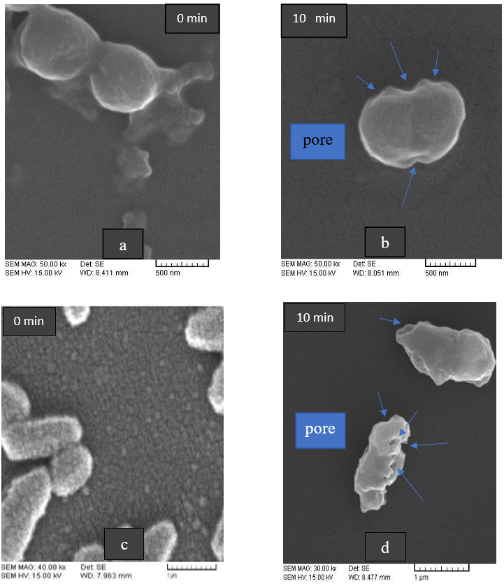
For the surface electron microscopic (SEM) analysis, we treated the microorganism suspensions with SLA for 10 minutes. To further examine alterations in the bacterial cell membranes, we scanned the treated and untreated bacteria using SEM of the SLA (Figure 4).
Timed Kill Evaluation
We only considered the MBC concentrations of SLA to study the time it took to kill the bacteria. For this purpose, 1000 µL/mL of CFS was poured into a Falcon tube. Then, 1000 µL/mL from the 24-hour culture of bacterial suspension (106 CFU/mL) was added to the tube series. The tubes were incubated at 37°C for 5, 10, 15 or 20 minutes. Next, 500 µL/mL was removed from each tube at the predetermined time and used for counting the bacterial colonies. For this purpose, we prepared dilution series up to 10-6 in tubes containing normal saline. Then, we removed a 100 µL/mL aliquot from each tube and cultured the bacterial samples on Muller Hinton agar. These plates were incubated at 37°C for 18-24 hours and the colony counting was performed based on CFU/mL, based on the following formula [20, 21]:

Number of Bacteria × Dilution / Volume = CFU

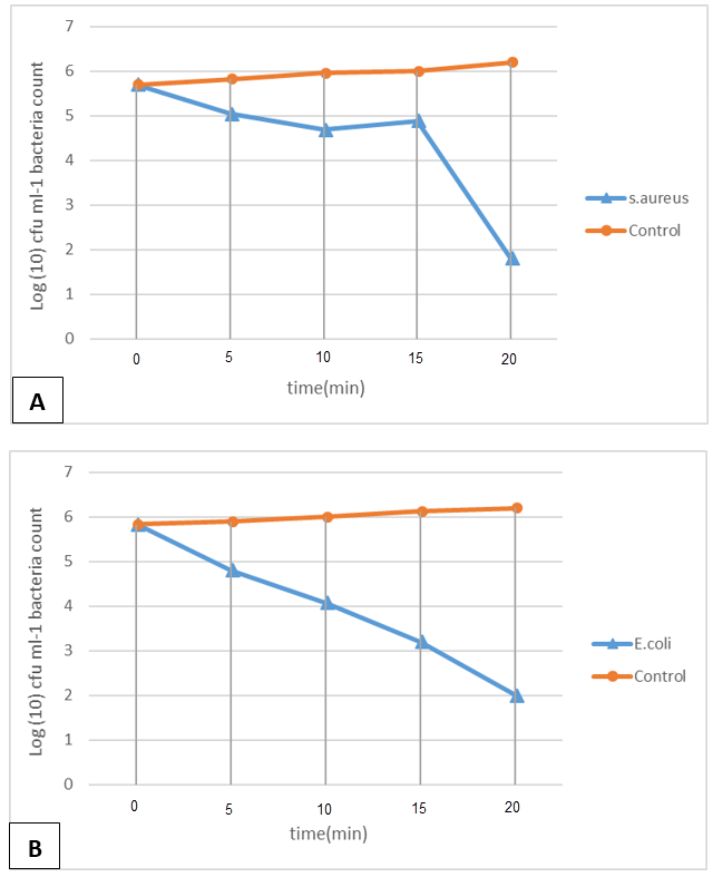
We used MRS broth for the negative control. The log CFA/mL of S. aureus (ATCC 25923) and E. coli (ATCC 25922) were considered at 0, 5, 10, 15 or 20 minutes after exposure to SLA. A control test was performed with the microorganisms but without the agent (Figure 5). The bactericidal or bacteriostatic property of the agent was determined by the assessment of the initial (log CFU/mL) and the final values (log CFU/mL) [22].
Determination of the Reduction Percentage
The cell counts from the plates in previous step were added to the following formula: Reduction Percentage = CFU0 - CFU1 / CFU0 × 100. The reduction percentages of E. coli and S. aureus were derived after exposure to the MBC concentration of SLA.
Cell Culture
The specific cell line samples were purchased from the Iranian Biological Resource Center. Samples of human normal fibroblasts (Hu02, IBRC C10309) were cultured in a T25 flask, containing DMEM (Dulbecco's modified eagle medium) with high glucose (Gibco, USA), 10% fetal bovine serum (FBS), powder penicillin and streptomycin (100 µg/mL; Sigma, USA) in an incubator (37°C, 5% CO2).
MTT Assay
The cytotoxic effect of the SLA was evaluated on Hu02 cells, using MTT assay after passaging and trypsinizing the cells. For this purpose, the cells were seeded on 96-well micro-titer plates with 10000 cells per well and incubated overnight. Freshly prepared SLA at the concentrations of 1/2 x MIC, MIC or 2 x MIC were added to the wells and incubated for 24, 48 or 72 hours (37°C, 5% CO2). Wells without SLA were considered as the negative control. Next, 20μL of 5 mg/mL MTT solution (3-[4,5-dimethylthiazole-2-yl]-2,5-diphenyltetrazolium bromide) was added to each well and the microplate was incubated in the dark at 37°C for 4-hrs. Thereafter, 150μL of DMSO (dimehtylsulfoxide) was added to each well. The microtitre plate was shaken for 5-10 minutes. After the formazan crystals had been dissolved, the supernatant’s absorbance was read on a microplate reader at 570 nm (Biotech, elx800, USA) [23]. The percent viability in each group was determined based on the following formula:
Percent Viability: (sample absorbance / average absorbance negative control) × 100 [24].
Statistical Analyses
To analyze the data, descriptive statistical processing, including central tendency indices (means and standard deviations) were used in the form of graphs on Excel and SPSS software, version 26. The mean rank difference in the inhibitions of the SLA was surveyed by Friedman test. The SLA toxicity was examined at varying concentrations and time points by one-way ANOVA and Tukey post-hoc tests.
Results
Qualitative Assessment - Agar Well Diffusion
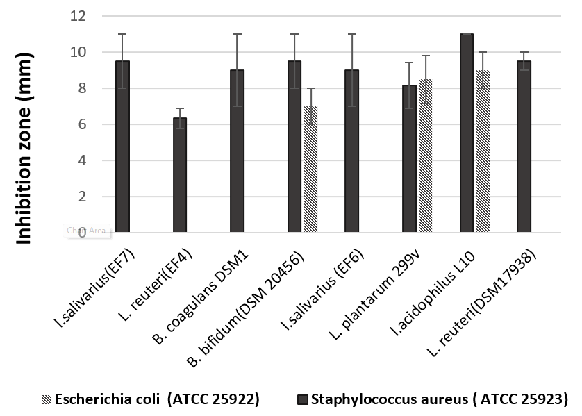
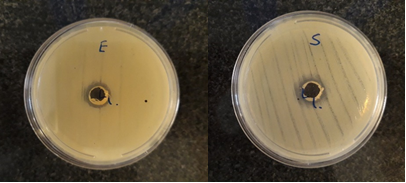
The antimicrobial activity of the strains were evaluated in triplicates against the E. coli and S. aureus batches. The inhibitory diameter of the probiotics CFS against the E. coli and S. aureus strains was measured (Figure 2) and the mean was calculated (Figure 1). The results indicated that SLA had the highest inhibition diameter against both E. coli (P=0.005) and S. aureus (P=0.019) strains based on the Friedman test.
Quantitative Assessment
In this step, the minimum inhibitory concentration (MIC) and minimum bactericidal concentration (MBC) were determined. Table 1 shows the MIC and MBC of SLA against E. coli and S. aureus. Based on the data shown in Table 1, the agent was considered bactericidal if the following relation applied: MBC/MIC ratio 4.


Table 1. Minimum Inhibitory Concentration (MIC), the Minimum Bactericidal Concentration (MBC) and MBC/MIC ratio of SLA against E. coli (ATCC 25922) and S. aureus (ATCC 25923).
SLA (µL/mL)
MIC, mean + SD MBC, mean + SD MBC/MIC Ratio
Escherichia coli (ATCC 25922) 12.5 ±0.00 12.5 ±0.00 1
Staphylococcus aureus (ATCC 25923) 12.5 ±0.00 25 ±0.00 2

Table 2. The reduction percentage of S. aureus ATCC 25923 and E. coli ATCC 25922 after exposure to SLA at different culture times.
Percent Reduction Vs Culture Time (min)
5 10 15 20
S. aureus (ATCC 25923) 15 53 77 99.98
E. coli (ATCC 25922) 25 96 97 99.99

Table 3. percentage viability of the Hu02 cells following 24, 48 and 72 hours of incubation at different concentrations (1/2 MIC, MIC, 2 MIC) of SLA, analyzed by one-way ANOVA and Tukey post-hoc test.
Viability Vs Time 6.25 12.5 25 P-value
Viability after 24hr 80.02 ± 5.03 50.45 ± 5.25 62.42 ± 7.15 0.002
Viability after 48hr 81.09 ± 5.05 60.94 ± 4.37 64.20 ± 5.01 0.005
Viability after 72hr 48.56 ± 1.87 31.71 ± 5.94 61.12 ± 15.31 0.026

Figure 1. Mean inhibitory diameter of CFS of probiotic strains at a concentration of 100 µL/mL, against E. coli and S. aureus (means ±SD’s).


Figure 2. The inhibition zone of SLA (100 μL/mL) against A: E. coli (9 mm) and, B:  S. aureus (11 mm) using well-diffused method.


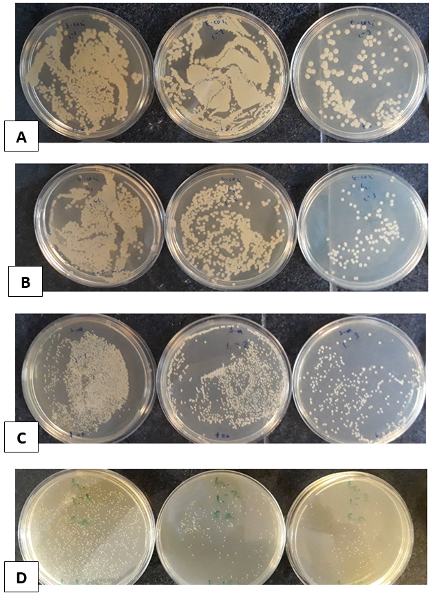
Figure 3. Microbial plates (10-1-10-3), evaluating the killing time effects of S. aureus (ATCC 25923) and E. coli (ATCC 25922) treated with SLA.
A: E. coli without exposure to SLA at t = 0 min. B: E. coli after exposure to SLA at t = 10 min. C: S. aureus without exposure to SLA at t = 0 min. D: S. aureus after exposure to SLA at t = 10 min.


Figure 4. The surface electron micrographs of bacteria, S. aureus and E. coli, treated versus untreated with SLA. (a): S. aureus without wall damage; (b): S. aureus with wall damages and numerous pores present in the treated cells; (c): E. coli without wall damage; (d) E. coli with numerous pores present in the treated cells.


Figure 5. The log counts of (A): S. aureus (ATCC 25923) and, (B): E. coli (ATCC 25922) at: 0, 5, 10, 15 and 20 minutes.


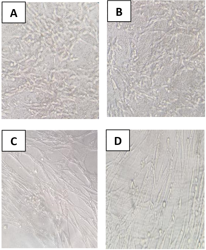
Figure 6. Normal skin fibroblasts (Hu02) before and after treatment with SLA (24 or 72 hrs).
A: Cells before treatment (control; 24 hrs). B: Cells before treatment (control; 72 hrs).
C: Cells treated with SLA at 12.5 µL/mL (24 hrs). D: Cells treated with SLA at 12.5 µL/mL (72 hrs).
Timed Killing
To evaluate the killing time of S. aureus and E. coli, we considered the MIC concentrations of SLA. Plates were counted based on CFU/mL (Figure 3a-3d), and the SEM micrographs as shown in Figure 4a-4d. The log counts of S. aureus and E. coli was considered at (t=0, 5, 10, 15 or 20 min) after exposure to the SLA (Figure 5).
According to Figure 3, microbial plates (10-1-10-3) treated and untreated with SLA were used to evaluate of the killing time of S. aureus (ATCC 25923) and E. coli (ATCC 25922). Figure 3 shows cells with and without cell wall damages.
According to Figure 5, SLA had bactericidal effect against S. aureus and E. coli, reducing the starting log CFU/mL by greater than 3 logs.
The Percent Reduction of Bacterial Cells
The percent reduction of E. coli and S. aureus after exposure to the MBC concentration of SLA was evaluated and the results are shown in Table 2. These findings demonstrated that 99.98% of S. aureus and 99.99% of E. coli were eliminated after exposure to SLA following a 20-min incubation.
MTT Assay
For MTT assay, the cytotoxic effect of SLA was evaluated on Hu02 cells (Figure 6 & Table 3), based on one-way ANOVA and Tukey post hoc test. Table 3 shows that there was a significant difference in percent viability of the Hu02 cells from the various SLA concentrations for the three exposure times (24, 48 or 72 hours). Increasing the concentration from 6.25 µL/mL to 12.5 µL/mL (P=0.002) and 25 µL/mL (P=0.025) significantly decreased the viability of the Hu02 cells after a 24-hr incubation. However, increasing the concentration from 12.5 to 25 µL/mL decreased the viability of the cells insignificantly after 24 hours of exposure. After the cells were exposed to SLA, increasing the concentration from 6.25 to 12.5 µL/mL (P=0.005) and 25 µL/mL (P=0.012) significantly decreased the percent viability of the cells over a 48-hr exposure. However, changing the concentration from 12.5 to 25 µL/mL decreased the viability of the cells insignificantly (48-hr exposure). Increasing the concentration from 12.5 to 25 µL/mL significantly increased the viability of the Hu02 cells after 72 hours (P=0.022).
Discussion
The current study explored the antibacterial and cytotoxic properties of the cell-free supernatant from Lactobacillus acidophilus (SLA) strain against gram-positive and gram-negative bacteria. This subject has not been investigated in previous studies.
This study showed that the strongest inhibition was produced by SLA. Earlier, Piatek, et al. have reported that a mixture of lactobacilli had a similar inhibition against E. coli [25]. The study conducted by Soleymanzadeh, et al. in 2020 showed that Lactobacillus strain and the product from its supernatant could be potential alternatives to existing antibiotics in controlling resistant infections [16]. Other studies have shown that another probiotic extract has also anti-pathogenic activity against S. aureus, E. coli, and P. aeruginosa [26, 27].
An earlier study conducted by Zahradnik in 2009 has shown that lactobacillus spp has a notable role in inhibiting microbial populations and their side effects [28], although the consumption of probiotic cells may cause few complications for patients with severe immune deficiencies [29]. In this context, we used the CFS of a probiotic species in this research. Other researchers have reported that the cell wall components in the broth of living or dead probiotics, and the bacteriocins component produced by probiotics, also contribute to the beneficial effects [30].
In the current study, the MIC and MBC of the SLA against E. coli were equal (12.5 μL/mL). Likewise, the MIC and MBC of L. acidophilus supernatant against Proteus strain was 25 mg/mL as reported earlier by Goodarzi, et al. [31]. In another study conducted by Sadatzadeh, et al. in 2018, the MIC of L. casei against Streptococcus spp was reported to be 12.5 mg/mL [26]. The results of these studies were fairly consistent with those of our study, in which SLA reduced 53% and 96% of S. aureus and E. coli colonies, respectively, after a 10-min exposure. In the current study, the inhibition rate approached essentially 100% against both S. aureus and E. coli after only a 20-min treatment. In another study, it was reported that L. casei has an inhibitory effect against E. coli and P. aeruginosa about 71% and 80%, respectively, while this rate approached about 75% for L. plantarum against S. aureus [32].
Anas, et al. have reported that the whole culture of L. plantarum has antimicrobial effects against S. aureus and E. coli [33]. In this regard, there have been reports about factors, such as organic acids, hydrogen peroxide, or bacteriocins produced by probiotics, inhibiting the growth of pathogenic bacteria [32, 34]. Another study has reported that Lactobacillus spp demonstrates strong inhibitory effects against S. aureus, which was consistent with our well diffusion study [32]. In recent years, researchers have shown that L. acidophilus has inhibitory effects against the growth of Enterobacteriaceae family [30, 35]. Indeed, most of the inhibitory effects have been related to the bacteriocin, the secondary metabolite from probiotics.  The highest amount of bacteriocins is generated in the initial stage of bacterial growth. All Lactobacilli tend to reduce the pH in the culture environment by secondary metabolites, such as organic acids after a 24-hr exposure. These organic acids are considered to be antimicrobials [31].
In the current study, the viability of Hu02 cells was 81.09% following a 48-hr incubation at a concentration of 6.25 µL/mL. In this context, the study by Dolati, et al. conducted in 2021 showed that the IC50 of Bacillus coagulans supernatant against cancer cells had low cytotoxic effect (only 23%) against a cell line from human foreskin fibroblasts (HFF) after a 48-hr exposure. But, the viability of HFF cells at 6 mg/mL was 60%, which later reached 100% at a concentration of 1-2 mg/mL [36].
We demonstrated that the viability of the Hu02 cells increased when the concentration was raised from 12.5 to 25 µL/mL following a 72-hr incubation (31.71% vs 61.12%). Metabolites produced by probiotic bacteria, such as organic acids and exopolysaccharides can affect cell proliferation. They can induce apoptosis by up-regulating the genes while down-regulating the anti-apoptotic genes [37, 38]. Nami, et al. have shown the effect of E. lactis metabolites on FHs-74 normal cells had not toxic effect and 95% of them grew well [39]. It seems that, probiotic supernatant at certain concentrations can be ineffective against growth inhibition or they may even enhance the growth.
Limitation of the Study
We did not use whole cultures of probiotic bacteria, although we used a wide range of supernatants from different probiotic strains. In future studies, we plan to use whole probiotic cultures, and examine the effect of the supernatants with or without microbial cells.
Conclusions
Considering the inhibitory effects of SLA and its low toxicity, we suggest that it is an effective and safe candidate for the inhibition of bacteria, such as S. aureus and E. coli. In addition, since the examined agent has low toxicity, its application is likely to reduce the side effects caused by the use of numerous antibiotics.
Conflict of Interests
The authors declare no conflict of interests with any entities.
Funding
This work was supported by a grant from the Iran University of Medical Sciences (grant No: 19107).
Acknowledgements
The authors would like to acknowledge the Department of Microbiology, Faculty of Advanced Sciences, Medical Sciences University Islamic Azad, Tehran, Iran and the Antimicrobial Resistance Research Center, Institute of Immunology and Infectious Diseases, Iran University of Medical Sciences, Tehran, Iran.
Compliance with Ethical Guideline
The concept and protocol of this study were reviewed and approved by the Institutional Review Board of Iran University of Medical Sciences, Tehran, Iran, prior to its conduction (Ethics review & approval No.: IR .IUMS.REC1399.863).
Authors’ Contributions
Project administration and supervision: Reza Hosseini Doust and Sara Minaean, Investigation and data curation: Ali majidpour, Mahdi Adabi, Microbial assey and writing, editing and approval of final manuscript: All authors.
Type of Study: Research | Subject: General

References
1. Wilson RM, Walker JM, Yin K. Different Concentrations of Lactobacillus acidophilus Cell Free Filtrate Have Differing Anti-Biofilm and Immunomodulatory Effects. Front Cell Infect Microbiol. 2021;11:737392. [DOI:10.3389/fcimb.2021.737392] [PMID] []
2. Dodds DR. Antibiotic resistance: A current epilogue. Biochem Pharmacol. 2017;134:139-46. [DOI:10.1016/j.bcp.2016.12.005] [PMID]
3. Santajit S, Indrawattana N. Mechanisms of Antimicrobial Resistance in ESKAPE Pathogens. Biomed Res Int. 2016;2016:2475067. [DOI:10.1155/2016/2475067] [PMID] []
4. Boucher HW, Talbot GH, Bradley JS, Edwards JE, Gilbert D, Rice LB, et al. Bad bugs, no drugs: no ESKAPE! An update from the Infectious Diseases Society of America. Clin Infect Dis. 2009;48(1):1-12. [DOI:10.1086/595011] [PMID]
5. Soleymanzadeh Moghadam S, Khodaii Z, Fathi Zadeh S, Ghooshchian M, Fagheei Aghmiyuni Z, Mousavi Shabestari T. Synergistic or Antagonistic Effects of Probiotics and Antibiotics- Alone or in Combination- on Antimicrobial-Resistant Pseudomonas aeruginosa Isolated from Burn Wounds. Archives of Clinical Infectious Diseases. 2018;13(3). [DOI:10.5812/archcid.63121]
6. Kadri SS. Key Takeaways From the U.S. CDC's 2019 Antibiotic Resistance Threats Report for Frontline Providers. Crit Care Med. 2020;48(7):939-45. [DOI:10.1097/CCM.0000000000004371] [PMID] []
7. Spizek J, Sigler K, Rezanka T, Demain A. Biogenesis of antibiotics-viewing its history and glimpses of the future. Folia Microbiol (Praha). 2016;61(4):347-58. [DOI:10.1007/s12223-016-0462-y] [PMID]
8. Peral MC, Martinez MA, Valdez JC. Bacteriotherapy with Lactobacillus plantarum in burns. Int Wound J. 2009;6(1):73-81. [DOI:10.1111/j.1742-481X.2008.00577.x] [PMID] []
9. Knackstedt R, Knackstedt T, Gatherwright J. The role of topical probiotics on wound healing: A review of animal and human studies. Int Wound J. 2020;17(6):1687-94. [DOI:10.1111/iwj.13451] [PMID] []
10. Yan F, Polk DB. Probiotics and immune health. Curr Opin Gastroenterol. 2011;27(6):496-501. [DOI:10.1097/MOG.0b013e32834baa4d] [PMID] []
11. Campana R, van Hemert S, Baffone W. Strain-specific probiotic properties of lactic acid bacteria and their interference with human intestinal pathogens invasion. Gut Pathog. 2017;9:12. [DOI:10.1186/s13099-017-0162-4] [PMID] []
12. Nagoba BS, Selkar SP, Wadher BJ, Gandhi RC. Acetic acid treatment of pseudomonal wound infections--a review. J Infect Public Health. 2013;6(6):410-5. [DOI:10.1016/j.jiph.2013.05.005] [PMID]
13. Loo AE, Wong YT, Ho R, Wasser M, Du T, Ng WT, et al. Effects of hydrogen peroxide on wound healing in mice in relation to oxidative damage. PLoS One. 2012;7(11):e49215. [DOI:10.1371/journal.pone.0049215] [PMID] []
14. Fijan S, Frauwallner A, Langerholc T, Krebs B, Ter Haar Nee Younes JA, Heschl A, et al. Efficacy of Using Probiotics with Antagonistic Activity against Pathogens of Wound Infections: An Integrative Review of Literature. Biomed Res Int. 2019;2019:7585486. [DOI:10.1155/2019/7585486] [PMID] []
15. Sadeghi-Bazargani H, Maghsoudi H, Soudmand-Niri M, Ranjbar F, Mashadi-Abdollahi H. Stress disorder and PTSD after burn injuries: a prospective study of predictors of PTSD at Sina Burn Center, Iran. Neuropsychiatr Dis Treat. 2011;7:425-9. [DOI:10.2147/NDT.S23041] [PMID] []
16. Soleymanzadeh Moghadam S, Mohammad N, Ghooshchian M, FathiZadeh S, Khodaii Z, Faramarzi M, et al. Comparison of the effects of Lactobacillus plantarum versus imipenem on infected burn wound healing. Med J Islam Repub Iran. 2020;34:94. [DOI:10.47176/mjiri.34.94] [PMID] []
17. Valdez JC, Peral MC, Rachid M, Santana M, Perdigon G. Interference of Lactobacillus plantarum with Pseudomonas aeruginosa in vitro and in infected burns: the potential use of probiotics in wound treatment. Clin Microbiol Infect. 2005;11(6):472-9. [DOI:10.1111/j.1469-0691.2005.01142.x] [PMID]
18. Divyashree S, Anjali PG, Somashekaraiah R, Sreenivasa MY. Probiotic properties of Lactobacillus casei - MYSRD 108 and Lactobacillus plantarum-MYSRD 71 with potential antimicrobial activity against Salmonella paratyphi. Biotechnol Rep (Amst). 2021;32:e00672. [DOI:10.1016/j.btre.2021.e00672] [PMID] []
19. Appiah T, Boakye YD, Agyare C. Antimicrobial Activities and Time-Kill Kinetics of Extracts of Selected Ghanaian Mushrooms. Evid Based Complement Alternat Med. 2017;2017:4534350. [DOI:10.1155/2017/4534350] [PMID] []
20. Mogana R, Adhikari A, Tzar MN, Ramliza R, Wiart C. Antibacterial activities of the extracts, fractions and isolated compounds from Canarium patentinervium Miq. against bacterial clinical isolates. BMC Complement Med Ther. 2020;20(1):55. [DOI:10.1186/s12906-020-2837-5] [PMID] []
21. Messick CR, Rodvold KA, Pendland SL. Modified time-kill assay against multidrug-resistant Enterococcus faecium with novel antimicrobial combinations. J Antimicrob Chemother. 1999;44(6):831-4. [DOI:10.1093/jac/44.6.831] [PMID]
22. Primavilla S, Pagano C, Roila R, Branciari R, Ranucci D, Valiani A, et al. Antibacterial Activity of Crocus sativus L. Petals Extracts against Foodborne Pathogenic and Spoilage Microorganisms, with a Special Focus on Clostridia. Life (Basel). 2022;13(1). [DOI:10.3390/life13010060] [PMID] []
23. Elikaei A, Vazini H, Javani Jouni F, Zafari J. Investigating Cytotoxic Effects of Juniperus Excelsa Extract on Esophageal Cancer Cell Line KYSE-30 and Normal Fibroblast Cell Line HU02. Medical Laboratory Journal. 2019;13(5):13-8. [DOI:10.29252/mlj.13.5.13]
24. Remzova M, Zouzelka R, Brzicova T, Vrbova K, Pinkas D, Rossner P, et al. Toxicity of TiO(2), ZnO, and SiO(2) Nanoparticles in Human Lung Cells: Safe-by-Design Development of Construction Materials. Nanomaterials (Basel). 2019;9(7). [DOI:10.3390/nano9070968] [PMID] []
25. Piatek J, Krauss H, Ciechelska-Rybarczyk A, Bernatek M, Wojtyla-Buciora P, Sommermeyer H. In-Vitro Growth Inhibition of Bacterial Pathogens by Probiotics and a Synbiotic: Product Composition Matters. Int J Environ Res Public Health. 2020;17(9). [DOI:10.3390/ijerph17093332] [PMID] []
26. Saadatzadeh A, Ameri A, Moghimipour E, Bahmani B, Gholipour S. Comparison of Antibacterial Efficacy of Probiotic Mouthwash with Chlorhexidine Against Common Oral Pathogens: An In-Vitro Study. Jundishapur Journal of Natural Pharmaceutical Products. 2018;13(1). [DOI:10.5812/jjnpp.65029]
27. Saadatzadeh A, Fazeli MR, Jamalifar H, Dinarvand R. Probiotic Properties of Lyophilized Cell Free Extract of Lactobacillus casei. Jundishapur J Nat Pharm Prod. 2013;8(3):131-7. [DOI:10.17795/jjnpp-8564] [PMID] []
28. Zahradnik RT, Magnusson I, Walker C, McDonell E, Hillman CH, Hillman JD. Preliminary assessment of safety and effectiveness in humans of ProBiora3, a probiotic mouthwash. J Appl Microbiol. 2009;107(2):682-90. [DOI:10.1111/j.1365-2672.2009.04243.x] [PMID]
29. Besselink MG, van Santvoort HC, Buskens E, Boermeester MA, van Goor H, Timmerman HM, et al. Probiotic prophylaxis in predicted severe acute pancreatitis: a randomised, double-blind, placebo-controlled trial. Lancet. 2008;371(9613):651-9. [DOI:10.1016/S0140-6736(08)60207-X] [PMID]
30. Quinto EJ, Jiménez P, Caro I, Tejero J, Mateo J, Girbés T. Probiotic Lactic Acid Bacteria: A Review. Food and Nutrition Sciences. 2014;05(18):1765-75. [DOI:10.4236/fns.2014.518190]
31. Goudarzi L, Kermanshahi RK, Moosavi-Nejad Z, Dalla MM. Evaluation of antimicrobial activity of probiotic lactobacillus strains against growth and urease activity of proteus spp. Journal of Medical Bacteriology. 2017;6(3-4):31-43.
32. Ershadian M, Branch D, Azad I. The Antimicrobial and Co-aggregation effects of probiotic lactobacilli against some pathogenic bacteria. Iranian Journal of Medical Microbiology. 2015;9(3):14-22.
33. Anas M, Eddine HJ, Mebrouk K. Antimicrobial activity of Lactobacillus species isolated from Algerian raw goat's milk against Staphylococcus aureus. World Journal of Dairy & Food Sciences. 2008;3(2):39-49.
34. Collado MC, Meriluoto J, Salminen S. Adhesion and aggregation properties of probiotic and pathogen strains. European Food Research and Technology. 2007;226(5):1065-73. [DOI:10.1007/s00217-007-0632-x]
35. Prashant, Tomar SK, Singh R, Gupta SC, Arora DK, Joshi BK, et al. Phenotypic and genotypic characterization of lactobacilli from Churpi cheese. Dairy Science and Technology. 2009;89(6):531-40. [DOI:10.1051/dst/2009029]
36. Dolati M, Tafvizi F, Salehipour M, Movahed TK, Jafari P. Inhibitory effects of probiotic Bacillus coagulans against MCF7 breast cancer cells. Iran J Microbiol. 2021;13(6):839-47. [DOI:10.18502/ijm.v13i6.8089] [PMID] []
37. Jafari-Nasab T, Khaleghi M, Farsinejad A, Khorrami S. Probiotic potential and anticancer properties of Pediococcus sp. isolated from traditional dairy products. Biotechnol Rep (Amst). 2021;29:e00593. [DOI:10.1016/j.btre.2021.e00593] [PMID] []
38. Dehghani N, Tafvizi F, Jafari P. Cell cycle arrest and anti-cancer potential of probiotic Lactobacillus rhamnosus against HT-29 cancer cells. Bioimpacts. 2021;11(4):245-52. [DOI:10.34172/bi.2021.32] [PMID] []
39. Nami Y, Haghshenas B, Haghshenas M, Abdullah N, Yari Khosroushahi A. The Prophylactic Effect of Probiotic Enterococcus lactis IW5 against Different Human Cancer Cells. Front Microbiol. 2015;6:1317. [DOI:10.3389/fmicb.2015.01317] [PMID] []

Add your comments about this article : Your username or Email:
CAPTCHA

Send email to the article author


Rights and permissions
Creative Commons License This work is licensed under a Creative Commons Attribution-NonCommercial 4.0 International License.

© 2025 CC BY-NC 4.0 | Iranian Journal of Toxicology

Designed & Developed by : Yektaweb