Write your message
Volume 18, Issue 2 (May 2024)                   IJT 2024, 18(2): 113-119 | Back to browse issues page

Ethics code: IR.MUI.MED.REC.1399.699


XML Print


Download citation:
BibTeX | RIS | EndNote | Medlars | ProCite | Reference Manager | RefWorks
Send citation to:

Samsaam Shariat S, Gheshlaghi F, Zoofaghari S. Effectiveness of Plasmapheresis in the Treatment and Clinical Outcome of Patients with Aluminum Phosphide Poisoning: A Randomized Controlled Clinical Trial. IJT 2024; 18 (2) :113-119
URL: http://ijt.arakmu.ac.ir/article-1-1296-en.html
1- Department of Clinical Toxicology, Clinical Toxicology Research Center, Isfahan University of Medical Sciences, Isfahan, Iran
2- Department of Clinical Toxicology, Isfahan University of Medical Sciences, Isfahan, Iran , Shafeajafar@med.mui.ac.ir
Full-Text [PDF 669 kb]   (230 Downloads)     |   Abstract (HTML)  (773 Views)
Full-Text:   (316 Views)

Introduction

Pesticide poisoning, with a mortality rate of 70%, accounts for more than 50% of all poisonings in some South Asian countries [1, 2]. Mortality due to aluminum phosphide (AlP) poisoning has been reported to be high in Iran during the last two decades, with a reported mortality rate of 30-100% [3, 4]. AlP, a deadly poison, is used to disinfect and protect grains, particularly rice, sold under such brand names as QuickPhos and Celphos, and known commonly in Iran as Rice tablets [5, 6]. The mortality caused by AlP poisoning, which has no specific antidote, has been reported to be about 70%, with the main cause being shock and multiple organ failure [7, 8].
Several treatment approaches have been taken for this poisoning, the most aggressive of which are intra-aortic balloon pump (IABP) and extracorporeal membrane oxygenation (ECMO). However, these treatments are both very invasive and expensive and require highly expert practitioners who are not readily available most of the time [9]. Phosphine, similar to cyanide, inhibits mitochondrial cytochrome oxidase and cellular oxygen utilization. The direct toxic effects of phosphine on cardiac myocytes, fluid loss, and adrenal glands can induce profound circulatory collapse. The direct corrosive effects of phosphide and phosphine on human tissues have been reported in the literature [10-12]. In AlP poisoning, phosphine gas dissolved in plasma is heavy and cannot be removed by hemodialysis. Therefore, studies have suggested that in these cases, phosphine gas can be removed from the blood by plasmapheresis [11-13].
Plasmapheresis has distinct advantages over hemodialysis and hemoperfusion. Primarily, there is no need for a central catheter for the patient because this procedure can be performed through two large peripheral vessels. Secondly, plasmapheresis, unlike hemodialysis and hemoperfusion, does not depend on the size of the molecules considered for detoxification or separation [14, 15]. Therapeutic plasma exchange (TPE), or plasmapheresis, is utilized to treat a wide range of diseases. The purpose of this procedure is to remove aggressive agents from the plasma that induce many clinical symptoms in patients [15]. TPE is recommended for treating patients with various diagnoses in different medical specialties. This procedure is employed to remove or reduce the concentration of proteins, fats, protein-bound hormones, antibodies, antigens, or immune complexes from the patients’ blood [14-18].
Numerous studies have reported the efficacy of plasmapheresis in the treatment of patients intoxicated with organophosphates (OP), amitriptyline, Amanita muscaria, propranolol, amlodipine, diltiazem, verapamil, carbamazepine, theophylline, levothyroxine, and toxic heavy metals, such as mercury [15, 19-25]. However, there are still conflicting views among experts as to the efficacy of plasmapheresis in the management of poisoning cases [22]. Considering the high prevalence of AlP poisoning and the incidence of deaths among patients, inadequate attention has been paid to plasmapheresis by clinicians, and contradictory findings reported by previous studies, additional research is warranted on the efficacy of plasmapheresis. This can provide evidence-based results for both researchers and healthcare practitioners. The available literature on plasmapheresis is mostly case reports [20, 21, 23-25]. Currently, there is only one in-depth study conducted on this treatment method, which was published by our research team in 2021 [14].
Aim of the Study: Given the above facts, this study was planned to evaluate the efficacy of plasmapheresis over a randomized controlled clinical trial in the treatment and clinical outcome of patients poisoned with AlP.

Materials and Methods

Study Population: The recruited population for this clinical trial included 90 patients with AlP poisoning who were referred to the Clinical Toxicology Department of Khorshid and Al-Zahra hospitals in Isfahan, Iran, between the years 2020 and 2022. Using a sample size formula, 30 patients were assigned to the intervention group, while 60 patients were allocated to the control group, at a 95% confidence level and 80% test power. Moreover, considering the case report nature of previous studies on this subject [20-24], the maximum ratio of patients with AlP poisoning in this study was 35%, with the error level being 0.25.
Ethical Guidelines: The study protocol was carefully reviewed and approved based on the university’s ethical guidelines by the Ethics Committee of Isfahan University of Medical Sciences. The study was awarded the approval code for ethics: IR.MUI.MED.REC.1399.699 and the clinical trial code: IRCT20200507047344N2 (https://en.irct.ir/trial/52254). Likewise, the written informed consent form for review and signature by the patients or their next of kin was also reviewed by the same committee and approved.
Inclusion and Exclusion Criteria: The criteria for including patients in the study were as follows: 1) being between 20 and 65 years old, 2) having no risk of blood coagulation disorder, and 3) having no history of chronic kidney disease.
The subjects were 90 eligible patients who voluntarily entered the study following a consecutive non-probability sampling, meaning that all eligible patients were identified and included in the study until the adequate sample size was met. The 90 patients were then divided into two groups of experimental (n=30) and control (n=60) patients using random allocation software. Patients were excluded from the study in cases of having bleeding or coagulation disorders that would be discovered during the intervention.
Patients’ Demographic and Laboratory Information: At the beginning of the study, patients’ gender, age, and the following clinical and laboratory parameters were evaluated and recorded: Pulse rate, systolic and diastolic blood pressure, the blood levels of calcium (Ca), potassium (K), magnesium (Mg), sodium (Na), glucose, and phosphorus (P), liver enzymes (ALT and AST), blood gases, cell counts and pH, partial pressure of carbon dioxide, bicarbonate (HCO3), partial pressure of oxygen, blood oxygen saturation, white and red blood cell count (WBC and RBC), hemoglobin (Hb), hematocrit (HCT), platelet count, lymphocytes (Lymph), and blood albumin (Alb) level.
Physical Monitoring: It should be noted that all patients underwent cardiac and hemodynamic monitoring. The patients were attended to by medical personnel if they were clinically restless and thirsty, or had signs of hypotension, tachycardia, cold, or perspired limbs, or if they had signs of metabolic acidosis, based on the venous blood gas data, pH≤7.2, and HCO3≤15 mEq/L.
Routine Treatments: The following routine treatment protocol was carried out for all patients: A continuous infusion of normal saline (in bolus and slowly) and norepinephrine was administered in addition to organic oil, such as almond or castor oil, by gavage. In case any sign of cardiac instability was observed, the rate and amount of fluid therapy administration were adjusted. The intravenous administration was performed of sodium bicarbonate at 2 meq/kg bolus every hour, vitamin C at 150 mg/h, n-acetyl cysteine at 300 mg/kg every 24 h, and magnesium sulfate and calcium gluconate at 1 g every six hours. Vitamin E was also given intramuscularly at 300 units every 12 h.
Group Treatments: The patients in the control group received only the above-mentioned routine treatments. Patients in the experimental group received plasmapheresis in addition to the routine treatment within the first six hours of the patients’ admission to the hospital.
Plasmapheresis Administration: The plasma volume for plasmapheresis was calculated in liters using the formula 0.07×body weight (kg)×(1-HCT) [23]. Approximately, 3 L of plasma was taken from the patient and replaced with 1.5 L of fresh frozen plasma (FFP), 1500 cc of normal saline, and 2-3 vials of albumin. This treatment lasted for 3-3.5 h. After the plasmapheresis, the levels of blood Ca, P, Na, K, and the complete blood cell count (CBC) were tested. Next, 12 h after the intervention, the following clinical and laboratory parameters were assessed and documented: PR, SBP, DBP, levels of Ca, K, Mg, Na, BS, P, ALT, AST, PCo2, HCO3, BE, PO2, O2sat, WBC, RBC, Hb, HCT, PLT, Lymph, and other tests, including Alb, and blood pH. Further, in cases of complications, such as tachypnea, dyspnea, respiratory distress, anaphylaxis, hypocalcemia, hypotension, metabolic alkalosis, hypokalemia, coagulation disorders, bleeding, mortality, recovery, and/or discharge from the hospital, the specific facts were recorded in the patients’ charts.
It is worth noting that in cases of acute dyspnea, a chest X-ray was requested with the suspicion of transfusion-related acute lung injury (TRALI). Moreover, appropriate treatment was provided in cases of any other complications. In order to comply with blinding conditions and prevent bias in data recording, the data collector and statistician had no knowledge of the type of intervention administered to either group.
Statistical Analyses: All of the collected data were entered into SPSS software (version 26) on a private and confidential computer under each patient’s code which were not identifiable except by the senior study team members. The data were presented as means±standard deviations (SD) or frequency and percentages. At the level of inferential statistics, an independent sample t-test was used to compare the means of variables between the two groups at the beginning of the study. Moreover, analysis of covariance (ANCOVA) was run to compare the variables between the two groups 12 h after the intervention by adjusting for the patients’ age, gender, and other variables at baseline. Furthermore, the paired sample t-test was used to compare the means of variables 12 h after the intervention and was compared to the baseline data for each of the two groups. In all analyses, the significance level between pairs of data was set at P≤0.05.

Results

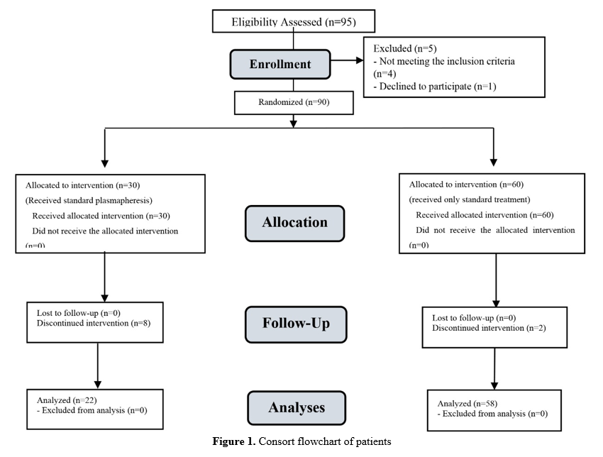
Patients’ Demographics: In this study, 8 out of 30 patients in the plasmapheresis group and 2 out of 60 patients in the control group were excluded from the study due to their unwillingness to participate in the study. Therefore, the data analyses were performed for 22 patients in the plasmapheresis group and 58 patients in the control group, as shown in Figure 1. The plasmapheresis group included 9 females (40.9%) and 13 males (59.1%) with a mean age of 29.54±11.59 years old, while the control group consisted of 21 females (36.2%) and 37 males (63.8%) with a mean age of 30.88±11.31 years old (P>0.05). See Table 1 for details.


Figure 1. Consort flowchart of patients

Table 1. Basic characteristics of patients with AlP poisoning in the two groups
Characteristics Plasmapheresis (n=22) Control (n=58) P-value
Gender:
Female 9 (40.9%) 21 (36.2%) 0.797
Male 13 (59.1%) 37 (63.8%)
Age: (year) 29.54±11.59 30.88±11.31 0.641
Comorbidity:
None 13 (59.1%) 38 (65.5%) 0.368
Diabetes 1 (4.5%) 5 (8.6%)
Hypertension 2 (9.1%) 6 (10.4%)
Mental diseases 6 (27.3%) 9 (15.5%)
Changes in Patients’ Blood Parameters: Examination of the patients’ clinical and laboratory parameters before admission and 12 h after the intervention showed that only the patients’ HCT in the plasmapheresis group was significantly lower than that of those in the control group 12 h after the intervention (P=0.046). Other parameters evaluated at baseline and 12 h after the intervention showed no significant differences between the two groups (P>0.05). Moreover, within-group comparisons indicated that the means for SpO2, HCO3, and INR increased, while the means for PLT, Hb, HCT, Alb, Ca, and K declined significantly in the plasmapheresis group 12 h after the intervention, compared to those before the intervention (P<0.05). The means of SpO2 and PO2 increased, while the means of PLT, RBC, Hb, Alb, Ca, and K decreased significantly in the control group 12 h after the intervention, compared to before the intervention (P<0.05, Table 2).
Clinical Outcomes: Evaluation of the clinical outcomes revealed that there were one case of bradycardia (4.5%), one case of itching (4.5%), and six cases of deaths (27.3%) recorded in the plasmapheresis group, while 14 cases of deaths (24.1%) occurred in the control group. There were no statistically significant differences in the number of deaths between the two groups (P>0.05, Table 3).

Table 2. Specification and comparison of clinical and laboratory parameters of patients with AlP poisoning in the two study groups
Variables Plasmapheresis group (n=22) Control Group (n=58) P-value1, 2
PR; bpm Baseline 99.59±26.72 88.14±15.82 0.070
12 h after the intervention 95.50±21.92 83.00±9.03 0.145
P-value3 0.609 0.099
SBP; mmHg Baseline 103.09±20.91 112.27±26.86 0.154
12 h after the intervention 135.00±21.21 117.07±13.85 0.129
P-value3 0.500 0.096
DBP; mmHg Baseline 62.41±21.01 69.51±16.21 0.113
12 h after the intervention 75.00±7.07 72.77±13.75 0.836
P=value3 0.989 0.613
SpO2; % Baseline 60.81±27.42 56.03±28.37 0.516
12 h after the intervention 69.72±29.45 74.16±28.95 0.666
P-value3 0.044 0.032
PCo2 Baseline 40.09±21.38 40.08±13.07 0.997
12 h after the intervention 41.65±11.20 37.58±15.51 0.242
P-value3 0.352 0.349
PO2; mmHg Baseline 43.25±36.10 37.73±25.40 0.454
12 h after the intervention 50.01±25.72 47.24±17.86 0.722
P-value3 0.161 0.031
HCO3; mEq/L Baseline 18.78±5.08 21.23±6.08 0.103
12 h after the intervention 23.71±9.78 22.47±5.06 0.661
P-value3 0.028 0.217
Ph Baseline 7.35±0.15 7.40±0.62 0.684
12 h after the intervention 7.27±0.31 7.35±0.18 0.382
P-value3 0.341 0.487
PLT count; ×109/L Baseline 227.62±81.34 210.32±89.70 0.443
12 h after the intervention 145.54±96.44 175.11±55.73 0.310
P-value2 0.006 0.039
INR Baseline 1.25±0.95 1.16±0.13 0.095
12 h after the intervention 1.59±0.22 1.56±0.93 0.921
P-value3 0.013 0.238
WBC count; ×109/L Baseline 10.50±5.36 11.91±12.87 0.622
12 h after the intervention 7.00±3.58 7.69±3.52 0.648
P-value2 0.178 0.149
Lymph count; ×109/L Baseline 28.54±13.19 30.45±21.17 0.704
12 h after the intervention 23.71±16.22 25.32±11.88 0.784
P-value3 0.660 0.534
RBC count; ×1012/L Baseline 4.89±0.81 5.21±0.56 0.095
12 h after the intervention 4.14±0.45 4.49±0.70 0.206
P-value3 0.161 0.001
Hb; g/dL Baseline 13.92±2.16 15.17±4.44 0.211
12 h after the intervention 11.31±1.51 12.55±1.89 0.114
P-value3 <0.001 0.005
HCT; % Baseline 41.71±5.10 43.60±6.39 0.222
12 h after the intervention 34.31±3.31 38.05±4.80 0.046
P-value3 0.001 0.356
Alb; g/dL Baseline 4.35±0.55 4.211±0.69 0.586
12 h after the intervention 3.63±0.25 3.24±0.43 0.420
P-value3 0.013 0.007
BS; mg/dL Baseline 157.33±76.24 133.20±61.88 0.160
12 h after the intervention 231.12±45.16 138.00±43.06 0.118
P-value3 0.116 0.162
AST; U/L Baseline 30.50±16.07 24.31±9.41 0.118
12 h after the intervention 38.60±19.70 20.00±8.08 0.102
P-value3 0.187 0.123
ALT; U/L Baseline 33.50±20.82 25.51±19.06 0.122
12 h after the intervention 42.60±22.87 24.00±27.09 0.190
P-value3 0.107 0.685
Ca; mg/dL Baseline 8.48±2.00 9.09±0.82 0.196
12 h after the intervention 7.67±0.84 8.11±0.89 0.203
P-value3 0.495 <0.001
P; mg/dL Baseline 3.45±0.40 5.50±7.72 0.386
12 h after the intervention 4.50±2.24 3.33±1.76 0.679
P-value3 0.425 0.441
Mg; mg/dL Baseline 2.24±0.59 2.51±2.68 0.668
12 h after the intervention 2.08±0.61 2.00±0.45 0.708
P-value3 0.734 0.597
K; mmol/L Baseline 4.16±0.62 3.91±0.46 0.860
12 h after the intervention 3.55±0.50 3.56±0.49 0.945
P-value3 0.005 0.001
Na; mEq/L Baseline 142.02±4.06 135.74±25.79 0.296
12 h after the intervention 131.29±41.33 134.53±32.20 0.808
P-value3 0.385 0.263
1: The significance level obtained from the independent samples t-test comparing the means of variables between the two groups at the beginning of the study.
2: The significance level obtained from Analysis of covariance (ANCOVA) comparing the means of the variables between the two groups 12 h after the intervention by adjusting age, gender, and variables at baseline.
3: The significance level obtained from the paired samples t-test comparing the means of variables 12 h after the intervention as compared to the baseline by adjusting the patients’ age and gender in each of the two groups.

Table 3. The patients’ outcome with AlP poisoning between the two study groups
Outcome Plasmapheresis group (n=22) Control Group (n=58) P-value
Bradycardia 1 (4.5%) 0 (0%) 0.071
Itching 1 (4.5%) 0 (0%) 0.071
Death 6 (27.3%) 14 (24.1%) 0.778

Disussion

The results of this study showed that plasmapheresis resulted in a significant increase in SpO2, HCO3, and INR and significant declines in PLT, Hb, HCT, Alb, Ca, and K levels. In the control group, the means of SpO2 and PO2 increased, while those of PLT, RBC, Hb, Alb, Ca, and K levels decreased significantly. However, these changes were not significant between the two groups upon between-group comparisons, and only the patients’ HCT in the plasmapheresis group was significantly lower than that of the patients’ in the control group 12 h after the intervention. This finding was justifiable as the blood plasma volume was obtained based on the HCT level in this study.
It is worth noting that although plasmapheresis caused a significant increase in HCO3, it did not reduce the patients’ acidosis significantly. This event might have been caused by concurrent respiratory acidosis in patients. In addition, metabolic acidosis was the main cause of death in these patients, which improved relatively following plasmapheresis. This was one of the advantages of plasmapheresis. We made a similar assertion in our previous article published in 2021 [14]. This suggestion was based on our clinical experience derived from our observations of the clinical outcomes in several patients with AlP poisoning that had lasted for only six hours. The patients’ condition improved, and the mortality rate decreased significantly [14].
To explain this, it is likely that the phosphine gas released from AlP is removed from the blood through plasma exchange due to its solubility in plasma. However, the gas may not be eliminated by dialysis due to its high volume of distribution. In contrast, Nenov et al. tried to remove dimethoate (an insecticide), but they failed to do so [22]. The discrepancy between the above finding and that of the present study can be attributed to differences in the type, time, amount of poisoning, and the duration of plasmapheresis.
In line with the current study, another investigation on the effect of plasmapheresis on a 19-year-old young woman poisoned with OP showed that a one-time plasmapheresis treatment led to an acceptable clinical outcome, and the patient regained consciousness upon performing plasmapheresis. In addition, the patient’s blood oxygen saturation and plasma cholinesterase level improved significantly. Therefore, we would recommend the use of plasmapheresis in toxicology clinics to manage serious cases of poisoning due to organophosphates [20]. Further, Ahila et al. have claimed that replacing cholinesterase with fresh frozen plasma could achieve desirable clinical outcomes in patients poisoned with similar compounds [19]. The findings from the study conducted by Rahimi et al. also indicate that the transfusion of fresh RBCs improves metabolic acidosis and increases the survival rate of patients poisoned with AlP [12].
In the current study, the oxygen saturation improved significantly in both study groups. However, the HCO3 level improved significantly only in the plasmapheresis group. Therefore, the decline in acidosis was more evident in the plasmapheresis group, but the mortality rate was not significantly different between the two groups. The potential of this new treatment to reduce acidosis is of great significance for researchers because acidosis is the most important cause of death in these patients. This finding can be associated with promising results with respect to patients’ clinical outcomes. Conceivably, the lack of a significant difference in the mortality rate in this preliminary study can be attributed to the small sample size. This point justifies the necessity of conducting further studies on this subject in the future.
Zamani et al. have suggested that removing damaged RBCs by any method in similar patients is likely to improve clinical outcomes. Although blood transfusion was associated with hemolysis, jaundice, fever, and hematuria, these authors believed that it saved their patients’ lives, as there was no treatment available at the time other than conservative management with antioxidants [26].
In the current study, 4.5% of the patients developed bradycardia and itching. Further, 27.3% of our patients died in the plasmapheresis group, while only 24.1% of them died in the control group. It may be stated that the complications that occur upon plasmapheresis are very rare, and the mortality rates are not significantly different between the two groups. In another study conducted by Disel et al., the patients suffered from hypoxia, which was attributed to respiratory muscle dysfunction and the accumulation of secretions in the lungs [20]. In two other studies, the plasmapheresis complications included anaphylactic reactions to IgA in the infused plasma, urticaria, TRALI, increased risk of infection, hypotension due to citrate in the infused plasma, vasovagal reaction, and coagulation disorder [27, 28].
It must be noted that some patients who were candidates for plasmapheresis were critically ill, and imminent death had been expected for them. Moreover, this method is the last-line treatment and the ultimate chance for these patients to survive; therefore, the above-mentioned complications are expected. As a result, paying attention to the patients’ dyspnea, bronchospasm, urticarial lesions, and hypotension is highly recommended. Furthermore, repeated evaluations of the patient’s blood parameters during and after plasmapheresis play important roles in controlling the patients’ condition and preventing the complications. 
Advantages and Limitations: The strong point of this study is that it was the first clinical trial of its kind. However, the small sample size and the lack of assessment for the patients’ employment and marital status, lifestyle, and previous poisoning incidents or suicide plans were among the study’s limitations. Lastly, since no significant differences were found in the patients’ mortality rate between the two groups, we recommend that additional studies be conducted in the future to establish the generalizability of the current study’s findings.
Conclusions
Based on the findings of this study, plasmapheresis is likely to increase the blood level of HCO3 significantly without impacting acidosis, despite the fact that in treatments with or without plasmapheresis, the levels of Hb, Alb, Ca, K, and PLT decreased and that of blood oxygen saturation (SpO2) increased significantly. Therefore, plasmapheresis significantly reduced acidosis in patients with AlP poisoning. Likewise, this treatment did not lower the mortality rate for patients in this intervention. Finally, conducting more studies on plasmapheresis is warranted, even in the form of case reports, so that the efficacy of this intervention can be evaluated more accurately.
Conflict of Interests
The authors declare that they had no conflict of interests.
Funding
This work was supported by the Office of Deputy Research and Technology, Isfahan University of Medical Sciences, Isfahan, Iran (Grant# 199358).
Acknowledgement
The authors would like to express their special gratitude to all patients for their participation in this study.
Compliance with Ethical Guidelines
Ethical issues, including plagiarism, data fabrication, and double publication, have been completely observed by the authors. The ethical approval code was also obtained from Isfahan University of Medical Sciences (Code: IR.MUI.MED.REC.1399.699), and the clinical trial code was approved and awarded (IRCT-20200507047344N2).
Authors' Contributions
Shiva Samsaam Shariat (SSS), Farzad Gheshlaghi (FG), and Shafeajafar Zoofaghari (SZ).
SZ designed the study, interpreted the data, and wrote the initial draft of the manuscript. FG designed the study and interpreted the data. SSS contributed to the statistical analysis and interpretation of the data. FG and SZ contributed to conducting the experiments and reviewed/edited the manuscript. All authors have read and agreed to the content of this article in advance of submission to the IJT.
Type of Study: Research | Subject: General

References
1. Eddleston M, Karalliedde L, Buckley N, Fernando R, Hutchinson G, Isbister G, et al. Pesticide poisoning in the developing world - a minimum pesticides list. Lancet.2002;360:1163-67. [doi:10.1016/S0140-6736(02)11204-9]
2. Eddleston M. Poisoning by pesticides. Medicine. 2020;48(3):214-17. [doi:10.1016/j.mpmed.2019.12.019]
3. Bagherian F, Kalani N, Rahmanian F, Abiri S, Hatami N, Foroughian M, et al. Aluminum phosphide poisoning mortality rate in Iran; A systematic review and meta-analysis. Arch Acad Emerg Med. 2021;9(1): 29-36. [doi: 10.22037/aaem.v9i1.1396] [pmid: 34870232]
4. Dorooshi G, Mirzae M, Fard NT, Zoofaghari S, Mood NE. Investigating the outcomes of aluminum phosphide poisoning in khorshid referral hospital, Isfahan, Iran: A retrospective study. Journal of Research in Pharmacy Practice. 2021;10(4):166. [doi:10.4103/jrpp.jrpp_88_21]
5. Garg KK. Review of aluminum phosphide poisoning. International Journal of Medical Science and Public Health. 2020;9(7):392-400. [doi:10.5455/ijmsph.2020.07118202020072020]
6. Singh S, Singh D, Wig N, Jit I, Sharma BK. Aluminum phosphide ingestion - a clinico-pathologic study. Journal of Toxicology: Clinical Toxicology. 1996;34(6):703-06. [doi:10.3109/15563659609013832]
7. Mathai A, Bhanu MS. Acute aluminium phosphide poisoning: Can we predict mortality?.Indian Journal of Anaesthesia. 2010; 54 (4): 302-07. [doi:10.4103/0019-5049.68372]
8. Hashemi-Domeneh B, Zamani N, Hassanian-Moghaddam H, Rahimi M, Shadnia S, Erfantalab P, et al. A review of aluminium phosphide poisoning and a flowchart to treat it. 2016;67:183-93. [Arhiv Za Higijenu Rada I Toksikologiju.]
9. Araújo RV, Santos SD, Igne Ferreira E, Giarolla J. New advances in general biomedical applications of PAMAM dendrimers. Molecules. 2018;23(11):2849. [doi:10.1515/aiht-2016-67-2784]
10. Gheshlaghi F, Lavasanijou MR, Moghaddam NA, Khazaei M, Behjati M, Farajzadegan Z, et al. N‑acetylcysteine, ascorbic acid, and methylene blue for the treatment of aluminium phosphide poisoning: Still beneficial? Toxicol Int. 2015; 22:40‑4. [doi:10.4103/0971-6580.172255]
11. Dorooshi G, Zoofaghari S, Mood NE, Gheshlaghi F. A newly proposed management protocol for acute aluminum phosphide poisoning. Journal of Research in Pharmacy Practice. 2018; 7:168‑69. [doi:10.4103/jrpp.JRPP_18_12]
12. Moghadamnia AA. An update on toxicology of aluminum phosphide. DARU J Pharm Sci . 2012;20:1-8. [doi:10.1186/2008-2231-20-25]
13. Ari HF, Turhan M, Baspinar H, Kirhan B, Ari M. Aluminum phosphide toxicity: A rare cause of multiorgan dysfunction syndrome. Indian Pediatrics Case Reports. 2022;2(2):91. [doi:10.4103/ipcares.ipcares_3_22]
14. Shariat SS, Zoofaghari S, Gheshlaghi F. Effectiveness of plasmapheresis in aluminum phosphate poisoning. Journal of Research in Pharmacy Practice. 2021;10(1):57. [doi:10.4103/jrpp.JRPP_21_27]
15. Ibrahim RB, Balogun RA. Medications in patients treated with therapeutic plasma exchange: prescription dosage, timing, and drug overdose. Seminars Dialysis. 2012;25:176e189. [doi:10.1111/j.1525-139X.2011.01030.x]
16. Samtleben W, Mistry‐Burchardi N, Hartmann B, Lennertz A, Bosch T. Therapeutic plasma exchange in the intensive care setting. Ther Apher. 2001;5(5):351-57. [doi:10.1046/j.1526-0968.2001.00383.x]
17. Yazdi MF, Baghianimoghadam M, Nazmiyeh H, Ahmadabadi AD, Adabi MA. Response to plasmapheresis in myasthenia gravis patients: 22 cases report. Romanian journal of internal medicine. 2012;50(3):245-47. [Link]
18. Yokoyama S, Kikkawa T, Hayashi R. Plasmapheresis: new trends in therapeutic applications. Therapy of hypercholesterolemia. In Proc 2nd International Symposium on Plasmapheresis: Artificial Organs. Supplement. 1984. [Link]
19. Ahila Ayyavoo DN, Muthialu N, Ramachandran P. Plasmapheresis in organophosphorus poisoning-intensive management and its successful use. J Clin Toxicol.2011;1:2161-95. [Link]
20. Disel NR, Acikalin A, Kekec Z, Sebe A. Utilization of plasmapheresis for organophosphate intoxication: A case report. Turkish Journal of Emergency Medicine. 2016;16(2):69-71. [doi:10.1016/j.tjem.2015.06.003]
21. Güven M, Sungur M, Eser B. The effect of plasmapheresis on plasma cholinesterase levels in a patient with organophosphate poisoning. Human & Experimental Toxicology. 2004;23(7):365-68. [doi:10.1191/0960327104ht462cr]
22. Nenov VD, Marinov P, Sabeva J, Nenov DS. Current applications of plasmapheresis in clinical toxicology. Nephrology dialysis transplantation. 2003;18(suppl_5):v56-8. [doi:10.1093/ndt/gfg1049]
23. Deng Y, Qiu L. Therapeutic plasma exchange: a second‐line treatment for brodifacoum poisoning following an anaphylactoid reaction to vitamin K. Clin Case Rep. 2017;5(1):35. [doi:10.1002/ccr3.756] [pmid: 28096987]
24. Sari I, Turkcuer I, Erurker T, Serinken M, Seyit M, Keskin A. Therapeutic plasma exchange in amitriptyline intoxication: case report and review of the literature. Transfusion and Apheresis Science. 2011;45(2):183-85. [doi:10.1016/j.transci.2011.07.015]
25. Patel N, Bayliss GP. Developments in extracorporeal therapy for the poisoned patient. Advanced Drug Delivery Reviews. 2015; 90:3-11. [doi:10.1016/j.addr.2015.05.017]
26. Zamani N, Hassanian-Moghaddam H, Ebrahimi S. Whole blood exchange transfusion as a promising treatment of aluminium phosphide poisoning. Arhiv Za Higijenu Rada I Toksikologiju. 2018;69(3):275-7. [doi:10.2478/aiht-2018-69-3129]
27. Kartal O, Gulec M, Caliskaner Z, Nevruz O, Cetin T, Sener O. Urticaria Case Reports: 587 Plasmapheresis in a Patient with "Refractory" Urticarial Vasculitis. World Allergy Organ J. 2012;5(Suppl 2):S203. [doi:10.1097/01.WOX.0000411702.50677.f2]
28. Basic‐Jukic N, Kes P, Glavas‐Boras S, Brunetta B, Bubic‐Filipi L, Puretic Z. Complications of therapeutic plasma exchange: experience with 4857 treatments. Therapeutic Apheresis and Dialysis. 2005;9(5):391-95. [doi:10.1111/j.1744-9987.2005.00319.x]

Add your comments about this article : Your username or Email:
CAPTCHA

Send email to the article author


Rights and permissions
Creative Commons License This work is licensed under a Creative Commons Attribution-NonCommercial 4.0 International License.

© 2025 CC BY-NC 4.0 | Iranian Journal of Toxicology

Designed & Developed by : Yektaweb