Write your message
Volume 18, Issue 2 (May 2024)                   IJT 2024, 18(2): 61-70 | Back to browse issues page

Ethics code: 01/21


XML Print


Download citation:
BibTeX | RIS | EndNote | Medlars | ProCite | Reference Manager | RefWorks
Send citation to:

Fernandes de Lima Neto M, Igo Teixeira de Assis E, Venância Nunes Azevedo A, Raiane Feitosa Melo Paulino L, Aragão Matos Donato M, Alves Peixoto C, et al . Extract of Actaea racemosa Protects Mice Ovarian Follicles Against Doxorubicin-induced Toxicity. IJT 2024; 18 (2) :61-70
URL: http://ijt.arakmu.ac.ir/article-1-1333-en.html
1- Laboratory of Biotechnology and Physiology of Reproduction–LABIREP, Federal University of Ceara, Sobral, CE, Brazil
2- Laboratory of Ultrastructure, CNPqAM/FIOCRUZ, Federal University of Pernambuco Recife, PE, Brazil
3- Nucleus of Biotechnology Applied to Ovarian Follicle Development, Federal University of São Francisco Valley, Petrolina, PE, Brazil
4- Nucleus of Research in Animal Experimentation–NUPEX, Federal University of Ceara, Sobral, CE, Brazil
5- Laboratory of Biotechnology and Physiology of Reproduction–LABIREP, Federal University of Ceara, Sobral, CE, Brazil , jrvsilva@ufc.br
Full-Text [PDF 1376 kb]   (242 Downloads)     |   Abstract (HTML)  (903 Views)
Full-Text:   (224 Views)

Introduction

Advances in early cancer diagnosis and therapeutic protocols have offered patients great chances of cure and survival. However, the side effects of chemotherapy have adverse consequences, such as gonadal toxicity and the loss of ovarian function [1, 2]. Doxorubicin (DOX), an antitumor antibiotic from the anthracycline class, is one of the antineoplastic agents used in clinical practice. Interfering with the synthesis and functioning of nucleic acids is the primary way in which it acts. In DNA, DOX intercalates and causes double strand breaks, interferes with the replication and transcription processes, and/or interrupts the repair process mediated by topoisomerase-II [3]. Damages to the ovaries include decreased ovarian reserve due to increased follicular atresia, oxidative stress, destruction of ovarian stromal cells, and increased apoptosis [4-6].
Some studies have shown that phytomedicines have great potential to protect the gonads against the deleterious effects of chemotherapy [7, 8]. Actaea racemosa (A. racemosa) bearing the botanical name Cimicifuga racemosa, or the common name Cimicifuga (Black cohosh), is an herbaceous plant of the Ranunculaceae family. The roots and rhizomes have attracted the greatest interest in pharmacology, where hundreds of other agents have been identified, including isoflavones, phenolic constituents, alkaloids, tannins, and triterpene glycosides [9]. A. racemosa has traditionally been used to treat pain and inflammation, cardiovascular disease, osteoporosis, and especially for the signs and symptoms of menopause [10, 11]. In addition, the extract of A. racemosa has strong antioxidant and anti-inflammatory effects [12]. Studies in postmenopausal women have found that the daily use of A. racemosa extract has beneficial effects on menopausal symptoms [13, 14] and improves endothelial function [15]. The extract also preserves mitochondrial integrity and ATP levels and reduces reactive oxygen species (ROS) formation and cell death in cultured neural and liver cells [16]. Recently, De Assis et al. [17] have shown that A. racemosa extract protects the ovarian tissue against the adverse effects of DOX in cultured mice ovaries. In rats with polycystic ovary syndrome, the A. racemosa extract has been shown to improve the antioxidant status, hormonal and lipid profiles, glucose levels, and liver functions. It has also induced Ki-67 expression in granulosa, theca, and stromal cells, which is a marker for cell proliferation [18]. In this way, by stopping cell apoptosis, the A. racemosa extract could be able to lessen ovarian damage brought on by DOX. However, no in vivo studies have been conducted to evaluate the effect of A. racemosa extract on protecting the ovaries against damage caused by DOX in animal or human models.
Aim of the Study: The purpose of this study was to determine if various dosages of A. racemosa extract could protect mice’s ovaries from damage caused by DOX. The effects of this extract were evaluated on follicle survival and growth, ovarian stromal cells, collagen fibers, cellular ultrastructure, levels of mRNA for Caspase-6 and TNF-α, and the expression of TNF-α protein.

Materials and Methods

Animals: This study is consistent with normative resolutions and the guidelines of the Ethics Committee on the Use of Animals of the Federal University of Ceara (Protocol No. 01/21). Adult Swiss female mice (Mus musculus) (n=42) were kept with free access to filtered water and mice feed (Nuvilab®). They were kept under 12-hour light/dark cycles at 22±2°C.
Evaluation of Estrous Cycle: The estrous cycle was evaluated for 10 days, as previously established [19]. Briefly, a vaginal smear was collected, placed on histological slides, and evaluated under light microscopy (Nikon, Eclipse, TS 100, Japan). The mice’s status was identified as proestrus, estrus, metestrus, or diestrus based on the cells that were observed. Only mice with a regular estrous cycle were used in the experiments.
Chemicals: The tested drugs were DOX (Libbs, Fauldoxo®, DOX hydrochloride), A. racemosa (L.) (MarjanFarma, Aplause®), and 20 mg of dry extract in a standardized 5-7% tablet equivalent to 1.0-1.4 mg of triterpene glycosides expressed as 23-epi-26-deoxyactein. The doses of A. racemosa extract were prepared by crushing the tablets and diluting them in a commercially available saline solution.
Experimental Design: Animals (n=42) were randomly divided into six groups, as shown in Figure 1. The mice from the control group (group 1) were pre-treated orally with saline solution (0.15 M NaCl), and after one hour, they received saline solution (0.15 M NaCl) intraperitoneally (i.p.). The mice in group 2 were pre-treated with saline solution (0.15 M NaCl), and one hour later, they received a single dose of DOX (10 mg/kg, i.p.). The animals in group 3 received 0.5 mg/kg A. racemosa extract, and after one hour, they were treated with saline solution (0.15 M NaCl, i.p.). In group 4, the mice were pre-treated with the 0.5 mg/kg A. racemosa extract, and one hour later, they received a single dose of DOX (10 mg/kg, i.p.). Animals in group 5 were pre-treated with the 5 mg/kg A. racemosa extract (5 mg/kg), and after one hour, they received saline solution (0.15 M NaCl, i.p.). Animals in group 6, received the 5 mg/kg A. racemosa extract, and one hour later, they received a single dose of DOX (10 mg/kg, i.p.). Mice in groups 3-6 were treated with A. racemosa extract once a day for 10 days.


Figure 1. Experimental Design. AR: Actaea racemosa extract; NaCl: saline solution; G: group; DOX: doxorubicin.
Morphological Assessment of Follicular Development: At the end of the treatment, the mice ovaries were removed and fixed in paraformaldehyde (4% in phosphate buffered saline [PBS], pH=7.4) for 48 h. They were then dehydrated in graded ethanol, clarified with xylene, and embedded in paraffin. They were serially sectioned at 7 μm thick and stained with hematoxylin and eosin (H&E).
The follicles were classified as follows: a) primordial: oocytes surrounded by a layer of granulosa cells in a squamous shape, b) primary: oocytes and a layer of granulosa cells in a cuboidal shape, c) secondary: oocytes surrounded by two or more granulosa cell layers in a cuboidal shape, and d) tertiary or antral: oocytes surrounded by many layers of granulosa cells with a visible antral cavity. Follicles with retracted oocytes, pycnotic nuclei, and/or surrounded by disorganized granulosa cells were classified as degenerated [20]. For the evaluation of follicular activation and growth, the follicles were classified as primordial, primary, secondary, or tertiary, as previously described [18]. The number of stromal cells was counted in an area of 100 μm2 of ovaries from all treatment groups, as previously described [18]. For each treatment sample, five optic fields were evaluated from various sections.
Analysis of the Extracellular Matrix and Density: To evaluate the collagen fibers, the ovarian sections were stained with Picrosirius Red (Abcam Kit), as described by Rittié [21]. The sections underwent a one-hour incubation period at room temperature in a Sirius Red solution (0.1%) after being dewaxed using xylene. A 0.5% acetic acid solution was then used to eliminate the extra color. For each treatment, the areas occupied by collagen fibers in five different optical fields were calculated using a camera mounted on the microscope. The images were analyzed by Image J software (version 1.51p, 2017). To obtain staining levels of collagen fibers, the average pixel intensity of the total area was measured after subtracting the background.
Immuno-histochemistry Assay: The ovarian sections were mounted on Starfrost glass slides (Knittel, Braunschweig, Germany), and TNF-α was analyzed, as described previously [22]. To retrieve antigenicity, the sections were incubated in citrate buffer at 95ºC in a decloaking chamber (Biocare; Concord, USA) for 40 min, while the endogenous peroxidase activity was blocked by incubation with peroxidase for 10 min. The sections were incubated with 10% normal goat serum for 10 min to block nonspecific binding sites. They were then incubated in a humidified chamber for 50 min at room temperature with anti-TNF-α antibody. Finally, they were incubated for 20 min with Easy Link One polymer, followed by DAB staining and counterstaining with hematoxylin for one minute. For negative controls, the tissues were incubated with a blocking buffer and without the primary antibody. The intensity of immune-staining was classified as absent, weak, moderate, or strong [23].
Ultrastructural Analysis by Transmission Electron Microscopy: The samples were fixed overnight in a solution containing 4% paraformaldehyde and 2.5% glutaraldehyde in 0.1 M cacodylate buffer. They were post-fixed in 1% osmium tetroxide, 0.8% potassium ferric cyanide, and 2 mM calcium chloride in 0.1 M cacodylate buffer at pH 7.2, dehydrated in acetone, and embedded in EMbed 812. The polymerization was performed at 60°C for three days. Ultrathin sections were counterstained with 5% uranyl acetate and lead citrate and examined under a transmission electron microscope (FEI Morgani 268D).
mRNA Analyses by Quantitative Real-time PCR (RT-qPCR): Utilizing a TRIzol® purification kit (Invitrogen; São Paulo, Brazil), total RNA extraction was carried out. A total of 20 μL was used for reverse transcription, made up of 10 μL of sample containing 1 mg of RNA, 4 μL of reverse transcriptase buffer (Invitrogen), 150 units of reverse transcriptase Superscript III, 0.036 U random primers, eight units RNAs in 10 mM dithiothreitol, and 0.5 mM of each dNTP (Invitrogen). The mixture was incubated at 42.1°C for one hour, subsequently at 80°C for five minutes, and then stored at -20°C. The negative controls were prepared with no reverse transcriptase added.
The SYBR Green was used to quantify the mRNA in a Step One Plus instrument (Applied Biosystems; Foster City, CA, USA). The reactions were composed of 1 μL of cDNA as a template in 9.4 μL of SYBR Green Master Mix (PE Applied Biosystems; Foster City, CA, USA), 0.5 μM of each primer, and 9.4 μL of ultrapure water. The primers amplified the mRNAs for Caspase-3
and TNF-α. The glyceraldehyde-3-phosphate dehydrogenase (GAPDH) was used as the reference gene (See
Table 1). The specificity of each primer pair was verified by analyzing the melting curve of the qPCR results. The first 10 min of denaturation and polymerase activation at 95°C were followed by 40 cycles of 15 sec each at 95°C, 30 sec at 58°C, and 30 sec at 72°C. This was the thermal cycling profile. The final extension was performed at 72°C for 10 min. The 2^ΔΔCt method was used to normalize the data.

Table 1. Primer pairs used for real-time polymerase chain reaction tests
Target Gene Primer Sequence
(5’→3’)
Forward (F)
Reverse (R)
GenBank Accession Nº.
GAPDH GAACGGATTTGGCCGTATTG GTGAGTGGAGTCATACTGGAAC F
R
GU214026.1
CASP3 ACATGGGAGCAAGTCAGTGG
CGTCCACATCCGTACCAGAG
F
R
XM_017312543.3
TNF-α CAGAAAGCATGATCCGCGAC
CCGCCTGGAGTTCTGGAAG
F
R
D84199.2
Statistical Analyses: The percentages of primordial and developing follicles and those of normal follicles were evaluated by chi-squared test (GraphPad Prism). Data for stromal cells, collagen fibers, and the levels of mRNA for Caspase-3 and TNF-α were analyzed using the Kruskal-Wallis test, followed by Dunn’s comparison. The results were presented as means±SEM. The differences were statistically significant at P<0.05.

Results

Protective Effect of A. racemosa Extract against Mice Ovarian Toxicity Induced by Doxorubicin: Following the administration of 0.5 or 5 mg/kg A. racemosa extract, the percentage of morphologically normal follicles was maintained similar to that of the control group (SHAM). However, DOX (10 mg/kg) drastically decreased the percentage of morphologically normal follicles. The mice that received both DOX and either a dosage of 0.5 or 5 mg/kg A. racemosa extract had higher percentages of normal follicles than those only receiving DOX. The percentage of normal follicles in these mice was comparable to those that received only A. racemosa extract at either 0.5 or 5 mg/kg (Figure 2). Figures 3A-3H represent morphologically normal or degenerated follicles. Regarding follicular growth, the DOX treatment reduced the percentage of secondary follicles but had no effect on the percentage of primary, tertiary, or primordial follicles (Figure 4).


Figure 2. Percentage of normal follicles in the ovaries of mice treated with DOX (10 mg/kg) alone or both DOX and 0.5 or 5 mg/kg A. racemosa extract. a, b, and c lowercase letters indicate statistically significant differences between treatments (P<0.05). AR: A. racemosa extract.

Figure 3. Mice ovaries showing morphologically normal (a-d) and degenerated (e-h) follicles at different stages of development. (a) Normal and (e) degenerated primordial follicles; (b) Normal and (f) degenerated primary follicles; (c) Normal and (g) degenerated secondary follicle; (d) Normal and (h) degenerated tertiary follicle. Granulosa cells (GC); Oocyte (O); Oocyte nucleus (N); Antrum (A); Cumulus oocyte complex (COC); Theca cells (TC) (400×, Scale bar: 100-μm).


Figure 4. Percentage of normal follicles per category (primordial, primary, secondary, or tertiary) in the ovaries of mice treated with only DOX (10 mg/kg) or both DOX and A. racemosa extract. Lowercase letters (a, b, and ab) denote statistically significant differences between pairs of treatment groups (P<0.05). AR:A. racemosa extract.
Evaluation of Collagen in the Ovarian Extracellular Matrix: Figure 5 reflects that neither DOX nor the combined DOX and A. racemosa extract influenced the organization of collagen fibers in the ovarian extracellular matrix (Figure 5A). Figure 5B shows that stained collagen fibers were more intensely observed in regions surrounding the follicles’ basement membrane.
Evaluation of Stromal Cell Density in the Mice Ovaries: The ovarian stromal cell density in mice treated with the extract at either dosage was similar to that of the control group (SHAM). Conversely, animals treated only with DOX showed a significant decrease in their ovarian stromal cells (Figure 6). Further, when mice received both A. racemosa extract and DOX, the stromal cell numbers were similar to those of the control group (SHAM). Figure 7 shows the histological sections of mice ovaries from different treatments.
Immunohistochemical Localization of TNF-α in the Mice Ovaries: Immunohistochemical analyses showed weak TNF-α staining in the follicles of the controls (Figure 8B). Moderate TNF-α staining was observed in oocytes and granulosa cells of follicles from mice treated with either dosage of A. racemosa extract combined with DOX (Figures 8D and 8E). The ovaries of mice treated with DOX showed strong staining for TNF-α mainly in the oocytes and stromal cells (Figure 8C).


Figure 5. Levels of collagen fibers (mean±SD) in mice ovaries treated with DOX (10 mg/kg) or both DOX and A. racemosa extract (0.5 and 5 mg/kg) (a). Representative images of collagen fibers labeled Picrosirius red (b). (b1) control group; (b2) DOX (10 mg/kg); (b3) A. racemosa extract (0.5 mg/kg);
(b4) A. racemosa extract (0.5 mg/kg)+DOX (10 mg/kg); (b5) A. racemosa extract (5 mg/kg); (b6) A. racemosa extract (5 mg/kg)+DOX (10 mg/kg). Scale bar: 100 μm (400×). AR: A. racemosa extract; O: oocyte; OS: ovarian stroma. Arrows indicate ovarian follicles.


Figure 6. Stromal cell density in mice ovaries treated with DOX (10 mg/kg) or both DOX and
A. racemosa extract (0.5 and 5 mg/kg). a, b, and c lowercase letters indicate statistically significant differences between treatments (P<0.05).


Figure 7. Ovarian stromal density in mice from the control group (a) or treated with DOX (10 mg/kg) (b); A. racemosa (0.5 mg/kg) (c); A. racemosa (0.5 mg/kg)+DOX (10 mg/kg) (d); A. racemosa (5 mg/kg); (e) and A. racemosa (5 mg/kg)+DOX (10 mg/kg) (f). Scale bar: 100 μm (400×).
AR: A. racemosa extract.


Figure 8. Immunohistochemical staining for TNF-α in mice ovaries. (a) negative control; (b) control group; (c) DOX (10 mg/kg); (d) A. racemosa (0.5 mg/kg); (e) A. racemosa (0.5 mg/kg)+DOX (10 mg/kg). Scale bar: 100 μm (400×). AR: A. racemosa extract. O: oocyte; GC: granulosa cells; OS: ovarian stroma. Arrows indicate ovarian follicles.
Ultrastructural Analyses of the Mice Ovarian Follicles: Ovaries from the control group (SHAM) showed well-preserved granulosa cells and zona pellucida with transzonal projections, homogeneous oocytes with well-defined mitochondria, and nuclear membranes (Figures 9A and 9B). The same structural characteristics were observed in the follicles from the mice treated with either A. racemosa extract alone (Figures 9E and 9F) or combined with DOX (Figures 9G and 9H). Lipid inclusions were also observed in the ovarian granulosa cells of mice in both the controls and those treated with the extract at 0.5 mg/kg (Figure 9E). In mice treated with DOX, smaller amounts of lipid inclusions were observed in granulosa cells, while the oocytes had the organelles that were not well recognizable and zona pellucida lacked transzonal projections (Figures 9C and 9D).
Levels of mRNA for Caspase-3 and TNF-α in the Mice Ovaries: The results showed that mice treated with DOX had increased mRNA levels for Caspase-3 (Figure 10). However, the levels did not differ among the mice treated with either the extract alone or combined with DOX, or those from the control group (SHAM). The mRNA levels for TNF-α were not influenced by either DOX alone or combined with the extract.


Figure 9. Ultrastructural images of follicles in the ovaries of mice from the control group (a,b) or treated with DOX (10 mg/kg) (c,d); A. racemosa (0.5 mg/kg) (e,f); both A. racemosa (0.5 mg/kg) and DOX (10 mg/kg) (g,h). AR: A. racemosa extract. M: mitochondria; Nu: nucleus; ZP: zona pellucida,
L: lipid; O: oocyte; Gr: granulosa cells; Re: endoplasmic reticulum; GC: golgi complex; Mv: microvillus.


Figure 10. Levels of mRNA for Caspase 3 and TNF-α in mice ovaries from the control group (SHAM), DOX (10 mg/kg), A. racemosa extract (0.5 mg/kg), and A. racemosa extract (0.5 mg/kg)+DOX (10 mg/kg). a and b lowercase letters indicate significant differences between treatments (P<0.05). AR: A. racemosa extract.

Discussion

This is the first study to provide experimental evidence that A. racemosa extract reduces DOX-induced damage to the mice ovarian follicles. The DOX treatment induced follicular atresia, while treating mice with the extract protected the ovarian follicles. The extract has several constituents with therapeutic properties, such as antioxidant, anti-inflammatory, and anti-apoptotic properties. The 23-epi-26-deoxyactein, one of the most abundant triterpene glycosides in the extract, stimulates mitochondrial biogenesis and activates factors involved in cell proliferation [24, 25]. The isoferulic acid constituent is believed to inhibit the synthesis of pro-inflammatory cytokines [26]. In vitro studies with neural and liver cells have shown that the A. racemosa extract preserves mitochondrial integrity and ATP levels, while preventing ROS formation and cell death [16]. In addition, the beneficial effects of the extract have already been demonstrated on the antioxidant, hormonal, and proliferative status of granulosa, theca, and stromal cells in rat ovaries [18].
In the current study, DOX reduced the percentage of secondary follicles. Previous studies have shown that acute DOX damage in the mice ovary is dependent on cells and follicle types [27]. A study has shown that DOX increases the number of developing follicles by over-activating primordial follicles. However, chemotherapy drugs have cytotoxic effects, culminating in the loss of follicular reserve [4]. DOX also decreases stromal cell density in mice ovaries. These cells support the tissue and provide a suitable micro-environment for follicular development. Previous studies have reported follicular death associated with vascular and stromal cell damage in human ovaries [6, 28]. The stromal cell death and the microvascular damage induce tissue hypoxia, which may contribute to the loss of ovarian follicles indirectly. In our study, the A. racemosa extract contributed to the maintenance of stromal cells and protected them against the damages caused by DOX.
In this study, the extract did not influence the ovarian extracellular matrix. Previous studies have not shown significant accumulation of collagen in the mice ovaries regardless of their age and the estrous cycle stage [29, 30]. Oktem et al. [31] have reported cellular death in human ovarian stroma after chemotherapy, but there was no association between increased follicular degeneration and changes in the extracellular matrix. Our findings are consistent with a recent in vitro study showing no significant changes in the collagen fibers in mice ovaries cultured with either A. racemosa extract or DOX alone [17].
The TNF-α was expressed in granulosa cells and oocytes from the secondary and tertiary follicles and in oocytes from the primordial and primary follicles of mice in all experimental categories. In this study, there was a moderate staining in the ovaries of mice treated with the extract at 0.5 mg/kg alone or combined with DOX. High TNF-α is associated with inflammation, apoptosis, cell growth, and differentiation [23]. Further, its effects on the ovarian cell death are dependent on the stages of follicular development. This may be associated with different levels of expression of the receptors [32]. The type-1 receptors are linked to the intracellular necrosis, which are essential for signaling pathways that lead to apoptosis, while type-2 receptors are more related to the expression of genes involved in cellular growth, survival, and differentiation. In mice ovaries, the sites of TNF-α include oocytes, granulosa cells, corpora lutea, and macrophages [33-35].
The ultrastructural analyses showed that ovarian follicles from mice treated with the extract at 0.5 mg/kg possessed well-preserved granulosa cells, zona pellucida, oocytes, and nuclear membranes. The mice treated with DOX showed lower lipid inclusions in their granulosa cells. In addition, no damage was observed in the organelles within the oocytes. The lipids in granulosa cells are important because they serve as the substrate for the synthesis of hormones [36]. In this context, our findings are consistent with those reported by an in vitro study on A. racemosa extract (5 ng/mL) and DOX (0.3 µg/mL) [17].
The DOX treatment increased the mRNA levels for Caspase-3 in the mice ovaries. Activated Caspase-3 induces cleavage of factors that cause apoptosis [37]. A recent study in mice has shown that DOX decreases ovarian reserve through apoptosis and over-activation of primordial follicles, involving DNA damages and Caspase-3 pathway [4]. De Assis et al. [17] have shown that adding A. racemosa extract at 5 ng/mL to the culture medium of mice ovaries increased the expression of antioxidant enzymes, such as superoxide dismutase and catalase, but decreased the expression of Caspase-3.
Conclusions
Based on our findings, the A. racemosa extract protects ovarian follicles in mice against the deleterious effects of DOX and helps preserve their ultrastructure. These effects are linked to the maintenance of normal follicle density, protection of stromal cells, and reduction in the immune-staining of TNF-α in the ovarian follicles.
Conflict of Interests
The authors had no conflict of interests to disclose with any internal or external entities.
Funding
This research was supported by a Brazilian grant provided by the National Council for Scientific and Technological Development (CNPq, Brazil, Grant No: 435524/2018-6).
Acknowledgement
The authors wish to thank the colleagues at the Laboratory of Biotechnology and Reproductive Physiology (LABIREP), the Nucleus of Research in Animal Experimentation (NUPEX) and the Laboratory of Pharmacology of Sobral (LAFS), Federal University of Ceará; Sobral, CE, Brazil.
Compliance with Ethical Guidelines
This study was conducted based on the guidelines and normative resolutions of the National Council for Control in Animal Experimentation (CONCEA). It was also consistent with the guidelines approved by the Ethics Committee on the Use of Animals (CEUA) of the Federal University of Ceara (approved under protocol No.: 01/21).
Authors' Contributions
Lima Neto, MF: contributed and participated in all phases of the research, from study design to final conception. Assis, EIT and Azevedo, VAN: participated in the organization of the experiments. Paulino, LRFM: assisted with gene analysis using the RT-qPCR technique. Donato, MAM and Peixoto, CA: assisted in the transmission electron microscopy analyses. Monte, APO and Matos, MHT: assisted in the immunohistochemistry analyses. Godinho, AN and Freire, JMO: guided all phases of the research and assisted in carrying out the experiments. Barberino, RS: assisted in the statistical analyses. Silva, AWB: and Silva, JRV: guided at all stages of research and reviewed the drafts of the manuscript. All authors have read and approved the final version of the manuscript.
 
Type of Study: Research | Subject: General

References
1. Yeo W, Pang E, Liem GS, Suen JJS, Ng RYW, Yip CCH, et al. Menopausal symptoms in relationship to breast cancer-specific quality of life after adjuvant cytotoxic treatment in young breast cancer survivors. Health Qual Life Outcomes. 2020;18(1):1-9. [doi:10.1186/s12955-020-1283-x]
2. Bellver J, Donnez J. Introduction: Infertility etiology and offspring health. FertilSteril. 2019; 111(6):1033-35. [doi:10.1016/j.fertnstert.2019.04.043]
3. Mobaraki M, Faraji A, Dolati P, Ataei M, Manshadi HRD. Molecular Mechanisms of Cardiotoxicity: A Review on the Major Side-effect of Doxorubicin. Indian J Pharm Sci. 2017; 79(3):335-44. [doi:10.4172/pharmaceutical-sciences.1000235]
4. Wang Y, Liu M, Johnson SB, Yuan G, Arriba AK, Zubizarreta ME, et al. Doxorubicin obliterates mouse ovarian reserve through both primordial follicle atresia and overactivation. Toxicol Appl Pharmacol. 2019;15(381):1-11. [doi:10.1016/j.taap.2019.114714] [pmid: 31437492]
5. Niringiyumukiza JD, Cai H, Chen L, Li Y, Wang L, Zhang M, et al. Protective properties of glycogen synthase kinase-3 inhibition against doxorubicin-induced oxidative damage to mouse ovarian reserve. Biomed Pharmacother. 2019; 116:108963. [doi:10.1016/j.biopha.2019.108963]
6. Soleimani R, Heytens E, Darzynkiewicz Z, Oktay K. Mechanisms of chemotherapy-induced human ovarian aging: double strand DNA breaks and microvascular compromise. Aging (Albany NY). 2011;3(8):782-93. [doi:10.18632/aging.100363] [pmid: 21869459]
7. Siddiqui AJ, Jahan S, Singh R, Saxena J, Ashraf SA, Khan A, et al. Plants in Anticancer Drug Discovery: From Molecular Mechanism to Chemoprevention. Biomed Res Int. 2022; 2022:1-18. [DOI:10.1155/2022/5425485]
8. Rad P, Safari F, Mohammadi J, Delaviz H. Preserved Ovarian Function After Toxicity WithDoxorrubicin in Rats: Protective Effect of Nasturtium Officinale Extract. Iran JToxicol. 2021;15(1):57-64. [doi:10.32598/IJT.15.1.747.1]
9. Campos LB, Gilglioni EH, Garcia RF, Brito Mdo N, Natali MR, Ishii-Iwamoto EL, et al. Cimicifuga racemosa impairs fatty acid β-oxidation and induces oxidative stress in livers of ovariectomized rats with renovascular hypertension. Free Radic Biol Med. 2012;53(4):680-89. [doi:10.1016/j.freeradbiomed.2012.05.043]
10. Morelli V, Naquin C. Alternative therapies for traditional disease states: menopause. Am Fam Physician. 2002;66(1):129-34. [Link]
11. Kim CD, Lee WK, Lee MH, Cho HS, Lee YK, Roh SS. Inhibition of mast cell-dependent allergy reaction by extract of black cohosh (Cimicifuga racemosa). ImmunopharmacolImmunotoxicol. 2004; 26(2):299-308. [doi:10.1081/IPH-120037728]
12. Guo Y, Yin T, Wang X, Zhang F, Pan G, Lv H, et al. Traditional uses, phytochemistry, pharmacology and toxicology of the genus Cimicifuga: A review. J Ethnopharmacol. 2017; 209:264-82. [doi:10.1016/j.jep.2017.07.040]
13. Ruhlen RL, Sun GY, Sauter ER. Black Cohosh: Insights into its Mechanism(s) of Action. Integr Med Insights. 2008. [doi:10.4137/117863370800300002]
14. Liske E, Hänggi W, Henneicke-von Zepelin HH, Boblitz N, Wüstenberg P, Rahlfs VW. Physiological investigation of a unique extract of black cohosh (Cimicifugaera cemosae rhizoma): A 6-month clinical study demonstrates no systemic estrogenic effect. J Womens Health Gend Based Med. 2002; 11(2):163-74. [doi:10.1089/152460902753645308]
15. Fernandes ES, Celani MFS, Fistarol M, Geber S. Effectiveness of the short-term use of Cimicifuga racemosa in the endothelial function of postmenopausal women: A double-blind, randomized, controlled trial. Climacteric.2020;23(3):245-51. [doi:10.1080/13697137.2019.1682542]
16. Rabenau M, Unger M, Drewe J, Culmsee C. Metabolic switch induced by Cimicifuga racemosa extract prevents mitochondrial damage and oxidative cell death. Phytomedicine. 2019;52:107-6. [doi: 10.1016/j.phymed.2018.09.177] [doi:10.1016/j.phymed.2018.09.177]
17. de Assis EIT, Azevedo VAN, De Lima Neto MF, Costa FC, Paulino LRFM, Barroso PAA, et al. Protective Effect of Cimicifuga racemosa (L.) Nutt Extract on Oocyte and Follicle Toxicity Induced by Doxorubicin during In vitro Culture of Mice Ovaries. Animals (Basel). 2022;13(1):1-18. [doi:10.3390/ani13010018]
18. Azouz AA, Ali SE, Abd-Elsalam RM, Emam SR, Galal MK, Elmosalamy SH, et al. Modulation of steroidogenesis by Actaea racemosa and vitamin C combination, in letrozole induced polycystic ovarian syndrome rat model: promising activity without the risk of hepatic adverse effect. Chin Med. 2021;16(1):1-17. [doi:10.1186/s13020-021-00444-z]
19. Marcondes FK, Bianchi FJ, Tanno AP. Determination of the estrous cycle phases of rats: some helpful considerations. Braz J Biol. 2002;62(4A):609-614. [doi:10.1590/S1519-69842002000400008]
20. Gouveia BB, Barberino RS, Dos Santos Silva RL, Lins TLBG, da Silva Guimarães V, do Monte APO, et al. Involvement of PTEN and FOXO3a Proteins in the Protective Activity of Protocatechuic Acid Against Cisplatin-Induced Ovarian Toxicity in Mice. Reprod Sci. 2021;28(3):865-76. [doi:10.1007/s43032-020-00305-4]
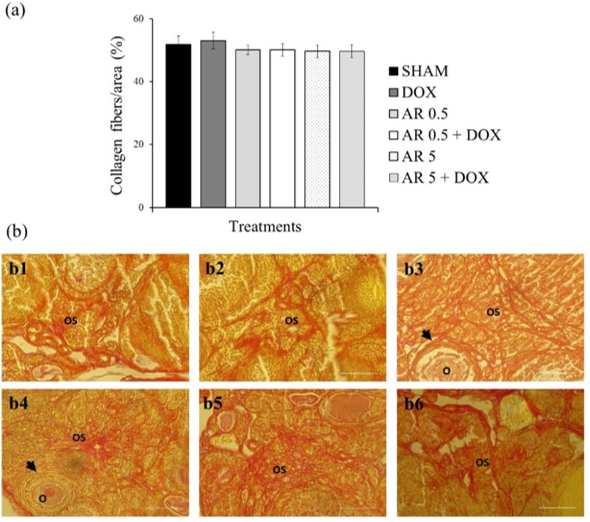
21. Rittié L. Method for Picrosirius Red-Polarization Detection of Collagen Fibers in Tissue Sections. Methods Mol Biol. 2017;1627:395-407. [doi:10.1007/978-1-4939-7113-8_26]
22. Barberino RS, Menezes VG, Ribeiro AEAS, Palheta RCJr, Jiang X, Smitz JEJ, et al. Melatonin protects against cisplatin-induced ovarian damage in mice via the MT1 receptor and antioxidant activity. Biol Reprod. 2017;96:1244-55. [doi:10.1093/biolre/iox053]
23. Kayedpoor P, Mohamadi S, Karimzadeh-Bardei L, Nabiuni M. Anti-inflammatory effect of silymarin on ovarian immunohistochemical localization of TNF-α associated with systemic inflammation in polycystic ovarian syndrome.Int J Morphol. 2017;35:723-32. [doi:10.4067/S0717-95022017000200054]
24. Suh KS, Choi EM, Jung WW, Kim YJ, Hong SM, Park SY, et al. Deoxyactein protects pancreatic β-cells against methylglyoxal-induced oxidative cell damage by the upregulation of mitochondrial biogenesis. Int J Mol Med;40(2):539-48. [doi:10.3892/ijmm.2017.3018]
25. Choi EM. Deoxyactein Isolated from Cimicifuga racemosa protects osteoblastic MC3T3-E1 cells against antimycin A-induced cytotoxicity. J Appl Toxicol. 2013;33(6):488-94. [doi:10.1002/jat.1784]
26. Schmid D, Woehs F, Svoboda M, Thalhammer T, Chiba P, Moeslinger T. Aqueous extracts of Cimicifuga racemosa and phenolcarboxylic constituents inhibit production of proinflammatory cytokines in LPS-stimulated human whole blood. Can J PhysiolPharmacol. 2009;87(11):963-72. [doi:10.1139/Y09-091]
27. Roti Roti EC, Leisman SK, Abbott DH, Salih SM. Acute doxorubicin insult in the mouse ovary is cell- and follicle-type dependent. PLoS ONE. 2012;7:42293. [doi:10.1371/journal.pone.0042293]
28. Ben-Aharon I, Meizner I, Granot T, Uri S, Hasky N, Rizel S, et al. Chemotherapy-induced ovarian failure as a prototype for acute vascular toxicity. Oncologist. 2012;17(11):1386-93. [doi:10.1634/theoncologist.2012-0172] [pmid:22956534]
29. Lliberos C, Liew SH, Zareie P, La Gruta NL, Mansell A, Hutt K. Evaluation of inflammation and follicle depletion during ovarian ageing in mice. Sci Rep.2021;11(1):1-15. [doi:10.1038/s41598-020-79488-4] [pmid:33432051]
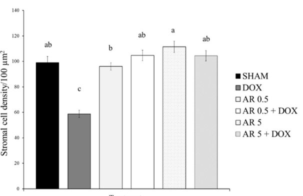
30. Briley SM, Jasti S, McCracken JM, Hornick JE, Fegley B, Pritchard MT, et al. Reproductive age-associated fibrosis in the stroma of the mammalian ovary. Reproduction. 2016;152(3):245-60. [doi:10.1530/REP-16-0129] [pmid: 27491879]
31. Oktem O, Oktay K. A novel ovarian xenografting model to characterize the impact of chemotherapy agents on human primordial follicle reserve. Cancer Res. 2007;67(21):10159-62. [doi:10.1158/0008-5472.CAN-07-2042]
32. Silva JRV, Lima FEO, Souza ALP, Silva AWB. Interleukin-1β and TNF-α systems in ovarian follicles and their roles during follicular development, oocyte maturation and ovulation. Zygote. 2020; 28(4):270-77. [doi:10.1017/S0967199420000222]
33. Marcinkiewicz JL, Krishna A, Cheung CM, Terranova PF. Oocytic tumor necrosis factor alpha: localization in the neonatal rat ovary and throughout follicular development in the adult rat. Biol Reprod. 1994;1251-60. [doi:10.1095/biolreprod50.6.1251]
34. Roby KF, Terranova PF. Localization of tumor necrosis factor (TNF) in the rat and bovine ovary using immune-cytochemistry and cell blot: evidence for granulosal production. In: Hirshfield, A.N. (eds) Growth Factors and the Ovary. Springer, Boston, MA. 1989:273-78. [doi:10.1007/978-1-4684-5688-2_31]
35. Sancho-Tello M, Tash J, Roby K, Terranova PF. Effects of lipopolysaccharide on ovarian function in the pregnant mare serum gonadotropin-treated immature rat. Endocrine Journal. 1993;503-11. [doi:10.2527/jas1987.6561768x]
36. Strauss JF, Fitz Gerald GA. Steroid Hormones and Other Lipid Molecules Involved in Human Reproduction. Yen and Jaffe's Reproductive Endocrinology. 2019;75-114. [doi:10.1016/b978-0-323-47912-7.00004-4]
37. Nagata S. Apoptosis and Clearance of Apoptotic Cells. Annu Rev Immunol. 2018; 36:489-517. [doi:10.1146/annurev-immunol-042617-053010]

Add your comments about this article : Your username or Email:
CAPTCHA

Send email to the article author


Rights and permissions
Creative Commons License This work is licensed under a Creative Commons Attribution-NonCommercial 4.0 International License.

© 2025 CC BY-NC 4.0 | Iranian Journal of Toxicology

Designed & Developed by : Yektaweb