Ethics code: mustansiriyah university ethics law


1- Lecturer, PhD genetics, medical genetics department / Iraqi center for cancer and medical genetics research /Mustansiriyah University/ Baghdad/ Iraq
2- Assist Prof, Assist of center Dean/ Iraqi center for cancer and medical genetics research/ Mustansiriyah University/ Baghdad/ Iraq , Zaynab.saad@iccmgr.org
3- Lecturer, PhD biotechnology, medical genetics department / Iraqi center for cancer and medical genetics research /Mustansiriyah University/ Baghdad/ Iraq
4- Bachelor, Molecular Biology Department, Iraqi Center for Cancer and Medical Genetics Research, Mustansiriyah University, Baghdad , Iraq
Full-Text [PDF 592 kb]
(242 Downloads)
|
Abstract (HTML) (1213 Views)
Full-Text: (104 Views)
Introduction
Currently, the majority of telecommunication sources, including radio and television broadcasts, as well as wireless technologies (e.g., mobile phones and local area networks), all operate at frequencies around 6 GHz [1].
Mobile phones utilizing 3G technology generally operate within a frequency range of 900 to 1800 MHz, whereas 4G networks function within a standard frequency range of 2 to 8 GHz. In comparison, 5G technology employs significantly higher frequencies, extending up to 60 GHz. Each country has established and implemented its own regulations for Wi-Fi channel bands, particularly concerning the frequencies utilized by Wi-Fi systems. Typically, Wi-Fi systems operate within the frequency range of 2.4 GHz to 60 GHz, covering the industrial, scientific, and medical bands. The human body is constantly exposed to electromagnetic fields (EMF) from mobile phone infrastructure, especially tower base stations [2]. The growing adoption of wireless networking technologies, especially Wi-Fi networks and WLAN hotspots, could subject individuals to elevated levels of radiofrequency electromagnetic radiation (EMR) [3]. In fact, in metropolitan locations throughout a variety of countries, significant levels of multi-source EMR, including radiofrequency EM spectrum, have been observed [4]. The expansion of cell phone towers for mobile connectivity brings up two issues: visual pollution and potential biological effects. In places with large population densities, the likelihood of the aforementioned two problems becomes more apparent. These two problems are interrelated in such settings. As a result, there is an increase in visual pollution and potential biological effects [5].
To understand the biological effect, several key points must be clarified. Oxidative stress is defined as the condition that results from an imbalance between the effectiveness of the antioxidant system in removing reactive oxygen species (ROS) and the rate at which ROS are produced. Superoxide and hydroxyl radicals are the two primary forms of ROS in living things. Although they are essential for many biological processes, their unchecked overproduction can cause DNA damage, such as single-and double-strand breaks and crosslinks [6]. In general, several internal or external stimuli, such as oxidative stress, may be brought on by gamma or UV radiation, which is thought to be the source of the creation of free radicals and molecular oxidation [7]. According to research, producing free radicals, especially hydroxyl radicals, which can cause DNA double-strand breaks, can be increased by EMF at incredibly low frequencies [8-10]. Extremely low-frequency EMFs have been shown by Mihai et al. to be capable of causing DNA strand breakage in healthy human cells [11]. Vero cells were exposed to an extremely low-frequency EMF (100 Hz, 5.6 mT). In comparison to the control group's untouched cells, a significant proportion of cells with damaged DNA (increased tail length) were found using the comet test and cell cycle analysis. Data from cell cycle analysis showed a rise in the number of cells in the S phase, which is a sign of DNA single-strand breakage. Mihai et al. proposed that the creation of ROS generated by EMF is the fundamental mechanism of reported DNA damage induction [11]. The biological effect of EMF is illustrated in Figure 1[12].

Figure 1. Electromagnetic waves' influence on biological processes and possible therapeutic uses [12].
Upon reviewing the latest research on the topic alongside previous studies, a total of 29 articles were identified that examine the genotoxic and epigenetic effects associated with the induction of DNA strand breaks (DNA-SB) by magnetic fields (MF). In total, 50% of the samples had MF effects, whereas 50% did not exhibit DNA-SB. However, when considering only genotoxic or epigenetic studies, 37.5% and 69.2% reported that MF induced DNA-SB, respectively. According to these findings, MF may contribute to DNA damage rather than acting as a direct genotoxic agent [13]. According to the documented effects, extremely low-frequency EMF has been classified as 'possibly carcinogenic to humans' by the International Agency for Research on Cancer [14]. However, many users and governmental organizations throughout the world continue to be concerned about the possible risk posed by the EM radiation that telecommunications equipment emits. Therefore, extensive parallel investigations on the effects of EMF on the critical functioning of biological systems and carcinogenicity should be conducted in conjunction with the development of EMF-based therapy systems. This brief study aimed to assess potential DNA damage in the blood of human volunteers following accumulated exposure to radiofrequency radiation from 3G and 4G mobile phone tower stations, using the comet assay.
Materials and Methods
Volunteers and Distance of Towers
This study investigated two categories of mobile phone towers, specifically 3G and 4G. A total of 14 volunteers took part, comprising 8 individuals who lived in proximity to mobile phone towers positioned directly above their residences, at elevations between 7 and 15 meters above the rooftops. The other 6 participants resided in areas far removed from any mobile phone towers, serving as the control group. According to [15], human blood samples were collected via venipuncture from the antecubital veins into vacutainer tubes containing Ethylenediaminetetraacetic acid. All patients signed a written informed consent form for anonymized evaluation and publication of their data. All reported investigations were carried out in conformity with national laws and the Helsinki Declaration of 1964.
Comet assay
The alkaline comet assay, primarily developed by Singh in 1988 [16] and further described by Dhawan in 2003 [17], was used to evaluate DNA damage. Some modifications were carried out in our Lab. Briefly, whole blood from volunteers was transported in an ice box to the laboratory and processed for the comet assay. Subsequently, 500 µl of 0.6% agarose with a low melting point was combined with 10 µl whole blood, and 90 µl of the blood/agarose combination was applied to two different locations of the comet slides. Slides were put on ice for 10 minutes, transferred to a lysis solution that had already been refrigerated, and then incubated at 4°C for 1:30 hours.
The slides were then added to an electrophoresis device that was horizontal and contained an alkaline electrophoresis buffer. After 20 minutes of DNA unwinding, electrophoresis was carried out at 4°C for 20 minutes at 0.8 V/cm. The slides were washed for 20 minutes in water and neutralization buffer, and then air dried before being stained with 50 µl of ethidium bromide solution (prepared immediately) [18]. Fluorescence microscopy and imaging software were used to examine 50 randomly chosen cells with comets per slide area (casp1.2.3b1). The "parameter DNA intensity in the tail" was used to objectively assess the degree of DNA damage (% tail DNA). Results are presented as the mean of the average calculated from the percentage of the tail of the entire slide area.
Statistical aspects
Quantitative data from the comet test, which scored 50 nucleoids per slide, were reported as mean±SD. GraphPad Prism 8 software [19] was used to perform a Student's t-test for independent samples with normally distributed analyses in order to compare numerical variables between the research groups. Blood samples from healthy individuals were compared with those from radiation-exposed volunteers. The same experiment yielded a mean±SD of 1.002±0.105 % tail DNA. Tail length, tail moment, and % tail DNA were measured and recorded using CASP software (version 1.2.3b1).
Results
The participants (n=14) expressed concerns about the potential health effects of mobile phone base stations and the duration of their exposure. Among them, 8 individuals who believed that mobile phone base stations could negatively impact health were living in close proximity to the towers, at distances of approximately zero meters and at heights ranging from 7 to 15 meters above their residences. The study results indicated significant genetic effects associated with accumulated exposure and close proximity to these towers. The remaining 6 participants, who served as the control group, lived in areas distant from any mobile phone towers.
In total, 32 to 40 cells were analyzed for each of the involved cases and controls. The classification of cell damage included five levels. G0 and G1 were considered normal, while G2, G3, and G4 were considered damaged levels with respect to tail DNA, % tail DNA, tail length, and olive tail moment as presented in Figure 2. By calculating cell frequency at each level, the type of damage was determined [17].
The following results of each case are presented in Figure 2:
Case 1: A 24-year-old male was exposed to a 3G communication tower for over three years, located 7 meters above him. His cells showed type 2 damage.
Case 2: A 26-year-old male was exposed to a 3G communication tower for over three years, located 7 meters above him. His cells were at damage type 3.
Case 3: A 20-year-old male was exposed to a 3G communication tower for three years, 7 meters above him. His cells were at damage type 3.
The first, second, and third cases, as previously noted, pertained to individuals who both worked and resided in the same vicinity, facing constant exposure (24/7) to a nearby 3G mobile phone tower. This tower was positioned roughly 7 meters above the roof of their building, with their bedrooms located on the second floor, merely 4 meters beneath the antenna. These individuals endured daily exposure for a duration of three years. Results from the comet assay indicated Type 2 and Type 3 DNA damage across all three cases. As depicted in Figure 2, the cells exhibited deviations from their typical condition.
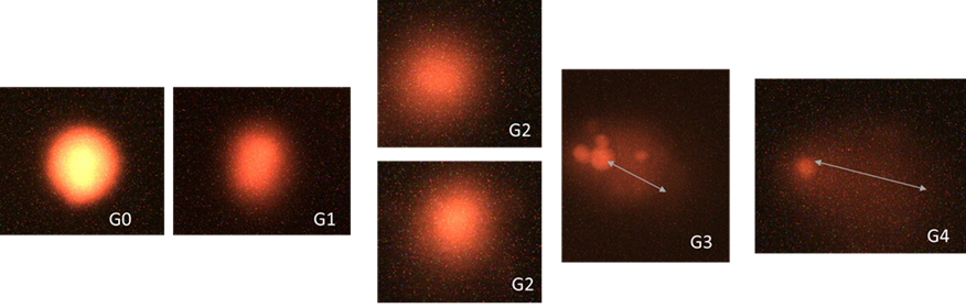
Figure 2: Shape movements of comet tails. Based on the intensity of the head and tail, there are five classifications. An integer between 0 and 4 was assigned to each category :( 0) = minimal damage and (4) = severe damage (i.e. almost all DNA in the tail) [17].
| volunteers |
Age/ Gender |
Zone* |
Distance from the cell tower (m) |
Estimation exposure time
(year) |
G3 |
G4 |
Tail moment |
Tail length |
Type of damage |
| 1 |
24/♂ |
0m |
7m |
3Y |
√ |
- |
2.67 ± 3.24 |
19.67 ± 10.51 |
2 |
| 2 |
26/♂ |
0m |
7m |
3Y |
√ |
- |
2.75 ± 3.48 |
17.79 ± 20.61 |
3 |
| 3 |
20/♂ |
0m |
7m |
3Y |
√ |
- |
2.17 ± 5.19 |
13.12 ± 19.68 |
3 |
| 4 |
22/♂ |
0m |
7m |
6Y |
√ |
- |
2.40 ± 3.11 |
18.28 ± 11.94 |
4 |
| 5 |
57y/♀ |
0 m |
9m vertical |
10y |
- |
¥ |
2.40 ± 3.11 |
18.28 ± 11.94 |
3 |
| 6 |
38/♀ |
0 m |
9 m |
10 y |
- |
¥ |
1.17 ± 1.86 |
14.88 ± 13.1 |
3-4 |
| 7 |
27/♀ |
0m |
9 m |
3Y |
- |
¥ |
2.17 ± 2.53 |
16 ± 12.05 |
3 |
| 8 |
26/♂ |
0m |
10 m |
6m |
- |
√ |
5.07 ± 13.5 |
23.94 ± 38.87 |
1 |
| 9 |
50/♀ |
- |
- |
- |
- |
- |
1.61 ± 1.58 |
16.89 ± 8.8 |
0 |
| 10 |
43/♀ |
- |
- |
- |
- |
- |
1.7 ± 1.4 |
15.8 ± 7.8 |
0 |
| 11 |
28/♂ |
- |
- |
- |
- |
- |
3.96 ± 7.19 |
22.94 ± 21.39 |
0-1 |
| 12 |
50/♀ |
- |
- |
- |
- |
- |
3.57 ± 4.95 |
18 ± 19.3 |
0-1 |
| 13 |
44/♂ |
- |
- |
- |
- |
- |
2.95 ± 4.95 |
13.31 ± 13.84 |
0-1 |
| 14 |
29/ ♀ |
- |
- |
- |
- |
- |
2.30 ± 5.95 |
12.23 ± 12.84 |
0 |
1-8 cases
9-14 control.
¥: exposed to G3 which updated to G4
Table 1. Parameters used to determine comet assay, and a comparison between two volunteer groups (apparently healthy group and exposure group).
Case 4: A 22-year-old male was exposed for six years to a 3G communication tower, 7 meters above him for 3 years, and was exposed previously for 2 years in another place. His cells were at damage type 4.
Case 5: A 57-year-old female was exposed to a vertically 9-meter 3G communication tower updated to 4G for 10 years in total. She revealed damage type 3.
Case 6: A 38-year-old female was exposed to a vertically 9-meter 3G communication tower updated to 4G for 10 years in total. She revealed damage types 3-4. The case was a heavy smoker.
Case 7: A 27-year-old female was exposed to a vertically 9-meter 4G communication tower for three years. She revealed damage type 3.
Case 8: A 26-year-old male who was exposed to a 10-meter vertical tower for 6 months showed type 1 cell damage.
All the control individuals who lived away from communication towers were either type zero or type 1 in which there was no serious damage.
Discussion
This study aimed to evaluate genetic damage in peripheral blood leukocytes of individuals living in proximity to mobile phone base stations through the single-cell gel electrophoresis (comet) assay. Moreover, it compared the findings with those of healthy control subjects. The findings of the present study emphasized two primary factors affecting DNA damage: the length of exposure and the distance of exposure, particularly for individuals living directly under mobile phone base stations (zero-meter zone). Both factors were determined to significantly influence the level of DNA damage noted in the exposed subjects.
At the molecular level, the comet test measures DNA single- and double-strand breaks. Such DNA damage has biological repercussions that include cell death, cancer growth, and mutations [20]. The extent of DNA damage in the blood of a participant (Case No. 8), who was exposed to EMF from mobile phone tower stations for a duration of six months, was determined to be similar to the baseline level seen in a healthy volunteer residing away from these towers. This implies that, at this point, the exposure had not caused notable DNA damage, suggesting that short-term exposure may not result in severe genotoxic consequences.
While the one case that was exposed for more than 3 years revealed type 3 damage forward type 4, both Cases 5 and 6 were exposed to the same type of towers and the same duration. However, Case No. 6 showed progression from Type 3 to Type 4 DNA damage. The individual was a heavy smoker, suggesting a possible synergistic effect between electromagnetic wave exposure and smoking in exacerbating genetic damage. A further example of Type 4 DNA damage was noted in Case No. 4, demonstrating the effects of prolonged exposure at a height of 7 meters over a six-year duration. In contrast, Cases 1, 2, and 3, which were subjected to the same height for only three years, displayed reduced levels of DNA damage (Types 2 and 3). This indicates that an extended duration of exposure may lead to a greater accumulation of genetic damage, possibly escalating the severity to Type 4.
In a previously conducted study [21], it was concluded that the mobile phone base stations provide communication; however, their constant radiation emissions have generated health concerns. In comparison to the area from which control samples were taken, the power density in the region between 50 and 100 meters from the base station exceeded the acceptable limits [21]. The participants (n = 8) in the current study, who lived within zero meters of a cell tower, illustrated the effect of direct exposure. DNA migration length, damage frequency, and damage index were all measured in the genetic sample groups.
According to a recent study by Zosangzuali, several oxidative stress indices (e.g., Glutathione, Glutathione S-transferase, Superoxide Dismutase) in the brains, hearts, kidneys, and livers of Swiss albino mice may be affected by exposure to mobile phone base stations that emit 1800-MHz radiofrequency EMR. Their findings showed that the formation of ROS had negative impacts on the mice's brains, as evidenced by increased lipid peroxidation and decreased antioxidant levels and activity [22].
Mobile phones, radio-based stations, phone towers, and high-voltage power lines emit electromagnetic radiation, which has been associated with a range of health issues, including the risk of cancer in people and negative impacts on animals, including birds. A recent literature review by Gupta et al. suggested that despite a dramatic growth in usage, there is still a dearth of awareness of the problems associated with mobile phone radiation [23].
Conclusion
In this original study, the comet assay was employed to detect the genotoxic effects of radiation from 3G and 4G mobile phone tower stations on the blood of individuals exposed to these emissions. The placement of towers above residential buildings, with heights ranging from zero meters to 7-15 meters, presents a risk of DNA damage accumulation, which may contribute to future health hazards for individuals.
The authors recommended conducting further genotoxic studies involving a larger sample of volunteers. They suggested placing the tower stations above uninhabited buildings to prevent continuous 24/7 exposure and raising the magnetic generators to heights above 15 meters to minimize their impact on genetic material. Moreover, telecom companies could establish these towers outside the cities in a wide area to protect people's lives from harm.
Ethical Considerations
This study was conducted in accordance with the ethics guidelines of Mustansiriyah University. This study was performed in agreement with the Declaration of Helsinki; samples used here are only for research purposes.
Authors' Contributions
Concept and design: Asma A. Data acquisition: Asma A., T.J. Amal A. Data analysis: Z.A. Manuscript preparation: Asma A. and ZA, Critical revision and finalizing of the manuscript: ZA and Asma A
Acknowledgement
The authors would like to thank Mustansiriyah University (https://uomustansiriyah.edu.iq/), Baghdad, Iraq, for its support in the present work. The study was also supported by the Iraqi Center for Cancer and Medical Genetics Research (ICCMGR). The authors would like to thank all the staff of both the Molecular Biology and Medical Genetics Departments in ICCMGR for their technical support.
Conflict of Interests
The authors declare that they have no conflict of interest.
Funding
This study was funded by the authors, and no external funding was received.
References
- Wu T, Rappaport TS, Collins CM. Safe for generations to come: considerations of safety for millimeter waves in wireless communications. IEEE Micro Mag. 2015; 16(2): 65–84. [DOI:10.1109/MMM.2014.2377587]
- Levitt BB, Lai H. Biological effects from exposure to electromagnetic radiation emitted by cell tower base stations and other antenna arrays. Environ Rev. 2011; 19: 495. [DOI:10.1139/a10-903]
- Fagua Fagua AL, Pinzón Abril RN, Rojas Casas JD. The adverse effects over human health caused by wireless wi-fi communication networks. Cultura Cientifica. 2016; 14:34-45. [Link]
- Sagar S, Adem SM, Struchen B, Loughran SP, Brunjes ME, Arangua L, et al. Comparison of radiofrequency electromagnetic field exposure levels in different everyday microenvironments in an international context. Environ Int. 2018; 114: 297-306. [DOI: 10.1016/j.envint.2018.02.036] [PMID: 29529581]
- Yahya SI. The use of camouflaged cell phone towers for a quality urban environment: Koya city as case study. UKH J Sci Eng. 2019; 3(1): 29-34. [DOI:10.25079/ukhjse. v3n1y2019.pp29-34]
- Xu ZZ, Fu WB, Jin Z, Guo P, Wang WF, Li JM. Reactive oxygen species mediate oridonin-induced apoptosis through DNA damage response and activation of JNK pathway in diffuse large B cell lymphoma. Leuk Lymphoma. 2016; 57(4): 888-98. [DOI: 10.3109/10428194.2015.1061127] [PMID: 26415087]
- de Oliveira GC, Maia GAS, Cortes VF, Santos HD, Moreira LM, Barbosa LA. The effect of gamma-radiation on the hemoglobin of stored red blood cells: the involvement of oxidative stress in hemoglobin conformation. Ann Hematol. 2013; 92(7): 899-906. [DOI: 10.1007/s00277-013-1719-z] [PMID: 23494204]
- Anderson LE. Biological effects of extremely low-frequency electromagnetic fields: in vivo studies. Am Ind Hyg Assoc J. 1993; 54(4): 186-96. [DOI: 10.1080/15298669391354540] [PMID: 8480634]
- Giorgi G, Marcantonio P, Bersani F, Gavoci E, Del Re B. Effect of extremely low frequency magnetic field exposure on DNA transposition in relation to frequency, wave shape and exposure time. Int J Radiat Biol. 2011; 87(6): 601-8. [DOI: 10.3109/09553002.2011.570855] [PMID: 21504343]
- Yokus B, Cakir DU, Akdag MZ, Sert C, Mete N. Oxidative DNA damage in rats exposed to extremely low frequency electro magnetic fields. Free Radic Res. 2005; 39(3): 317-23. [DOI: 10.1080/10715760500043603] [PMID: 15788236]
- Mihai CT, Rotinberg P, Brinza F, Vochita G. Extremely low-frequency electromagnetic fields cause DNA strand breaks in normal cells. J Environ Health Sci Eng. 2014; 12: 15. [DOI: 10.1186/2052-336X-12-15] [PMID: 24401758]
- Saliev T, Begimbetova D, Masoud BA, Matkarimov B. Biological effects of non-ionizing electromagnetic fields: Two sides of a coin. Prog Biophy Mol Biol. 2019; 141: 25-36. [DOI: 10.1016/j.pbiomolbio.2018.07.009] [PMID: 30030071]
- Ruiz-Gómez MJ, Martínez-Morillo M. Electromagnetic fields and the induction of DNA strand breaks, Electromag Biol Med. 2009; 28(2): 201-14. [DOI: 10.1080/15368370802608696] [PMID: 19811402]
- IARC (International Agency for Research on Cancer). IARC monographs on the evaluation of carcinogenic risks to humans: Preamble. 2006. [Link]
- Al-Salmani K, Abbas HH, Schulpen S, Karbaschi M, Abdalla I, Bowman KJ, et al. Simplified method for the collection, storage, and comet assay analysis of DNA damage in whole blood. Free Radic Biol Med. 2011; 51(3): 719–25. [DOI: 10.1016/j.freeradbiomed.2011.05.020] [PMID: 21658444]
- Singh NP, McCoy MT, Tice RR, Schneider EL. A simple technique for quantitation of low levels of DNA damage in individual cells. Exp Cell Res. 1988; 175(1): 184-91. [DOI: 10.1016/0014-4827(88)90265-0] [PMID: 3345800]
- Dhawan A, Bajpayee MM, PandeyAK, ParmarD. Protocol for the single cell gel electrophoresis/comet assay for rapid genotoxicity assessment. Sigma. 2003: 1-10. [Link]
- Saad Z, Barakat N, Tawfeeq A, Yaseen NY, Jaffer TH, Kamel S, et al. Determination of DNA damage induced after bitter orange (Citrus aurantium) essential oil administrated in vivo. Iraqi J Cancer Med Genetics. 2016; 9(2):176-80. [DOI:10.29409/ijcmg.v9i2.190]
- GraphPad (online). [Link]
- Mayer C, Popanda O, Zelezny O, von Brevern MC, Bach A, Bartsch H, et al. DNA repair capacity after gamma-irradiation and expression profiles of DNA repair genes in resting and proliferating human peripheral blood lymphocytes. DNA Repair (Amst). 2002; 1(3): 237–50. [DOI: 10.1016/s1568-7864(01)00019-2] [PMID:12509255]
- Gandhi G, Kaur G, Nisar U. A cross-sectional case control study on genetic damage in individuals residing in the vicinity of a mobile phone base station. Electromagn Biol Med. 2015; 34(4):344-54. [DOI: 10.3109/15368378.2014.933349] [PMID: 25006864]
- Zosangzuali M, Lalremruati M, Lalmuansangib C, Nghakliana F, Pachuau L, Bandara P, et al. Effects of radiofrequency electromagnetic radiation emitted from a mobile phone base station on the redox homeostasis in different organs of Swiss albino mice. Electromagn Biol Med. 2021; 40(3): 393-407. [DOI: 10.1080/15368378.2021.1895207] [PMID: 33687298]
- Gupta S, Sharma RS, Singh R. Non-ionizing radiation as possible carcinogen, Int J Environ Health Res. 2022; 32(4): 916-40. [DOI: 10.1080/09603123.2020.1806212] [PMID: 32885667]
Type of Study:
Research |
Subject:
General