Ethics code: IR.SU.REC.1402.10


Adib S, Keramati K, Ahmadi-hamedani M, Mohammadnezhad Nasrabadi L. Investigation of the Potential of Thymex Plus in Hematological and Platelet Modulation in Myocardial Infarction Rats. IJT 2026; 20 (1) :1-7
URL:
http://ijt.arakmu.ac.ir/article-1-1550-en.html
1- DVM, Veterinary Medicine, Semnan University, Semnan, Iran.
2- Department of Basic Sciences, Faculty of Veterinary Medicine, Semnan University, Semnan, Iran. , K.keramati@semnan.ac.ir
3- Department of Clinical Sciences, Faculty of Veterinary Medicine, Semnan University, Semnan, Iran.
4- Legal Medicine Research Center, Legal Medicine Organization, Tehran, Iran.
Full-Text [PDF 580 kb]
(118 Downloads)
|
Abstract (HTML) (472 Views)
Full-Text: (58 Views)
Introduction
Myocardial infarction (MI) is a cardiovascular disease (CVD) that is an imbalance between myocardial oxygen demand and coronary blood supply. This disproportion contributes to the destruction of the myocardial tissue and the eventual destruction of cardiomyocytes, which together result in death [1]. Therefore, MI is recognized as one of the serious diseases and a potential cause for a high number of deaths across several countries [2]. Myocardial oxidative stress (OS) is associated with endothelial dysfunction, inflammation, and an imbalance in antioxidants, leading to impaired cardiac function. Inflammation and OS contribute to apoptosis, making them vital factors in the development of MI [3]. Excessive production of myocardial adrenergic receptors leads to inflammation of the heart muscle and the death of heart cells (myocytes). During this process, the levels of cyclic adenosine monophosphate (cAMP) rise, activating protein kinase A. As a consequence of this activation, L-type calcium channels are phosphorylated, which eventually increases and leads to the occurrence and development of MI [4]. Some mechanisms in the pathogenesis of MI include OS, apoptosis, and inflammation. The negative impact of OS on cardiac function occurs through decreased activity of antioxidant enzymes, such as catalase, superoxide dismutase, and glutathione peroxidase. Reactive oxygen species (ROS) induce lipid peroxidation, which generates malondialdehyde, as well as the elution of pro-apoptotic toxins onto the mitochondrial membrane [5]. Heart failure includes various complex clinical syndromes resulting from abnormal alterations in cardiac structure and/or function, which lead to dysfunction of either ventricular systolic or diastolic function [6]. The main symptoms include dyspnea, fatigue, and fluid retention, such as pulmonary congestion, systemic blood stasis, and peripheral edema [7]. The OS and inflammation increase in chronic heart failure. Dysfunction of cardiac mitochondria is an indicator of heart failure and one of the leading causes of OS. It negatively impacts cellular components, including the mitochondria, creating a vicious cycle [8,9]. A complete blood count is a commonly used laboratory test that includes a white blood cell (WBC) count, red blood cell (RBC) count, platelet count (PLT), and morphological indices, like red cell distribution width (RDW) [10]. It was stated that inflammatory processes and activation of neurohormonal and adrenergic systems profoundly affect erythrocyte maturation due to disturbances in erythrocyte membrane homeostasis, which increases the levels of RDW [11].
The RDW is a quantitative measure that indicates the variation in the volume of erythroid cells and is typically used in hematology to assist in diagnosing anemia [12]. Population-based studies suggest that RDW can predict mortality in CVDs and other acute or chronic conditions, like MI and heart failure [13,14]. Platelets play a crucial role in the pathophysiology of MI, as they activate endothelial cells and promote fibrin formation, ultimately leading to the formation of a thrombus within the coronary vasculature. They are involved in thrombogenesis and release mediators that amplify local inflammation. Additionally, platelet count has been linked to the severity of MI, as well as to increased risks of death and reinfarction [15-17]. The RDW-to-PLT ratio (RPR) combines two independent parameters and is a new index that reflects the severity of inflammation [18]. RPR has been established as a reliable prognostic marker in CVDs [19]. Iron deficiency anemia is a common complication in chronic diseases [20]. Anemia is a significant comorbidity in heart failure, impairing exercise tolerance, lowering quality of life, and contributing to worse clinical outcomes. Iron deficiency is considered a primary cause of anemia, as iron is crucial for optimal hematopoiesis (blood cell production) [21]. Erythroblasts and reticulocytes are the main contributors to hemoglobin (Hb) synthesis [22]. Iron deficiency causes resistance to hematopoietic growth factors, including erythropoietin, and interferes with the differentiation and maturation of all types of hematopoietic cells [23]. Isoproterenol (ISO) is a synthetic catecholamine and ß-adrenergic agonist that is commonly used for the management of bronchial asthma, allergic emergencies, ventricular bradycardia, status asthmaticus, cardiac arrest, and in glaucoma [24]. High ISO concentrations produce cytotoxic reactive free radicals during autoxidation, which injure and necrose cardiac muscle [25]. Subcutaneous ISO injection is commonly used in studies of MI to explore the cardioprotective effects of natural and synthetic agents. The ISO induces lesions in rat hearts, serving as a reliable model for studying heart attacks in humans [26]. The β-adrenoreceptors found in reticulocytes and mature erythrocytes are of the β2 subtype. ISO significantly enhances adenylate cyclase activity in reticulocytes, increasing the synthesis of cAMP. Reticulocytes are more sensitive to ISO than mature erythrocytes, showing a 6 to 9-fold increase in adenylate cyclase activity. As these cells mature into erythrocytes, they become unable to generate cAMP in response to ISO, despite the presence of β-adrenergic receptors, indicating a functional uncoupling of the receptor from the enzyme during erythroid maturation [27,28].
The individual ingredients of Thymex have been well established to have antioxidant and anti-inflammatory activities. So far, the synergistic effect of these ingredients, in relation to MI, has not been thoroughly investigated. The synergistic antioxidant action of thymol and honey, with anti-inflammatory and wound-healing effects, could represent a novel approach to regulating hematological variables in CVDs. According to recent studies, the combined effects of thymol and honey may enhance cardioprotection by reducing OS and inflammation, both of which are major pathophysiological drivers of MI [29, 30].
The present research aimed to investigate the possible protective role of a natural combination, Thymex Plus (TYM), created from thymol and honey, against blood-related alterations, such as total reticulocyte and PLT, and the RPR, in MI rats. The ISO model is considered the conventional method for inducing MI in rat models [31]. This study primarily emphasizes the hematological alterations that result from implementing this model, rather than directly assessing cardiac damage within the context of this research.
Materials and Methods
The rats used in the experiment were 18 male Wistar rats, aged 6-8 months and weighing 200-250g. They were obtained at the breeding institution in the Laboratory Animal Research Institute. Rats were maintained in the same environmental conditions in terms of temperature (20-24°C), humidity, and the light/dark cycle of 12/12 hours. These animals had free access to food and water and received regular lab animal pellets. All injections and blood sampling were performed at the same time of day (between 8:00 and 10:00 a.m.) to minimize the influence of circadian variations on hematological parameters. The rats were randomly divided into three experimental groups (six rats per group) to acclimate them before the start of the study.
Ethical considerations
All animal procedures were performed following the institutional and national guidelines (ARRIVE guidelines) for animal care and approved by the Animal Research Ethical Committee of Semnan University of Veterinary Medicine, Iran (Ethics Code: IR.SU.REC.1402.10).
Study design
Plasma biochemical indices, including urea, uric acid, Rats were randomly divided into three groups as follows:
Group I: Control group; in this experimental group, no intervention was performed.
Group II: ISO group; the rats received subcutaneous injections of ISO (Sigma-Aldrich) at a dosage of 85 mg/kg of body weight on days 1 and 2, with a 24-hour interval (Murugesan et al., 2011). Subsequently, on days 3, 4, 5, 6, and 7, they were administered 1.5 ml of normal saline orally.
Group III: ISO and TYM Group (ISO-TYM); the rats received subcutaneous injections of ISO at a dosage of 85 mg/kg of body weight on days 1 and 2, with a 24-hour interval. Following this, on days 3, 4, 5, 6, and 7, they were orally given 1.5 mL of TYM by gavage. The TYM is formulated from thyme extract (Thymus vulgaris) and honey. Each 5 mL of the syrup contains approximately 1.1 mg of total phenolic compounds (calculated as thymol), 0.2 g of honey, and 653 mg of thyme extract.
Cardiac blood sampling
Animals were subjected to anesthesia through intraperitoneal administration of ketamine at a dosage of 80 mg/kg body weight, in conjunction with xylazine at a dosage of 10 mg/kg body weight. Euthanasia was confirmed by observing the cessation of both cardiac activity and respiratory movements, per ARRIVE guidelines. Blood samples were collected on day 7, immediately after the final treatment administration, under anesthesia and euthanasia procedures as described. Following this procedure, 4 ml of blood was extracted from the heart of each rat utilizing a syringe. Of the collected blood, 2 ml was placed in a clotting activator test tube, while the remaining 2 ml was placed in a test tube containing EDTA. In this study, a cell counter (Celltac α VET; MEK-6550) was utilized to measure blood cellular indices, and the test tubes containing EDTA were employed for this purpose. The clotting activator test tubes were centrifuged for 10 min at 3000 rpm (Behsan, HB-200) to measure biochemical blood indices. Afterward, the separated plasma was transferred to a microtube and stored in a freezer. Finally, biochemical indices were assessed manually using the appropriate kits.
Hematological examination
Hematological indices were measured, including Hb, WBC count, RBC count, hematocrit (HCT), PLT count, mean corpuscular volume (MCV), mean corpuscular hemoglobin (MCH), mean corpuscular hemoglobin concentration (MCHC), RDW, and the RPR. Reticulocytes were manually counted on peripheral blood smears stained with new methylene blue. Their percentage per 100 RBCs was calculated, and the absolute count was determined using the formula: Absolute reticulocyte count (× 103/μL) = Reticulocyte% × RBC count (× 106/μL) × 10.
Data analysis
The selection of a sample size of six animals per group was based on findings from previous research investigating hematological alterations in rat models of MI induced by ISO [8]. A post-hoc power analysis confirmed that this sample size provided over 80% power to detect statistically significant differences in key parameters (p<0.05). The Shapiro-Wilk test was used to assess the normality of the data. For data that did not follow a normal distribution, group comparisons were performed using the Kruskal-Wallis test, with results reported as median and interquartile range (IQR). A p-value of less than 0.05 was considered statistically significant. All parameters passed the normality test (Shapiro-Wilk); therefore, one-way ANOVA followed by Tukey's post hoc test was employed for statistical comparison. To evaluate the robustness of our statistical findings, effect sizes (Cohen's d) were calculated for the key group comparisons. Furthermore, a post hoc power analysis was performed to evaluate the statistical power to detect moderate to significant effects based on the determined effect sizes. This analysis was based on a sample size of six rats per group.
Results
Figure 1 presents box plots of hematological parameters of three groups of the study: control, ISO, and ISO-TYM. p-values for statistical comparisons between these groups are provided above each box, indicating that the observed differences are statistically significant. Hb (p=0.012), RBC count (p=0.038), and HCT (p=0.050) all showed significant or borderline significant decreases in the ISO group compared to the control group. Although these values improved significantly in the TYM-treated group, none of the changes reached statistical significance compared with the ISO group. The PLT levels also dropped significantly in the ISO group compared with the control (p=0.041); however, TYM treatment did not result in a statistically significant recovery (p=0.057). Reticulocyte count decreased significantly in the ISO group compared to the control (p=0.014), and this rise was even more pronounced in the ISO-TYM group, which demonstrated significantly higher values than both the ISO (p=0.021) and control (p=0.003) groups. The RDW and the RPR were both significantly elevated in the ISO group compared to the control (p<0.05). Treatment with TYM significantly lowered these values compared to the ISO group (p<0.05). To further evaluate these findings, the effect sizes (Cohen's d) were calculated. The post-hoc power analyses were performed for the key comparisons: ISO vs Control, with an effect size of 1.20 (large) and a power of 0.47. ISO-TYM vs ISO, with an effect size of -0.32 (small-to-negligible), with a power of 0.08. A comparison between ISO-TYM and the control has created an effect size of 1.42 (considerable effect); power was 0.60. These findings suggest that the research had sufficient power to measure large effect sizes, especially in the comparison between ISO-TYM and the control, regardless of the small sample size.
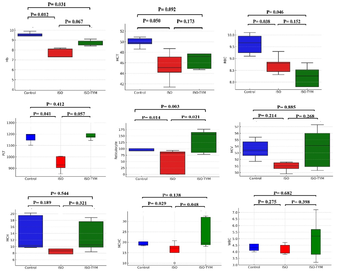
Figure 1. Box plots illustrating Hb, HCT, RBC, RET, and PLT indices, and RPR across the three study groups. In the box plot, the data points represent individual measurements, while the box indicates the interquartile range (IQR), with the median marked by the central line of the box. The p-values indicate the statistical significance of differences between groups. All parameters passed the normality test (Shapiro-Wilk); therefore, one-way ANOVA followed by Tukey’s post hoc test was employed for statistical comparison.
Table1. Comparison of mean RDW and RPR across the three study groups
| Group |
RDW (%) |
RDW/PLT |
| Mean |
SD |
Mean |
SD |
| Control |
11.51b |
0.23 |
0.98b |
0.05 |
| ISO |
12.10a |
0.51 |
1.28a |
0.12 |
| ISO-TYM |
11.66b |
0.48 |
1.03b |
0.19 |
Different superscript letters (a, b) indicate statistically significant differences between groups (p<0.05). Statistical comparisons were performed using one-way ANOVA followed by Tukey's post hoc test. Specifically, 'a' denotes a significant difference versus control, and 'b' denotes a significant difference versus the ISO group. All parameters passed the normality test (Shapiro-Wilk); therefore, one-way ANOVA followed by Tukey’s post hoc test was used for statistical comparison.
Discussion
In the present study, the hematological effects of TYM, a combination of Thymus vulgaris and honey, were investigated in a rat model of ISO-induced MI. The findings show that TYM significantly reversed ISO-induced anemia, increasing Hb, HCT, and RBC levels, which were otherwise decreased in the rats. These results are consistent with previous studies demonstrating the protective roles of thymol and carvacrol, major constituents of Thymus vulgaris, in reducing OS and inflammation, which have central effects on the pathophysiology of MI [29]. Studies on carvacrol in people with cardiac disease have reported its therapeutic benefits against MI, cardiac hypertrophy, hypertension, arrhythmias, and various cardiovascular pathologies, which are explained by its antioxidant and anti-inflammatory effects [33]. Additionally, research on the industrial dye tartrazine showed that thymol and carvacrol can prevent the production of toxins resulting from heme degradation and oxidation processes [34]. Thymol and carvacrol inhibit heme degradation and the breakdown of the porphyrin ring, thereby preserving the structure of Hb by maintaining its secondary and tertiary structures, which helps prevent oxidative damage. In the present study, TYM (containing thymol) compensated for the ISO-induced reduction in Hb, increasing Hb levels in treated rats [35]. Moreover, analysis of fish fed thymol revealed significant increases in Hb, HCT, RBC, and white blood cell counts, indicating that thymol increases blood oxygen-carrying capacity and has an immunomodulatory effect [36]. These findings align with the results obtained in the current experiment, supporting the thesis that thymol can be used to treat anemia.
The anemia induced by ISO in rats, reflected by decreases in Hb, HCT, and RBC, was improved with TYM treatment, leading to an increase in Hb and HCT, along with higher reticulocyte counts. Thymol may help reduce the adverse effects of erythrocyte damage by inhibiting OS, reducing cell volume, and preventing energy depletion that leads to RBC death. It allows thymol to prevent microcirculatory disturbances and may play a role in supporting recovery from anemia [37]. Unlike earlier studies that showed higher reticulocyte counts in anemic heart patients, reticulocytosis was not observed in the MI rats after a week. However, TYM induced reticulocytosis in group III, suggesting it could help improve hematological parameters [38]. Studies suggest that honey can also help raise Hb levels in pregnant women who lack iron, especially when combined with iron supplements [39]. Acacia honey appears to aid the body in producing more RBCs and increasing Hb levels [40]. In rats with lead-induced anemia, honey slowed the progression of anemia by increasing iron levels in the blood and stimulating the bone marrow [41]. However, a study in Pakistan found that rats fed acacia honey had lower Hb levels [42]. Nevertheless, honey's beneficial qualities suggest that it could help alleviate anemia, particularly in patients with heart conditions.
Research has demonstrated that elevated RDW is an independent predictor of poor prognosis in coronary heart disease and is associated with higher mortality in acute myocardial infarction (AMI) patients [43]. The RDW reflects inflammation, which plays a critical role in the onset and progression of AMI [44]. In this study, RDW was increased in rats with MI; however, TYM treatment significantly lowered it. Our findings revealed that TYM effectively modulates hematological parameters in a rat model of ISO-induced MI. It aligns with previous studies on thymoquinone, a key component of thymol, which have shown that it reduces OS, inflammation, and myocardial necrosis [45, 46]. Thymol enhances heart contraction by reducing OS, lowering heart rate, and protecting the myocardium. It even lowers the risk of CVDs by balancing lipids and preventing further harm to the heart. Furthermore, thymol augmented myocardial protection by inducing the expression of Bcl-2 (an antiapoptotic gene) and suppressing the expression of Bax (a pro-apoptotic gene) in rats' hearts, which could also contribute to minimizing cell apoptotic death [47]. Although the present study revealed numerous findings regarding the impact of TYM on hematological parameters in an MI model, we focused only on these parameters. Although assessment of alterations in hematological indices is informative, combining the analysis of cardiac-related biomarkers and histopathology assays may provide deeper insights into the cardioprotective effect of TYM. This exploration was limited to the changes in hematology as a preliminary circumstance of the potential of TYM. It would also be important for future research to integrate cardiac biomarkers, such as troponins and creatine kinase-MB, with histopathological analysis to assess myocardial injury, the inflammatory and repair mechanisms that follow.
Conclusions
The TYM demonstrated significant protective effects in hemogram indices, such as PLT, RDW, RPR, and reticulocyte count, on ISO-induced MI in rats. These effects included some improvements. The results of this study enhanced the potential of TYM as a compound with hematological modulating capabilities. However, to clarify the precise mechanisms of these effects, further studies are recommended.
Limitations
The present study mainly examined hematological changes induced by ISO. Direct assessments of myocardial injury, such as cardiac histopathology, measurement of cardiac biomarkers (troponins, CK-MB), and cardiac function tests, were not performed. Therefore, findings on MI and the protective effects of TYM are indirect and require further specialized cardiac studies. Although plasma samples were collected for biochemical analyses, data on OS and inflammatory markers are omitted from this report. Future research is needed to clarify the mechanistic pathways of TYM’s cardioprotective effects.
Data Access and Responsibility
The authors confirm that this article contains original
work and accept full responsibility for its content.
Ethical Considerations
All animal procedures were performed following the institutional and national guidelines (ARRIVE guidelines) for animal care and approved by the Animal Research Ethical Committee of Semnan University of Veterinary Medicine, Iran (Ethics Code: IR.SU.REC.1402.10).
Authors' Contributions
Mahmood Ahmadi-hamedani: Writing – original draft, Project administration, Formal analysis, Data curation, Conceptualization. Keivan Keramati: Review & editing, Project administration, Supervision, Conceptualization. Sina Adib: Methodology, Conceptualization. Leila Mohammadnezhad Nasrabadi: Methodology.
Acknowledgement
The authors thank all participants for their cooperation and sample contribution. This research was supported by the Semnan University, Semnan, Iran.
Conflict of Interests
The authors declare that there is no conflict of interest.
Funding
This work has been supported by the Deputy of Research and Technology, Semnan University, Semnan, Iran.
References
- Frangogiannis NG. Pathophysiology of myocardial infarction. Compr physiol. 2011;5(4):1841-75. [DOI: 10.1002/cphy.c150006] [PMID: 26426469]
- Rohani C, Jafarpoor H, Mortazavi Y, Esbakian B, Gholinia H. Mortality in patients with myocardial infarction and potential risk factors: a five-year data analysis. ARYA Atherosclerosis. 2022;18(1):1-8. [DOI:10.48305/arya.v18i0.2427] [PMID:36815954]
- Garg M, Khanna D, Kalra S, Balakumar P. Chronic oral administration of low-dose combination of fenofibrate and rosuvastatin protects the rat heart against experimentally induced acute myocardial infarction. Fundamental Clin Pharmacol. 2016;30(5):394-405. [DOI: 10.1111/fcp.12204] [ PMID: 27148865]
- Euteneuer F, Mills PJ, Rief W, Ziegler MG, Dimsdale JE. Association of in vivo β-adrenergic receptor sensitivity with inflammatory markers in healthy subjects. Psychosom Med. 2012;74(3):271-7. [DOI: 10.1097/PSY.0b013e318245d762] [PMID: 22366585]
- Hosseini A, Rajabian A, Sobhanifar MA, Alavi MS, Taghipour Z, Hasanpour M, et al. Attenuation of isoprenaline-induced myocardial infarction by Rheum turkestanicum. Biomed Pharmacother. 2022;148:112775. [DOI:10.1016/j.biopha.2022.112775] [PMID:35240528]
- Van der Meer P, Gaggin HK, Dec GW. ACC/AHA versus ESC guidelines on heart failure: JACC guideline comparison. J Am Coll Cardiol. 2019;73(21):2756-68. [DOI: 10.1016/j.jacc.2019.03.478] [PMID: 31146820]
- Bozkurt B, Coats AJ, Tsutsui H, Abdelhamid M, Adamopoulos S, Albert N, et al. Universal definition and classification of heart failure: a report of the heart failure society of America, heart failure association of the European society of cardiology, Japanese heart failure society, and writing committee of the universal definition of heart failure. J Card Fail. 2021;27(4):387-413. [DOI:10.1002/ejhf.2115] [PMID: 33663906]
- Tahery S, Ahmadi-Hamedani M, Keramati K, Javaheri Vayghan A, Naeimi S. Protective effect of Flunixin meglumin on changes induced by isoproterenol in serum biochemical profile, malondialdehyde, and heart histology of adult male rats. Iran J Toxicol. 2019;13(4):33-7. [DOI: 10.32598/IJT.13.4.605.1]
- Aimo A, Castiglione V, Borrelli C, Saccaro LF, Franzini M, Masi S, et al. Oxidative stress and inflammation in the evolution of heart failure: from pathophysiology to therapeutic strategies. Eur J Prev Cardiol. 2020;27(5):494-510. [DOI:10.1177/2047487319870344] [PMID: 31412712]
- Yayla ME, İlgen U, Okatan İE, UsluYurteri E, Torgutalp M, Keleşoğlu Dinçer AB, et al. Association of simple hematological parameters with disease manifestations, activity, and severity in patients with systemic sclerosis. Clin Rheumatol. 2020;39(1):77-83. [DOI: 10.1007/s10067-019-04685-0] [PMID: 31317426]
- Salvagno GL, Sanchis-Gomar F, Picanza A, Lippi G. Red blood cell distribution width: a simple parameter with multiple clinical applications. Crit Rev Clin Lab Sci. 2015;52(2):86-105. [DOI:10.3109/10408363.2014.992064] [PMID: 25535770]
- Hu B, Cao J, Hu Y, Qin Z, Wang J. Relationship between red blood cell distribution width and all-cause mortality in disseminated intravascular coagulation patients: a retrospective analysis. Int J Gen Med. 2021;14:8301-9. [DOI:10.2147/IJGM.S329296] [PMID: 34815702]
- Huang S, Zhou Q, Guo N, Zhang Z, Luo L, Luo Y, et al. Association between red blood cell distribution width and in-hospital mortality in acute myocardial infarction. Med. 2021;100(15):e25404. [DOI:10.1097/MD.0000000000025404] [PMID: 33847638]
- Salvatori M, Formiga F, Moreno-Gónzalez R, Chivite D, De Amicis MM, Cappellini MD, et al. Red blood cell distribution width as a prognostic factor of mortality in elderly patients firstly hospitalized due to heart failure. Kardiol Pol. 2019;77(6):632-8. [DOI: 10.33963/KP.14818] [PMID: 31066720]
- Falk E, Nakano M, Bentzon JF, Finn AV, Virmani R. Update on acute coronary syndromes: the pathologists’ view. Eur Heart J. 2013;34(10):719-28. [DOI:10.1093/eurheartj/ehs411] [PMID:23242196]
- Roh JW, Lim S, Hwang Y, Lee KY, Choo EH, Choi IJ, et al. Ischemic and bleeding events associated with thrombocytopenia and thrombocytosis after percutaneous coronary intervention in patients with acute myocardial infarction. J Clin Med. 2020;9(10):3370. [DOI:10.3390/jcm9103370] [PMID:33096782]
- Quan XQ, Ji HY, Jiang J, Huang JB, Zhang CT. Prognostic utility of the combination of platelet count with neutrophil-to-lymphocyte ratio in aged patients with acute myocardial infarction undergoing percutaneous coronary intervention. Emerg Med Int. 2021;2021:4023472. [DOI:10.1155/2021/4023472] [PMID:33981459]
- Lehmann F, Schenk LM, Bernstock JD, Bode C, Borger V, Gessler FA, et al. Elevated red cell distribution width to platelet ratio is associated with poor prognosis in patients with spontaneous, deep-seated intracerebral hemorrhage. Front Neurol. 2021;12:751510. [DOI:10.3389/fneur.2021.751510] [PMID:34867736]
- Karagöz E, Tanoğlu A, Ülçay A, Erdem H, Turhan V, Kara M, et al. Mean platelet volume and red cell distribution width to platelet ratio for predicting the severity of hepatic fibrosis in patients with chronic hepatitis C. Eur J Gastroenterol Hepatol. 2016;28(7):744-8. [DOI: 10.1097/MEG.0000000000000647] [PMID: 27101403]
- Zafon C, Lecube A, Simó R. Iron in obesity: an ancient micronutrient for a modern disease. Obesit rev. 2010;11(4):322-8. [DOI: 10.1111/j.1467-789X.2009.00638.x]
- Nanas JN, Matsouka C, Karageorgopoulos D, Leonti A, Tsolakis E, Drakos SG, et al. Etiology of anemia in patients with advanced heart failure. J Am Coll Cardiol. 2006;48(12):2485-9. [DOI:10.1016/j.jacc.2006.08.034] [PMID: 17174186]
- Van der Putten K, Braam B, Jie KE, Gaillard CA. Mechanisms of disease: erythropoietin resistance in patients with both heart and kidney failure. Nat Clin Pract Nephrol. 2008;4(1):47-57. [DOI:10.1038/ncpneph0655] [PMID: 18094727]
- Camaschella C, Pagani A. Iron and erythropoiesis: a dual relationship. Int J Hematol. 2011;93(1):21-6. [DOI: 10.1007/s12185-010-0743-1] [PMID: 21170616]
- Amano S, Arai M, Goto S, Togari A. Inhibitory effect of NPY on isoprenaline-induced osteoclastogenesis in mouse bone marrow cells. Biochim Biophys Acta. 2007;1770(6):966-73. [DOI:10.1016/j.bbagen.2007.02.009] [PMID: 17383824]
- Zhou R, Xu Q, Zheng P, Yan L, Zheng J, Dai G. Cardioprotective effect of fluvastatin on isoproterenol-induced myocardial infarction in rat. Euro J Pharmacol. 2008;586(1-3):244-50. [DOI:10.1016/j.ejphar.2008.02.057] [PMID: 18384769]
- Khalil MI, Ahmmed I, Ahmed R, Tanvir EM, Afroz R, Paul S, et al. Amelioration of isoproterenol-induced oxidative damage in rat myocardium by Withania somnifera leaf extract. Biomed Res Int. 2015;2015:624159. [DOI:10.1155/2015/624159] [PMID: 26539517]
- Chatzinikolaou PN, Margaritelis NV, Paschalis V, Theodorou AA, Vrabas IS, Kyparos A, et al. Erythrocyte metabolism. Acta Physiol. 2024;240(3):e14081. [DOI:10.1111/apha.14081] [PMID: 38270467]
- Porzig H, Moudry R, Montandon JB. Analysis by cell hybridization of mechanisms that regulate β‐Adrenergic responses in reticulocytes and in differentiating erythroid cells. J Cell Physiol. 1991;147(3):439-46. [DOI: 10.1002/jcp.1041470309]
- Tohidi B, Rahimmalek M, Arzani A, Sabzalian MR. Thymol, carvacrol, and antioxidant accumulation in Thymus species in response to different light spectra emitted by light-emitting diodes. Food Chem. 2020;307:125521. [DOI:10.1016/j.foodchem.2019.125521] [PMID: 31655264]
- Dżugan M, Tomczyk M, Sowa P, Grabek-Lejko D. Antioxidant activity as biomarker of honey variety. Mol. 2018;23(8):2069. [DOI: 10.3390/molecules23082069] [PMID: 30126199]
- Tran HT, Mai TP, Nguyen LH, Nguyen VH, Nguyen TH, Bui SS, et al. Myocardial infarction model induced by isoproterenol in rats and potential cardiovascular protective effect of a nattokinase-containing hard capsule. Phytomed Plus. 2023;3(3):100472. [DOI:10.1016/j.phyplu.2023.100472]
- Ahmadi-Noorbakhsh S, Mirabzadeh Ardakani E, Sadighi J, Aldavood SJ, Farajli Abbasi M, Farzad-Mohajeri S, et al. Guideline for the care and use of laboratory animals in Iran. Lab Anim. 2021;50(11):303-5. [DOI:10.1038/s41684-021-00871-3] [PMID:34621075]
- Mansi, Garg V, Sahu BD. Carvacrol and its effect on cardiovascular diseases: From molecular mechanism to pharmacological modulation. Food Biosci. 2024;57:103444. [DOI: 10.1016/j.fbio.2023.103444]
- Eltobshy SAG, Hussein AM, Elmileegy AA, Askar MH, Khater Y, Metias EF, et al. Effects of heme Oxygenase-1 upregulation on isoproterenol-induced myocardial infarction. Korean J Physiol Pharmacol. 2019;23(3):203-17. [DOI: 10.4196/kjpp.2019.23.3.203] [PMID: 31080351]
- Fakharian P, Taghavi F, Kianmehr Z, Atashian M. Inhibitory effects of thymol and carvacrol on heme degradation and oxidative products due to tartrazine: In silico and in vitro studies. Heliyon. 2024;10(2):e24576. [DOI:10.1016/j.heliyon.2024.e24576] [PMID:38312565]
- Hafsan H, Saleh MM, Zabibah RS, Obaid RF, Jabbar HS, Mustafa YF, et al. Dietary thymol improved growth, body composition, digestive enzyme activities, hematology, immunity, antioxidant defense, and resistance to Streptococcus iniae in the rainbow trout (Oncorhynchus mykiss). Aquac Nutr. 2022;2022:3288139. [DOI:10.1155/2022/3288139] [PMID: 36860433]
- Mahmud H, Mauro D, Föller M, Lang F. Inhibitory effect of thymol on suicidal erythrocyte death. Cell Physiol Biochem. 2009;24(5-6):407-14. [DOI:10.1159/000257433] [PMID:19910681]
- Kendall RG, Mellors I, Hardy J, McArdle B. Patients with pulmonary and cardiac disease show an elevated proportion of immature reticulocytes. Clin Lab Haematol. 2001;23(1):27-31. [DOI: 10.1046/j.1365-2257.2001.00353.x] [PMID: 11422227]
- Afroz R, Tanvir EM, Karim N, Hossain MS, Alam N, Gan SH, et al. Sundarban honey confers protection against isoproterenol-induced myocardial infarction in Wistar rats. Biomed Res Int. 2016;2016:6437641. [DOI:10.1155/2016/6437641] [PMID:27294126]
- Widowati R, Akati V, Arlym LT. Acacia honey consumption increases hemoglobin level of pregnant women with anemia. BIO web confer. 2024;127:8. [DOI: 10.1051/bioconf/202412703002]
- Fihri AF, Al-Waili NS, El-Haskoury R, Bakour M, Amarti A, Ansari MJ, et al. Protective effect of Morocco carob honey against lead-induced anemia and hepato-renal toxicity. Cell Physiol Biochem. 2016;39(1):115-22. [DOI:10.1159/000445610] [PMID: 27322825]
- Aliyu M, Odunola OA, Owumi SE, Gbadegesin MA, Choudhary MI, Farooq AD, et al. Daily consumption of honey: Effects on male Wistar albino rats. Int J Food Nutr Saf. 2012;1(2):66-74. [Link]
- Parizadeh SM, Jafarzadeh‐Esfehani R, Bahreyni A, Ghandehari M, Shafiee M, Rahmani F, et al. The diagnostic and prognostic value of red cell distribution width in cardiovascular disease; current status and prospective. Biofact. 2019;45(4):507-16. [DOI:10.1002/biof.1518] [PMID: 31145514]
- Fang L, Moore XL, Dart AM, Wang LM. Systemic inflammatory response following acute myocardial infarction. J Geriatr Cardiol. 2015;12(3):305-12. [DOI:10.11909/j.issn.1671-5411.2015.03.020] [PMID: 26089856]
- Rathod S, Agrawal Y, Sherikar A, Nakhate KT, Patil CR, Nagoor Meeran MF, et al. Thymoquinone produces cardioprotective effect in β-receptor stimulated myocardial infarcted rats via subsiding oxidative stress and inflammation. Nutr. 2022;14(13):2742. [DOI:10.3390/nu14132742] [PMID: 35807920]
- Ojha S, Azimullah S, Mohanraj R, Sharma C, Yasin J, Arya DS, et al. Thymoquinone protects against myocardial ischemic injury by mitigating oxidative stress and inflammation. Evid Based Complement Alternat Med. 2015;2015:143629. [DOI:10.1155/2015/143629] [PMID: 26101531]
- Nagoor Meeran M, Jagadeesh GS, Selvaraj P. Thymol attenuates altered lipid metabolism in β-adrenergic agonist induced myocardial infarcted rats by inhibiting tachycardia, altered electrocardiogram, apoptosis and cardiac hypertrophy. JFF. 2015;14:51-62. [DOI: 10.1016/j.jff.2015.01.013].
Type of Study:
Research |
Subject:
Special