Introduction
Natural products represent ample potential sources of therapeutic agents and have been used for centuries for the maintenance of health and the treatment or management of microbial and parasitic infections, diabetics, oxidative stress associated diseases, among others. Decades of scientific research has also validated the medicinal effects of many natural products against several diseases [1-4].
Herbal remedies remain the major source of healthcare need for more than half of the global population. Today, a large number of conventional drugs have phytochemical backbones [5]. Because plants are natural product, they are often perceived as being “safe”. However, scientific reports have shown that, in addition to the therapeutic properties, there are also potential risks associated with the consumption of herbal products [2, 5-7]. Therefore, there is a need for thorough scientific investigations of medicinal plants to evaluate their potential toxicity [7].
Cordyla pinnata (C. pinnata), commonly known as Bush mango, is a flowering plant that belong to Fabaceae family. It is native to Western Tropical Africa from Senegal to Nigeria. The plant is harvested from the wild and used locally as wood, food and traditional medicine [8]. In African traditional medicine, the extract of the leaves, roots and barks of C. pinnata have been used as tonic, water purifier, diuretic, oxytocic, cholagogue, aphrodisiac, and to treat diarrhea, gastro-intestinal discomfort, worms, lumbago, syphilis and schistosomiasis [9].
However, despite the widespread traditional uses of this plant, scientific evidence of its efficacy is scanty while its safety or even toxicity is missing in scientific literatures. The purpose of this study was to investigate the acute and the subacute effect of the methanol extract of C. pinnata on the hematological and biochemical biomarkers of organs integrity.
Materials and Methods
Sample collection and extraction: Fresh leaves of C. pinnata were collected in March 2019 in Minna, Nigeria. The leaves were thoroughly washed under running tap water to remove the contaminants, after which they were cut into pieces, dried for 2 weeks (37° C) and finely grounded using a grinder mill. A 50 g sample of the leaves was extracted with 200 mL of methanol, using soxhlet apparatus and the resulting extract was concentrated using a rotary evaporator.
Chemicals and reagents: Methanol was obtained from Sigma-Aldrich (St Louis, USA). Randox Liquizyme assay kits for Aspartate Transaminase (AST), Alanine transaminase (ALT), Alkaline phosphatase (ALP), total proteins, albumin, urea and spectrodiagnostic kits for sodium, chloride and bicarbonates were used to determine the biochemical parameters. All chemicals and reagents were of analytical grades and were obtained from Sigma-Adrich (St Louis, USA).
Experimental animals: Healthy albino rats weighing 125±3 g were obtained from the animal holding unit of Federal University of Technology, Minna, Niger State, Nigeria. The rats were maintained under laboratory condition of temperature and humidity, and 12 h light-dark sequences. They were allowed free access to rat pellets and water ad-libitum.
Acute toxicity study: The median lethal dose (LD50) of the extract of C. pinnata leaves was determined by administering it to six groups of rats at doses of 10, 100, 1000, 1600, 2900 or 5000 mg/kg body weight, respectively, according to the method described by Amos et al. [10]. A control group was also set up, comprising of 3 rats, receiving 2 ml/kg normal saline. The extract was administered to the rats once orally, using an esophageal cannula. The animals were observed for the adverse effects and mortality within 24 hours of treatment and after a week.
Sub-chronic study: Twenty albino rats were divided into 4 groups (A-D) and were treated with 0.2 ml normal saline (control), 150 mg/kg, 300 mg/kg or 600 mg/kg of the extract, respectively. All treatments were administered daily through oral route for 21 days.
Collection and preparation of blood, serum and organs: After 21 days of administering the extract, the animals were fasted overnight and then sacrificed under ether anesthesia. Blood samples were collected in ethylenediaminetetraacetic acid (EDTA) bottle for hematological analyses. Another set of blood samples was collected in EDTA free sample bottle and was allowed to cloth. They were centrifuged at 3000 rpm for 10 minutes and the plasma collected [11], and were kept in a freezer at minus 20° C for biochemical analyses. The rats’ liver, kidneys, spleen and heart were excised, weighed and the relative organ-body weight ratios were determined.
Analysis of biochemical parameters: Standard methods were used for the estimation of aspartate aminotransferase and alanine aminotransferase [12], and the concentrations of total protein [13], albumin and direct bilirubin in the sera [14]. The chloride and sodium concentrations were determined, using standard procedures [15] while urea and creatinine concentrations were evaluated based on the methods of Burtis et al. [16], and Heinegard and Tinderstrom [17].
Hematological analyses: Blood samples were analyzed for hematological parameters, which included Hemoglobin (HB), Packed Cell Volume (PCV), Mean Cell Volume (MCV), Mean Corpuscular Hemoglobin (MCH), Mean Corpuscular Hemoglobin Concentrations (MCHC), White Blood Cell (WBC) and Red Blood Cell (RBC) and platelets, using an automated Sysmex KX-21 (Tokyo, Japan) hematology analyzer as described by Dacie and Lewis [18].
Statistical analyses: Data were analyzed using statistical analysis system (SAS) and presented as mean±SEM. Comparisons among various groups were carried out by one-way analysis of variance (ANOVA) followed by Duncan’s multiple regression test (DMRT). The level of significance was set at P<0.05 [19].
Results
Acute toxicity of C. pinnata in rats: The safe dose and LD50 of the C. pinnata in rats were found to be 1600 mg/kg and >5000 mg/kg, respectively. No animal death was recorded throughout the study period. Animals administered 2900 and 5000 mg/kg of the extract showed some behavioral changes including; hair erection, accelerated heart rate, hyperactivity; however, no death occurred among the rats (Table 1).

Biochemical parameters: All of the extract doses administered to rats significantly reduced (p<0.05) the serum level of alanine aminotransferase compared to that seen in the controls. The level of serum aspartate aminotransferase was not significantly altered by the extract at all concentrations tested (Figure 1). The serum concentrations of albumin, bilirubin and total proteins were not significantly altered by the extract at the tested doses compared with those in the controls (Figure 2).

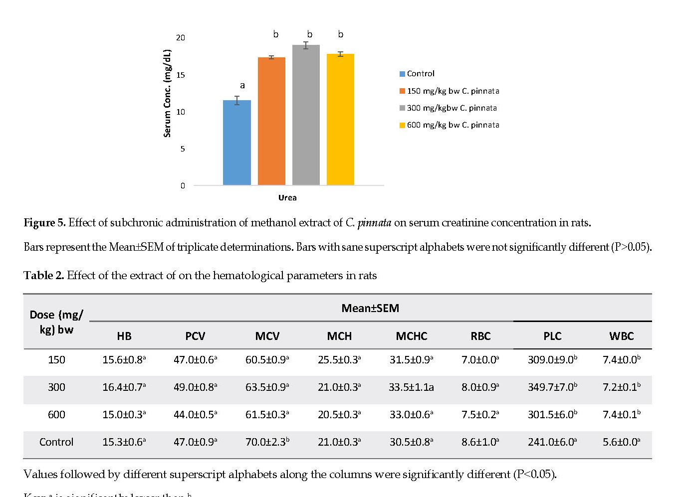
All doses of extract administered to rats significantly reduced (p<0.05) the sodium concentration in the sera, compared to that found for the controls. The concentration of chloride; however, was not significantly altered (p>0.05) by the extract at all concentrations tested (Figure 3). The urea concentration was significantly higher (p<0.05) than that of the controls (Figure 4) while the serum creatinine level in the groups treated with the extract were similar (p>0.05) to that found in the controls (Figure 5).
Hematological parameters: The hematological parameters, which included HB, PCV, MCH, MCHC and RBC remained unchanged (p>0.05) in rats administered 150, 300 or 600 mg/kg of the extract compared to those detected for the control group. The WBC and platelet levels were significantly raised in rats treated with 600 mg/kg of the extract compared with that of the controls (Table 2).
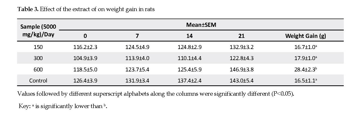
Weight gain vs. relative organ weight: There was a significant increase (p<0.05) in weight gain (Table 3), relative liver, kidney, lung and heart-body weight ratio (Table 4) of the rats treated with 600 mg/kg of the extract compared with those recorded in the controls. However, the body weight gains and relative organ weight in rats treated with 150 and 300 mg/kg of the extract were similar to those obtained for the controls (p<0.05).

Discussion
As more pharmacological and clinical information on medicinal plants become available, the toxicological database for these agents is also refined. Like their synthetic counterparts, toxicological studies must be performed for medicinal plant to validate their safety [20]. Unfortunately, there is limited scientific evidence reported pertinent to the safety of C. pinnata. In the present study, there was no test substance related to the mortality observed at 5000 mg/kg. Therefore, no acute toxicity was found in rats treated with the extract of C. pinnata and the approximate acute toxicity lethal value (LD50) were determined to be higher than 5000 mg/kg, and as such, it could be generally regarded as safe (GRAS).
This proves that the C. pinnata extract could be safely administered in acute treatments up to 5000 (mg/kg)/day. This finding is in agreement with Clarke and Clarke, who reported that any compound or drug with the oral LD50 greater than 1000 mg/kg of body weight could be considered low in toxicity and safe for use in rats [21]. However, Becker suggested that variables, such as animal species, strain, age, gender, diet, bedding, ambient temperature, caging conditions, and time of the day can influence the LD50 values obtained, and as such, there are considerable uncertainties in extrapolating the LD50 obtained for one species over others [20]. This finding suggests that LD50 may not be considered as a biological constant.
The negative influence of toxic compounds on the body weight of the laboratory animals is recognized and well documented in published literature [22]. The toxic nature of the administered product is generally correlated with its ability to produce a 10% or more decrement in body weight or growth rate of the selected test animals [23]. The results of the present study however, suggest that C. pinnata extract at doses of 150 and 300 mg/kg has no negative impact on the body weight of rats.
Relative organ weight may serve as the indication of the pathological and physiological status in humans and animals. Toxic substances induce abnormal metabolic reactions that affect such vital organs as heart, liver, spleen, kidneys and lungs [22]. The present findings also suggest that 150 and 300 mg/kg doses of the extract of C. pinnata are non-toxic on the vital organs tested as it did not induce organ swelling, atrophy or hypertrophy. Therefore, this extract is considered safe for maintaining the normal function of the organs at 150 and 300 mg/kg. However, the higher body weight gains in rats treated with 600 mg/kg can best be explained by the enhanced physiological processes in test animals.
Hematological assessment is useful to determine the extent of toxic effects of plant extracts on the blood constituents of an animal [23]. The analysis of blood parameters is closely related to risk evaluation because when tests involve rodents, the hematological system has a higher predictive value of the abnormal toxicity signs and symptoms in humans [24]. The present study revealed no noticeable hemolytic changes of HB, PCV, MCH, MCHC and RBC following treatment with the extract of C. pinnata for 21 days. These findings exclude the possibility of such occurrences as anemic condition or other RBC related disorders (e.g. thalassemia, polycythemia, liver disease and hypothyroidism).
Increases in the indices of WBC and differentials are generally considered as markers of stress and defense mechanisms triggered by the immune system against various inflammatory conditions, such as polymyalgia rheumatica, bacterial infections, hemorrhage and leukemia. The rise in the WBC in rats treated with 600 mg/kg of the extract may indicate its effect on inducing the immune response to increase the production of leukocytes [6]. This is however, advantageous as it will increase the animal’s ability to fight off infections. The higher platelet counts in 600 mg/kg treated rats suggest the thrombopoietic effects of the extract.
Elevation in the levels of serum transaminase enzymes is highly indicative of the hepatic impairment in the animals [25]. The insignificant changes in the plasma AST levels in response to 150, 300 or 600 mg/kg of the extract suggest that it caused no reaction in the rat liver. ALT is a cytoplasmic enzyme that increases in plasma, signifying cellular injuries caused by toxins in the liver. Liver injury is characterized by the predominant elevation of ALT and increased level of mitochondrial enzyme AST in plasma, reflecting severe tissue injuries [26].
However, the extract reduced ALT level (Figure 1), which may account for its protective effect on the liver. The level of serum albumin decreases in response to inflammation [27]. Recent studies have shown that not only the albumin concentration but also its function are reduced in liver disorders, such as cirrhosis [14, 28]. The insignificant variation in the levels of Total proteins (TP), albumin, and bilirubin supports the non-toxic effect of the extract on the liver and negates a link with liver dysfunction.
One of the major roles of kidneys is to filter out metabolites, such as creatinine, urea and electrolytes from the plasma through the glomeruli. Since creatinine and urea are normally filtered from the plasma and only reabsorbed or secreted by the proximal tubule to a minor extent, both have been used as indices of renal clearance [29]. During renal impairment, the excretion of these metabolites by the kidneys is altered and thus they accumulate in the serum [11].
Consequently, the observed significant increase in the serum urea concentrations in rats treated with 150, 300 or 600 mg/kg is an indication of renal impairment. The extract must have either altered the metabolism of urea, leading to increased synthesis or decreased the tubular excretion [30]. This finding corroborated with the findings reported by Aldler et al. [31] and Judykay et al. [32], which demonstrated that the raised serum urea levels in patients may indicate a pre-renal problem. The decrease in the sodium concentration also indicates compromised nephrotic integrity by the extract. However, the insignificant alterations in the chloride concentration suggest that the integrity of renal tubules was not compromised with respect to the excretion and maintenance of the normal levels of this electrolyte in the animal systemic environment [11].
Conclusions
The acute toxicity test suggests that oral single dose administration of C. pinnata up to 5000 mg/kg is practically non-toxic to Wistar rats. The sub-chronic toxicity study suggests that extract does not cause a significant effect in hematological and biochemical indices of liver integrity, when administered orally at doses of 150 to 600 mg/kg to Wistar rats. However, significant alterations of the markers of kidneys integrity call for exercising caution when using this extract as an oral remedy in the long term.
Ethical Considerations
Compliance with ethical guidelines
The authors fully observed the principles governing the use of laboratory animals, as laid out by Committee on Ethics for Medical and Scientific Research, the Federal University of Technology, also the current internationally accepted principles for laboratory animal use and care as contained in the Canadian Council on Animal Care Guidelines and Review Protocol. All available data are presented in the manuscript.
Funding
This research did not receive any specific grant from funding agencies in the public, commercial, or not-for-profit sectors.
Author's contributions
All authors contributed in preparing this article.
Conflict of interest
The authors declared no conflict of interest.
Acknowledgements
The authors appreciate the technical staff of the Department of Biochemistry, Federal University of Technology, Minna, Nigeria, for their kind assistance throughout the conduction of this study.
References
Lawal B, Shittu OK., Oibiokpa FI, Berinyuy EB, Mohammed H. African natural products with potential antioxidants and hepatoprotectives properties: a review. Clin Phytosci. 2016; 2:23. [DOI:10.1186/s40816-016-0037-0]
Bashir L, Shittu OK, Sani S, Busari MB, Adeniyi KA. African natural products with potential anti-trypanosomal properties: A review. Int J Biochem Res Rev. 2015; 7(2):45-79. [DOI:10.9734/IJBCRR/2015/16039]
Lawal B, Shittu OK, Kabiru AY, Jigam AA, Umar MB, Berinyuy EB, et al. Potential antimalarials from African natural products: A review. J Intercult Ethnopharmacol. 2015; 4(4):318-43. [DOI:10.5455/jice.20150928102856] [PMID] [PMCID]
Upendra Rao M, Sreenivasulu M, Chengaiah B, Jaganmohan Reddy K, Madhusudhana Chetty C. Herbal medicines for diabetes mellitus: A review. Int J PharmTech Res. 2010; 2(3):1883-92.
Saad B, Azaizeh H, Said O. Tradition and perspectives of Arab herbal medicine: a review. Evid Based Complement Alternat Med. 2005; 2(4):475-9. [DOI:10.1093/ecam/neh133] [PMID] [PMCID]
Debelo N, Afework M, Debella A, Makonnen E, Ergete W, Geleta B. Assessment of hematological, biochemical and histopathological effects of acute and sub-chronic administration of the aqueous leaves extract of Thymus schimperi in rats. J Clin Toxicol. 2016; 6(2):1000286. [DOI:10.4172/2161-0495.1000286]
Uddin N, Hasan MR, Hasan MM, Hossain MM, Alam MR, Hasan MR, et al. Assessment of toxic effects of the methanol extract of Citrus macroptera Montr. Fruit via biochemical and hematological evaluation in female Sprague-Dawley rats. PLoS One. 2014; 9(11):e111101. [DOI:10.1371/journal.pone.0111101] [PMID] [PMCID]
Kirkbride Jr JH. Dupuya, a new genus of Malagasy Legumes (Fabaceae). Novon. 2005; 15(2):305-14.
Nyunaï N. Cordyla pinnata (Lepr. Ex A. Rich.) Milne-Redh [Internet]. 2011 [Updated 2018 May 20]. Available from: https://www.prota4u.org/database/protav8.asp?fr=1&g=pe&p=Cordyla+pinnata+(Lepr.+ex+A.Rich.)+Milne-Redh
Amos TN, Bashir L, Saba SE, Saba MA, Mohammed BM, Abdulsalam IH, et al. Phytochemicals and acute toxicity profile of aqueous and methanolic extracts of Crateva adansonii leaves in Swiss albino rats. Asian J Biochem. 2015; 10(4):173-9. [DOI:10.3923/ajb.2015.173.179]
Bashir L, Shittu OK, Busari MB, Sani S, Aisha MI. Safety evaluation of giant African land snails (Archachatina maginata) haemolymph on hematological and biochemical parameters of albino rats. J Adv Med Pharm Sci. 2015; 3(3):122-30. [DOI:10.9734/JAMPS/2015/16393]
Reitman S, Frankel S. A colorimetric method for the determination of serum glutamic oxalacetic and glutamic pyruvic transaminases. Am J Clin Pathol. 1957; 28(1):56-63. [DOI:10.1093/ajcp/28.1.56] [PMID]
Gornall AG, Bardawill CJ, David MM. Determination of serum proteins by means of biuret reaction. J Biol Chem. 1949; 177(2):751-66. [PMID]
Doumas BT, Watson WA, Biggs HG. Albumin standards and the measurement of serum albumin with bromocresol green. Clin Chim Acta. 1971; 31(1):87-96. [DOI:10.1016/0009-8981(71)90365-2]
Tietz NW. Clinical guide to laboratory tests. 3rd ed. Philadelphia, PA: WB Saunders Company; 1995.
Tietz NW. Tietz textbook of clinical chemistry and molecular diagnostics. Burtis CA, Ashwood ER, Bruns DE, editors. Philadelphia: Elsevier Saunders; 2006.
Heinegård D, Tiderström G. Determination of serum creatinine by a direct colorimetric method. Clin Chim Acta. 1973; 43(3):305-10. [DOI:10.1016/0009-8981(73)90466-X]
Dacie SJV, Lewis SM. Practical haematology. 8th ed. London: Churchill Livinstone; .1995.
SAS institute Inc. SAS/STAT.User’s Guide (Version 9). SAS institute Inc. CARY,NC. [Internet] 2019. [Updated: 2019 Nov 24]. Available from: https://support.sas.com/documentation/onlinedoc/91pdf/sasdoc_91/stat_ug_7313.pdf
Becker RA, Plunkett LM, Borzelleca JF, Michael Kaplan A. Tiered toxicity testing: Evaluation of toxicity-based decision triggers for human health hazard characterization. Food Chem Toxicol. 2007; 45(12):2454-69. [DOI:10.1016/j.fct.2007.05.030] [PMID]
Clarke EGC, Clarke ML. Veterinary toxicology. London: Bailliere Tindall & Cassell Ltd; 1967.
Berinyuy EB, Lawal B, Olalekan AA, Olalekan IA, Yusuf AA, Sakpe S, et al. Hematological status and organs/body-weight parameters in wister rats during chronic administration of Cassia occidentalis. Intl Blood Res Rev. 2015; 4(3):1-7. [DOI:10.9734/IBRR/2015/22021]
Schilter B, Andersson C, Anton R, Constable A, Kleiner J, O'Brien J, et al. Guidance for the safety assessment of botanicals and botanical preparations for use in food and food supplements. Food Chem Toxicol. 2003; 41(12):1625-49. [DOI:10.1016/S0278-6915(03)00221-7]
Lawal B, Shittu OK, Abubakar AN, Haruna GM, Saidu S, Ossai PC. Haematopoetic effect of methanol extract of Nigerian honey bee (Apis mellifera) propolis in mice. J Coast Life Med. 2015; 3(8):648-51. [DOI:10.12980/JCLM.3.2015J5-89]
Lawal B, Shittu OK, Oibiokpa FI, Mohammed H, Umar SI, Haruna GM. Antimicrobial evaluation, acute and sub-acute toxicity studies of Allium sativum. J Acute Dis. 2016; 5(4):296-301. [DOI:10.1016/j.joad.2016.05.002]
Yusuf AA, Lawal B, Yusuf MA, Omonije YO, Adejoke AO, Raji FH, et al. Free radical scavenging, antimicrobial activities and effect of sub-acute exposure to Nigerian Xylopia aethiopica seed extract on liver and kidneys functional indices of albino rat.Iran J Toxicol. 2018; 12(3):51-8.
Shittu OK, Lawal B, Alozieuwa BU, Haruna GM, Abubakar AN, Berinyuy EB. Alteration in biochemical indices following chronic administration of methanolic extract of Nigeria bee propolis in Wister rats. Asian Pac J Trop Dis. 2015; 5(8):654-7. [DOI:10.1016/S2222-1808(15)60907-0]
Shittu OK, Lawal B, Abubakar NA, Berinyuy EB, Musa BB, Olalekan IA. Toxicological implications of methanol extract from Nigerian Bee propolis on some selected rat tissues. J Pharm Biomed Sci. 2015; 5(7):524-31.
Soufane S, Bouzidi A, Mahdeb N, Krache S. Evaluation of acute and subacute toxicity of fruit methanolic extract from Citrullus colocynthis in male albino rats. Int J Pharmacogn Phytochem Res. 2017; 9(4):567-80. [DOI:10.25258/phyto.v9i4.8130]
Yusuf AA, Lawal B, Abubakar AN, Berinyuy EB, Omonije YO, Umar SI, et al. In-vitro antioxidants, antimicrobial and toxicological evaluation of Nigerian Zingiber officinale. Clin Phytosci. 2018; 4:12. [DOI:10.1186/s40816-018-0070-2]
Aldler AI, Stevens RJ, Manley SE, Bilous RW, Cull CA, Holman RR, et al. Development and progression of nephropathy in type 2 diabetes: The United Kingdom Prospective Diabetes Study (UKPDS 64). Kidney Int. 2003; 63(1):225-32. [DOI:10.1046/j.1523-1755.2003.00712.x] [PMID]
Judykay T. Nutrition for reducing urea and creatinine in the blood Diabetes Care. 2007; 27:2191-2. https://scholar.google.com