Write your message
Volume 18, Issue 1 (January 2024)                   IJT 2024, 18(1): 14-20 | Back to browse issues page

Ethics code: 00650


XML Print


Download citation:
BibTeX | RIS | EndNote | Medlars | ProCite | Reference Manager | RefWorks
Send citation to:

Amedu N, Adeleye R, Abdur-Rahman H, Abolarin P, Omotoso G. Impact of Sodium Benzoate on Motor Coordination, Cerebellar Purkinje Cell Layer, and Oxidative Stress in Wistar Rats’ Brain. IJT 2024; 18 (1) :14-20
URL: http://ijt.arakmu.ac.ir/article-1-1281-en.html
1- Department of Anatomy, Faculty of Basic Medical Sciences, Adeleke University, PMB 250, Ede, Osun State, Nigeria , amedunath11@gmail.com
2- Department of Anatomy, Faculty of Basic Medical Sciences, Adeleke University, PMB 250, Ede, Osun State, Nigeria
3- Department of Anatomy, Faculty of Basic Medical Sciences, Chrisland University, PMB 2131, Abeokuta, Ogun State, Nigeria
4- Department of Anatomy, Faculty of Basic Medical Sciences, University of Ilorin, PMB 1515, Ilorin, Kwara State, Nigeria
Full-Text [PDF 909 kb]   (78 Downloads)     |   Abstract (HTML)  (158 Views)
Full-Text:   (43 Views)
Introduction
The cerebellum plays a crucial role in various forms of motor learning [1]. It has the ability to integrate, modify, or impact a broad spectrum of neural activities that are still not fully understood [2]. In fact, the cerebellum participates in the coordination and integration of both motor and cognitive processes. Structurally, the cerebellar cortex comprises three tiers: the basal granular layer houses granule cells and interneurons; the central layer contains the Purkinje cell bodies, and the uppermost layer consists of Purkinje cell dendrites and parallel fibres. Also, within the outer layer, inhibitory interneurons such as stellate and basket cells are located, utilizing GABA to inhibit the Purkinje cell dendrites [3].
In the cerebellar circuitry, two prominent types of neurons hold key roles: Purkinje and granule cells [1]. Climbing fibres, axons originating externally, and entering the cerebellum, connect with Purkinje cells and send branches directly to the deep nuclei. Among all cell types in the brain, Purkinje cells receive the most synaptic inputs. It has been suggested that the number of spines on a solitary human Purkinje cell can reach as high as 200,000 [3]. The cerebellum is the main target of drug exposure, drug abuse and addiction, intoxication, and environmental toxins [4-6].
Sodium benzoate (NaB) serves as a popular food additive and preservative, effectively inhibiting the proliferation of microorganisms, such as bacteria, yeast, and molds in a wide range of foods and beverages [7]. Further, NaB has application in addressing such conditions as depression, pain, schizophrenia, autism spectrum disorders, and some neurodegenerative diseases [8]. Also, major regulatory bodies consider NaB safe for public consumption [7]. Nevertheless, there have been instances of adverse effects linked to NaB. For instance, experimental rats exposed to NaB have exhibited unfavorable shifts in various blood biochemical indicators [9]. The use of NaB may potentially lead to DNA damage and increased formation of micronuclei [10]. Notably, NaB can interact with ascorbic acid in beverages, resulting in the formation of carcinogenic compound, such as benzene [11].
Although, NaB effects have been reported in numerous studies previously, there are gaps in our understanding of its impact on aspects of motor coordination and the cerebellar Purkinje cell layer. This layer is of particular interest because the cells are the principal output neurons in the cerebellum. They receive input from various sources, including the cerebellar cortex and other brain regions, and play a critical role in the integration and modulation of motor signals before transmitting them to deep cerebellar nuclei and to other parts of the brain. This study was planned to investigate the impact of NaB on motor coordination, cerebellar Purkinje cell layer, and oxidative stress in Wistar rats.
Materials and Methods
Animals
A total of 18 adolescent male Wistar rats with an average weight of 217.6 grams were obtained from Temilola animal husbandry located in Ogbomoso, Oyo State, Nigeria. They were placed in the animal facility of the Faculty of Basic Medical Sciences at Adeleke University in Ede, Nigeria. They were kept in standard plastic cages with iron lids. The rats were allowed a period of ten days to adapt to the environment before they were divided in groups and administered with the assigned substances. Throughout this adaptation phase and for the entire duration of the study, the rats were subjected consistently to 12 hours of alternating light and dark cycles. Also, the rats had free access to both foods and potable water. The study's procedures and treatments were approved by the Research Ethics Committee of Adeleke University in Ede, Nigeria (Registration Code: N0:00650).
Experimental Design
After the adaptation period, the rats were randomly divided into three groups of six rats each. For a continuous span of 28 days, the treatments were given by oral gavage as follows: Group 1 (control) was given 0.5 ml of distilled water; Group 2 received sodium benzoate at 100 mg/kg of body weight; and Group 3 was administered sodium benzoate at 300 mg/kg. The dosage and duration of the assigned treatments were based on preliminary studies carried out by the authors.
Behavioral Tests
Hanging Wire Test: The test was conducted for four days after the completion of NaB administration, based on the modified method of Aartsma-Rus & van Putten [12] and that of Amedu & Omotoso [13]. The apparatus consisted of a 55-cm wide metallic wire, 2-mm thick, secured between two vertical supports. The gaps between the metallic wire and the soft bedding beneath it measured 37-cm. In this experiment, each rat was held by its tail and allowed to grip the 2-mm thick metallic wire, using its front paws exclusively. Subsequently, a stopwatch was started to measure the duration until the rat fell off the wire. This was the recorded duration of hanging for each animal. The same procedure was used for all animals, with each rat undergoing a maximum of three hanging attempts before calculating the average hanging duration.
Footprint Test: The method used for this test was adapted based on procedures described by two earlier studies [13, 14]. The test itself took place five days after the administration phase had been ended. The test apparatus was constructed as an open-top runway connected to a cage with openings at both ends. The runway spanned a length of 60-cm and a width of 11-cm. Moreover, the runway was bordered by two walls, each standing at a height of 12-cm. The rats were allowed 30 minutes to be familiarized with the testing environment. In order to capture the footprints, the rats’ front paws were coated with non-toxic red ink, while the hind paws were soaked in blue ink. The rats were then encouraged to walk down the runway covered with white paper. Six rats from each group participated, and every rat underwent three trials. Once the footprints dried up, the subsequent parameters were measured using a ruler and pencil. We measured the base width, overlap width, stride length for the front limbs, and stride length for the hind limbs. The average value for each parameter was subsequently computed for each group.
Brain Sample Harvesting
After the completion of the behavioral evaluation, the two rats selected for histopathological examinations were injected with ketamine intraperitoneally (50mg/ml; Pakson Pharma, PVT Ltd., India). Following this step, a perfusion and fixation process was carried out, using a buffered solution of 10% normal saline [15]. Subsequently, the rats were decapitated, and their cerebella were carefully harvested. The excised section of the cerebella from each rat was then subjected to post-fixation in a buffered 10% normal-saline solution for 24 hours.
Histopathological Analyses
After being post-fixed in buffered 10% normal-saline for 24 hours, the tissue samples were processed and embedded in paraffin wax blocks. The blocks were subsequently sliced into thin sections and stained with Haematoxylin and Eosin (H&E), and cresyl fast violet (CFV). The staining was performed following the guidelines provided by Bancroft & Layton [16] and Wolfe [17] to enable the standard histological and Nissl substance examinations of the cerebellar tissue samples. The stained sections were examined using a light binocular microscope (Olympus, New Jersey, USA), and the images were photographed on the attached Amscope camera (MD500, CA, USA). For each section, six visual fields of the Purkinje cell layer were photographed and used for the subsequent quantification of cell numbers, utilizing the ImageJ software (National Institute of Health, USA).
Immuno-histochemical Assay
The tissue sections went through paraffin wax removal and antigen retrieval processes, followed by exposure to Phosphate Buffered Saline (PBS) and hydrogen peroxide to inhibit the activity of natural peroxidase. To prevent background staining, normal goat serum was utilized, after which the sections were exposed to polyclonal rabbit anti-GFAP serum (diluted 1:500) for a period of 24 hours at 4°C. Subsequently, a series of applications including biotinylated mouse anti-rabbit solution and the Avidin-Biotin complex were performed sequentially. The immune complexes were then visualized, using 3,3’-Diaminobenzidine. To provide contrast, Haematoxylin counterstaining was performed, and the stained sections were examined under a light binocular microscope (Olympus, New Jersey, USA) and photographed on the Amscope camera (MD500, CA, USA).
Malondialdehyde Assay
The cerebellar tissue samples (n=4), were rinsed with PBS and subsequently homogenized, using a Teflon Potter–Elvehjem homogenizer on ice to create the homogenate solution. This homogenate was then subjected to centrifugation at 13,000g for five minutes, and the supernatant was used to measure the MDA level in each sample. The evaluation of MDA concentration was conducted consistent with the procedure provided by assay kits received from Sigma-Aldrich (St. Louis, MO, USA).
Statistical Analyses
The data analysis was carried out on GraphPad Prism software, version 9.0. Statistical comparisons were made using one-way analysis of variance (ANOVA) coupled with Tukey's multiple post-hoc tests. All results were presented as the means ± standard deviations, and the statistical significance of the differences were set at the level of P<0.05.
Results
Effect of Sodium Benzoate on Motor Coordination
As shown in Figure 1, the outcomes from the hanging wire experiment demonstrated that there was no significant difference in the duration for the animals to drop from the wire, in those treated with the NaB100mg/kg (75±2.6 s) compared to the control (80 ±1.5s). However, the animals given NaB300mg/kg exhibited notably shorter times (65 ±2.6s; P<0.05) to fall from the wire, as compared to both the control (80±1.5 s) and those treated with NaB100mg/kg (75 ±2.6 s). The representative photograph of the footprints (Figure 5) illustrated that the controls displayed stances with closely positioned forelimb (marked in red) and hind limb footprints (marked in blue), indicative of a stable proximity.

Figure 1.
Time spent hanging to wire. Data expressed as mean ± SD (n=6).

NaB=Sodium benzoate. aP<0.05 vs Control group; bP<0.05 vs NaB 100mg/kg treated group.


Figure 2. Footprint analysis of stride length in cm. Data expressed as mean ± SD (n=6).
NaB=Sodium benzoate. All P values were >0.05.


Figure 3.
Footprint analysis of base width. Data expressed as mean ± SD (n=6).

NaB=Sodium benzoate.  All P values were >0.05.

Figure 4.
Footprint analysis of overlap length. Data expressed as mean ± SD (n=6).

NaB=Sodium benzoate. All P values were >0.05

Figure 5. Representative footprint photograph.

Conversely, the NaB100mg/kg group showed broader overlapping stances and smaller strides. In contrast, the footprint pattern in the NaB300mg/kg group revealed extended strides compared to the control.
As shown in Figure 3, the analysis of base width indicated that there was no significant difference (P>0.05) in the base width between the NaB100mg/kg and 300mg/kg treated groups (1.5 ±0.23 cm Vs. 1.5 ±0.20 cm) in contrast to that of the control (2.0 ±0.26 cm). Likewise, the findings for the stride length (Figure 2) and overlap length (Figure 4) revealed no significant differences (P>0.05) between the NaB100mg/kg and 300mg/kg treated groups as compared to the control.
Impact of Sodium Benzoate on Cerebellar Purkinje cells, Nissl Substance, and GFAP Expression
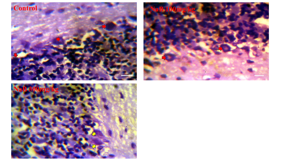
The image displayed in Figure 6 depicts a photomicrograph of the rat cerebellum sections stained with H&E. In the group treated with NaB at 300mg/kg, the layer containing Purkinje cells appeared distorted, and fewer cells were present compared to those of other groups, particularly the control. The Purkinje cells count, as illustrated in Figure 8, was significantly lower (P<0.05) in the NaB300mg/kg treated group (40±1.7) compared to that of the control (51±6.9). Figure 7 presents photomicrographs of the rat cerebellar sections stained with CFV. In both the control and the NaB100mg/kg treated groups, there was intense staining of Nissl substances (red arrows), and only few cells exhibited chromatolysis. However, in the NaB300mg/kg treated group, the Nissl substances in numerous cells were faintly stained, and displayed chromatolysis (yellow arrows). As shown in Figure 9, sections of the cerebellum processed with GFAP-immunostaining showed no noticeable immune reactivity in the control group. However, both the NaB100mg/kg and NaB300mg/kg treated groups displayed cells that were immunopositive for GFAP (red arrows). The NaB300mg/kg treated group exhibited a higher number of GFAP-immunopositive cells.
Figure 6. Representative photomicrograph of H&E-stained section of rats` cerebellum.
Control group, NaB 100mg/kg group, and NaB 100mg/kg treated group. Scale bar =180 μm.
Blue arrows = Purkinje cell layer; Yellow arrows = Distorted Purkinje cell layer.


Figure 7. Representative photomicrograph of CFV-stained section of rats’ cerebellum.
Control group, NaB 100mg/kg group, and NaB 100mg/kg treated group. Scale bar=180 μm.
Red arrows = Intense Nissl stain of purkinje cells; Yellow arrows = lightly stained Nissl substance of purkinje cell.


Figure 8.
The number of Purkinje cells per field counted in rat Purkinje cell layer.

Data expressed as mean ± SD. NaB=Sodium benzoate. a P < 0.05 vs Control group.


Figure 9. Representative photomicrograph of GFAP-Immunoreactive section of rats’ cerebellum.
Control group, NaB 100mg/kg group, and NaB 100mg/kg treated group. Scale bar = 45 μm.
Red arrows = immuno-positive cells.


Figure 10.
Malondialdehyde level in rats` cerebellum. Data express as mean ± SD.

NaB=Sodium benzoate; MDA= Malondialdehyde. a P<0.05 vs Control group; b P<0.05 vs NaB 100mg/kg treated group.

Influence of Sodium Benzoate on Indicators of Oxidative Stress
The MDA concentration, as shown in Figure 10, was considerably elevated in the group treated with NaB at 300mg/kg compared to the control (0.73±0.12 vs. 0.37±0.058; P˂0.05). Conversely, the MDA level in the NaB100mg/kg treated group did not display a significant difference as compared to the control (0.43±0.12 vs. 0.37±0.058; P>0.05).
Discussion
The role of cerebellum is critical in motor learning [1]. It can integrate, or influence a wide range of neural activities, which are not yet fully understood [2]. Indeed, cerebellum is involved in the coordination or integration of motor and cognitive processes. In animal models, gait and motor coordination are tested using footprint patterns, hanging from wire, and beam walking, among other tests [13, 14].
In this study, the data obtained from the hanging wire test indicated that there was no substantial disparity in the length of time it took for the animals to fall from the wire comparing the controls with those in NaB100mg/kg group. However, the animals in the NaB300mg/kg group displayed a significantly shorter duration to drop off of the wire compared to both the control and the NaB100mg/kg groups. This finding suggests that NaB at the higher dosage (300mg/kg) had a greater impact on the animals' ability to maintain their hold on the wire, thus leading to sooner falls.
The examination of the base widths (the width between the feet during movement) revealed that there were no significant differences between the groups treated with NaB100mg/kg and 300mg/kg, compared to the control. The lack of significant difference suggests that sodium benzoate treatment did not noticeably affect the base width of the feet in rats during walking. Similarly, the stride length (the distance covered in one step) and overlap length (the degree of overlap between successive steps) were also assessed. The results indicated that there were no significant differences in stride and overlap lengths between the groups treated with NaB at 100 or 300mg/kg, as compared to the control. This observation implies that sodium benzoate treatment did not result in significant alterations in the animals' stride length or the degree of overlap between their steps.
The large, spherical Purkinje cell bodies are densely arranged within a slim layer in the cerebellar cortex, referred to as the Purkinje layer [3]. These cells are the prominent neurons of the cerebellar circuitry that receive the most synaptic inputs [1]. In the current study, the cohort group administered with NaB at 300mg/kg exhibited noticeable distortions in the Purkinje cells layer with reduced cell counts as compared to those in other groups, especially the control. The quantification of Purkinje cells, as shown in Figure 8, revealed a substantial reduction in the NaB 300mg/kg treated group in contrast with the controls. This implies that the NaB at a higher dose had negative effects on the Purkinje cells, both in terms of their number and arrangement in the cerebellum. As Purkinje cell axons are the sole output in the cerebellar cortex, losses or deficits in their number could result in such pathologies as ataxia, multiple sclerosis, and other abnormal neurological conditions [18].
The Nissl stain is widely used to examine the overall morphology of the brain, identify neuronal abnormalities, and analyse alterations in neurons due to various factors, such as injury, disease, or experimental treatments [19]. In both the control and the NaB100mg/kg treated groups, there was intense staining of the Nissl bodies, and only a few cells exhibited chromatolysis, indicative of disintegration or breakdown of Nissl bodies. However, in the NaB300mg/kg treated group, the Nissl bodies in numerous cells were faintly stained, and displayed chromatolysis. These findings imply that the treatment with NaB at 300mg/kg had a more pronounced effect on cellular structure and function compared to those observed for the lower NaB dosage.
Assessment of GFAP staining provides valuable insights into the extent of astrocyte activation and the degree of CNS damage, or disease progression. In cases of brain injury, neurodegenerative diseases, and similar neurological disorders, the expression of GFAP can increase as brain tissue responses to damage or stress [20]. The increased expression of GFAP is often used as an indicator of gliosis, which is the process of astrocyte activation in response to neural tissue injury. In this study, we found that treatment with NaB led to a rise in the expression of GFAP in the cerebellum. This effect was more prominent in the group treated with the higher dose of NaB (300mg/kg) compared to the lower dose (100mg/kg), and the untreated controls. This finding implies that various doses of NaB trigger the activation of astrocytes, leading to increased GFAP expression, i.e., gliosis within the cerebellum. Further, the finding also suggests that the effect is linked to the NaB dosage, with the higher dose exhibiting a stronger response. This finding also implies that the group treated with NaB at 300mg/kg displayed substantially greater neurological alterations and responses to the injury, possibly associated with neurodegenerative processes.
Malondialdehyde (MDA) molecule is formed as a result of lipid peroxidation, which occurs due to oxidative stress and the interaction of reactive oxygen species with lipid molecules in the cell membranes [21]. This molecule serves as a marker for oxidative damage and is often measured to assess the levels of lipid peroxidation and oxidative stress in biological systems [22]. In this study, the concentration of MDA was notably elevated in the group treated with NaB at 300mg/kg compared to that of the control. The consequences of the elevated MDA concentration in the group treated with NaB at 300mg/kg could include increased oxidative stress, cellular damages, potential inflammation, and risk of various diseases. The evidence for oxidative stress and cellular damage was already established based on the results of this study, as presented in Figures 6, 7 & 10.
Conclusions
This study investigated the effects of sodium benzoate on motor coordination, cerebellar histology, and oxidative stress in rats’ cerebella. The group treated with NaB300mg/kg exhibited alterations in the Purkinje cell layers, increased oxidative stress, as indicated by elevated MDA levels, and motor incoordination. However, the group treated with NaB100mg/kg showed minimal differences compared to the control. The study findings underscore dose-dependent outcomes and suggest a complex interplay between sodium benzoate administration and various physiological parameters. Further research is warranted to fully understand the implications of these findings and their potential significance in clinical contexts.
Conflict of Interests
Authors declares no conflicts of interest with any entities.
Funding
This research did not receive any funding from funding agencies in the public, commercial or non-profit sectors.
Acknowledgements
The authors wish to express their gratitude to Abimbola Ajose, Elizabeth Ajayi and Ezra Lambe for their valuable technical assistance they provided during this study.
Compliance with Ethical Guidelines
The study protocol and treatment procedures were approved by the Research Ethics Committee of Adeleke University, in Ede, Osun State, Nigeria (No: 00650) in agreement with the recommendations of the National Research Council Guidelines for Care and Use of Laboratory Animals (NRC Publication: 2011).
Authors' Contributions
NOA and GO: participated in the design and interpretation of the study results, data analysis, and the review of the manuscript. NOA, PA, HA, and RA: conducted the experiments, collected the tissue samples and contributed to the data analyses. NOA and RA: performed histopathological and immunochemical analyses. All authors participated in writing the drafts of the manuscript, and reviewed, and approved the final article prior to submission.
Type of Study: Research | Subject: General

References
1. Wright M, Skaggs W, Årup Nielsen F. The Cerebellum. WikiJournal of Medicine. 2016;3(1). [DOI:10.15347/wjm/2016.001]
2. Haines DE, Dietrichs E. The cerebellum - structure and connections. Handb Clin Neurol. 2012;103:3-36. [DOI:10.1016/B978-0-444-51892-7.00001-2] [PMID]
3. Shepherd GM. The synaptic organization of the brain. New York: Oxford university press; 2003. [DOI:10.1093/acprof:oso/9780195159561.001.1]
4. Manto M. Toxic agents causing cerebellar ataxias. Handb Clin Neurol. 2012;103:201-13. [DOI:10.1016/B978-0-444-51892-7.00012-7] [PMID]
5. Alekseeva N, McGee J, Kelley RE, Maghzi AH, Gonzalez-Toledo E, Minagar A. Toxic-metabolic, nutritional, and medicinal-induced disorders of cerebellum. Neurol Clin. 2014;32(4):901-11. [DOI:10.1016/j.ncl.2014.07.001] [PMID]
6. Manto M, Perrotta G. Toxic-induced cerebellar syndrome: from the fetal period to the elderly. Handb Clin Neurol. 2018;155:333-52. [DOI:10.1016/B978-0-444-64189-2.00022-6] [PMID]
7. Bruna GOL, Thais ACC, Lígia ACC. Food additives and their health effects: A review on preservative sodium benzoate. African Journal of Biotechnology. 2018;17(10):306-10. [DOI:10.5897/AJB2017.16321]
8. Walczak-Nowicka LJ, Herbet M. Sodium Benzoate-Harmfulness and Potential Use in Therapies for Disorders Related to the Nervous System: A Review. Nutrients. 2022;14(7). [DOI:10.3390/nu14071497] [PMID] []
9. Zulfiqar A, Riaz H, Muhammad R, Tariq I, Sabir SM, Ali SW, et al. Toxicological evaluation of sodium benzoate on hematological and serological parameters of wistar rats. International Journal of Agriculture and Biology. 2018;20(11):2417-22.
10. Saatci C, Erdem Y, Bayramov R, Akalın H, Tascioglu N, Ozkul Y. Effect of sodium benzoate on DNA breakage, micronucleus formation and mitotic index in peripheral blood of pregnant rats and their newborns. Biotechnology & Biotechnological Equipment. 2016;30(6):1179-83. [DOI:10.1080/13102818.2016.1224979]
11. Piper JD, Piper PW. Benzoate and Sorbate Salts: A Systematic Review of the Potential Hazards of These Invaluable Preservatives and the Expanding Spectrum of Clinical Uses for Sodium Benzoate. Compr Rev Food Sci Food Saf. 2017;16(5):868-80. [DOI:10.1111/1541-4337.12284] [PMID]
12. Aartsma-Rus A, van Putten M. Assessing functional performance in the mdx mouse model. J Vis Exp. 2014(85). [DOI:10.3791/51303] [PMID] []
13. Amedu NO, Omotoso GO. Influence of Vitexin on ataxia-like condition initiated by lead exposure in mice. Toxicology and Environmental Health Sciences. 2020;12(4):305-13. [DOI:10.1007/s13530-020-00041-x]
14. Carter RJ, Morton J, Dunnett SB. Motor coordination and balance in rodents. Curr Protoc Neurosci. 2001;Chapter 8:Unit 8 12. [DOI:10.1002/0471142301.ns0812s15] [PMID]
15. Gage GJ, Kipke DR, Shain W. Whole animal perfusion fixation for rodents. J Vis Exp. 2012(65). [DOI:10.3791/3564]
16. Bancroft JD, Layton C. The hematoxylins and eosin. Bancroft's Theory and Practice of Histological Techniques2019. p. 126-38. [DOI:10.1016/B978-0-7020-6864-5.00010-4]
17. Wolfe AJ, Brubaker L. Urobiome updates: advances in urinary microbiome research. Nat Rev Urol. 2019;16(2):73-4. [DOI:10.1038/s41585-018-0127-5] [PMID] []
18. Redondo J, Kemp K, Hares K, Rice C, Scolding N, Wilkins A. Purkinje Cell Pathology and Loss in Multiple Sclerosis Cerebellum. Brain Pathol. 2015;25(6):692-700. [DOI:10.1111/bpa.12230] [PMID] []
19. Paul CA, Beltz B, Berger-Sweeney J. The nissl stain: a stain for cell bodies in brain sections. CSH Protoc. 2008;2008:pdb prot4805. [DOI:10.1101/pdb.prot4805] [PMID]
20. Yang Z, Wang KK. Glial fibrillary acidic protein: from intermediate filament assembly and gliosis to neurobiomarker. Trends Neurosci. 2015;38(6):364-74. [DOI:10.1016/j.tins.2015.04.003] [PMID] []
21. Gawel S, Wardas M, Niedworok E, Wardas P. [Malondialdehyde (MDA) as a lipid peroxidation marker]. Wiad Lek. 2004;57(9-10):453-5.
22. Singh Z, Karthigesu IP, Singh P, Rupinder K. Use of malondialdehyde as a biomarker for assessing oxidative stress in different disease pathologies: a review. Iranian Journal of Public Health. 2014;43(Supple 3):7-16.

Add your comments about this article : Your username or Email:
CAPTCHA

Send email to the article author


Rights and permissions
Creative Commons License This work is licensed under a Creative Commons Attribution-NonCommercial 4.0 International License.

© 2024 CC BY-NC 4.0 | Iranian Journal of Toxicology

Designed & Developed by : Yektaweb