Write your message
Volume 18, Issue 1 (January 2024)                   IJT 2024, 18(1): 21-28 | Back to browse issues page

Ethics code: All animal experiments were approved by the ethical committee of FSBSI ESIMER (identification code,


XML Print


Download citation:
BibTeX | RIS | EndNote | Medlars | ProCite | Reference Manager | RefWorks
Send citation to:

Vokina V, Sosedova L, Yakimova N, Kapustina E. Exposure of Parental Rats to Mercury Chloride during Progenesis Affects the CNS Parameters of the Adult Offspring. IJT 2024; 18 (1) :21-28
URL: http://ijt.arakmu.ac.ir/article-1-1282-en.html
1- Federal State Budgetary Scientific Institution, East-Siberian Institute of Medical and Ecological Research, 12A Microdistrict 3, Angarsk, Russian Federation , vokina.vera@gmail.com
2- Federal State Budgetary Scientific Institution, East-Siberian Institute of Medical and Ecological Research, 12A Microdistrict 3, Angarsk, Russian Federation
Full-Text [PDF 831 kb]   (73 Downloads)     |   Abstract (HTML)  (167 Views)
Full-Text:   (35 Views)
Introduction
According to the World Health Organization (WHO), mercury refers to the heavy metal that adversely affects environmental conditions and poses severe hazards to living organisms. Mercury can be found in three main forms: elemental, inorganic and organic compounds. The main amount of elemental mercury is contained in the earth's crust and oceans’ waters. Oxidized in the air, elemental mercury forms inorganic compounds that enter waters and soil by means of rain, snow, industrial and household wastes. Modified under the influence of bacteria, fungi and phytoplankton, inorganic mercury becomes organic compounds, accumulates in food chains and enters the human body through seafood and other edible products [1]. Upon contact with mercury, toxicity can occur mainly through the respiratory tract when contaminated food and drinks are consumed. The toxicity can also occur through the skin when bathing in polluted waters [2, 3]. The accumulation of mercury is most pronounced in the kidneys, brain, and liver, and is known to cause mutations in eukaryotic cells [4]. Experiments in cultured mammalian cells have revealed chromosomal aberrations, sister chromatid exchanges, and single-strand DNA breaks in fibroblasts from rats and mice [5-9].
Animal studies have shown the effects of mercury on the reproductive organs, including infertility, stillbirth, congenital malformations, and spontaneous abortions [10-13]. In addition, animal experiments have found that HgCl2 causes alterations in the estrous cycle, myometrium activities, and abnormal development of ovarian follicles [11-13]. Under the influence of mercury in the male body, failure of the reproductive functions has also been discovered. This can be expressed as dysfunctional spermatogenesis, fluctuations in testosterone levels, changes in the number and motility of spermatozoa, and accumulation of mercury in testicular tissues [14-18]. Currently, the long-term effect of chemicals on the subsequent generations is very relevant but remains unclear. The mercury load in parents may lead to the transfer of genetic and epigenetic disorders to the embryo. This may be manifested not only in the form of lethal mutations, but also as various pathological alterations in the offspring’s organs and systems, and their genetic instability. Literature reviews suggest that when animal parents come into contact with various toxic substances, the risk of developing severe cancers and CNS lesions increases in them and their offspring [19, 20].
The reproductive toxicity and mutagenic effects of mercury on the organs of mammals can give rise to mercury exposure in the animals’ offspring during prognosis when the gametes in juvenile organisms are being matured.  The above facts indicate the need to conduct research on the effects of mercury and its compounds on the reproductive organs of animal parents, and the health of their offspring. Therefore, the aim of this study was to investigate the effect of mercury exposure on the CNS and peripheral systems of the first-generation offspring from parental rats.
Materials and Methods
Animals and Treatments
The male and female Wistar rats used in this study were born and raised in the East Siberian Institute of Medical and Ecological Research (ESIMER). The animals consisted of 44 males; 90 females and 235 tested offspring (total = 369). These animals were kept on standard diet (BioPro; Russia) and potable water ad libitum.  The animals were also kept under 12 hours of light and dark cycles in a well-ventilated environment at 22-25ºC and 55-60% humidity. For six weeks, 15 male and 15 female rats were subcutaneously injected with a solution of mercury chloride (99.5% HgCl2; Himkomplekt, Ufa, Russia) at 0.35 mg/kg/day. The control rats (15 male and 15 female) were injected subcutaneously with normal saline solution.
Experimental Procedures
Mating Outcomes
Twenty-four hours after administration of HgCl2, the treated rats mated with intact partners at a ratio of one male to two females. Two days before the expected date of birth, the females were kept in separate cages. The rat pups from all groups were weaned from their mothers on the 30th day after birth. Stillbirths, neonatal mortality, and pup weights were recorded and assessed on the 4th, 7th, 14th, and 21st postnatal days (PND-4, PND-7, PND-14 & PND-21).
The percentage of stillbirths was calculated as follows: Stillbirths Percentage = Number of stillbirths divided by the total number of births in the group multiplied by 100. The death of newborn pups (except for the stillbirths) within seven days after their birth was taken into account as neonatal mortality. The percentage of neonatal mortality was calculated as follows: Percent Neonatal Mortality = Number of neonatal deaths / Total number of births in the group multiplied by 100.
Neurobehavioral Development
We evaluated the rats’ offspring for neurobehavioral development by observing the surface-righting reflex and motor activities in open field tests on postnatal days 4 to 21 (PND-4 to PND-21). The open field apparatus for testing was made of plexiglass (15 cm × 15 cm with 15 cm walls), divided by transverse lines into 1x1-cm squares. We placed each animal in the center of the platform, recorded the number of squares it crossed by one paw during movement (PND-4), and the duration of locomotion (PND-7). These observations were carried out for 30 seconds. The open field test setup on PND-14 was a 30x30-cm area with 20-cm walls, divided into squares with a side of 6-cm.  Next, each animal was placed in the center of the platform, the duration of locomotion was recorded for 1-min. For adult offspring, they were tested in the open field, followed by resident-intruder test, rotarod test, the development of a food-procuring reflex on PND-90, and lastly, we performed an electroneuromyography test.
Behavioral Experiments
The open field apparatus was a round arena 97-cm in diameter, with a wall height of 42-cm, and a hole in the floor with 2-cm diameter. The animals were observed for three minutes. The durations of locomotion and immobility, the number of rearing, grooming and freezing were recorded.  To study the social interactions, the resident-intruder test was used, which examined the modeling of intraspecific behavior of the animals. The experimental rat, "resident", was alone in the cage for 1-hour, after which a second animal, "intruder", was placed next to it for 5-minutes. The small "stranger rats" created the condition for the zoo-social dominance of the latter. During the 5-minute joint stay of the "resident" and the "stranger", we recorded the aggressive behavior of the "resident", i.e., the total number of attacks (vertical threats).
Next, we performed the Rota-rod test by placing each rat on a 4-cm diameter horizontal rod, rotating at a speed of 25 rpm. First, a training session of retention on the rod was performed (1-min), and then the rats were re-placed on the rod and the number of rats that remained on the rod for 3-min was recorded. During this test, we recorded the length of time each animal was able to stay balanced while walking on the drum without falling.
Learning Test
The ability of rats to learn was examined using a modified shuttle chamber adapted to work with positive food reinforcement. The chamber (25x25x30 cm) consisted of two compartments separated by a transparent gate. A rat was placed in the first compartment while the second compartment was equipped with two feeders and LED lamps above them. Food was added to the feeder only when the light turned on. The light stimulus was turned on for 20-sec, after which the transparent gate was opened and the rat was allowed to make transition to the feeders. With a correct attempt, the animal received one portion of food reinforcement, but with an incorrect one, the rat was returned to the first compartment. The development of the conditioned reflex was carried out for 10 days with 20 attempts per day. The number of correct attempts was then recorded for each rat.
Peripheral Nerves Test
To assess the functional state of the peripheral nerves in adult rats on PND-90, stimulation electroneuromyography (ENMG) was performed using needle electrodes. For local anesthesia and immobilization during the application of the electrodes and ENMG registration rats were subcutaneously injected with a 0.1% solution of a synthetic anesthetic, medetomidine hydrochloride (Apicenna, Russia), at a dose of 0.01 mg/kg. The reference and stimulating electrodes were inserted into the biceps femoris muscle, the ground electrode was fixed on the foot. The ENMG study was carried out, using an electro-neuromyograph "Neuro-EMG-Micro" ("Neurosoft", Russia), and the following parameters were analyzed: muscle response (M-response) amplitude, latency period (latency), M-response duration, involvement area [3, 7].
Ethical Approval
The study was conducted in accordance with the guidelines of the humane treatment of the animals based on the requirements of the International Guidelines for Biomedical Research in Animals (WHO, Geneva, Switzerland, 1985), the UK Animal Law (Scientific Procedures) (UK, 1986) and the guidelines of the National Institutes of Health on the Care and Use of Laboratory Animals (NIH Publication No. 8023, revised 1978). All animal experiments were approved by the Ethics
Committee of the East Siberian Institute for Medical and Ecological Research (identification code: E41/20; approval date: October 5, 2020. Procedural changes were approved every 6 months.

Statistical Analysis
All data were analyzed using Statistica software, version 6.1 (TIBCO, Tulsa, USA). The normal distribution of the data was verified by the Shapiro-Wilk’s test. To compare the quantitative parameters with non-normal distribution in unrelated samples, we used Mann-Whitney U-test. The quantitative parameters with normal distribution in unrelated samples were compared using Student t-test. The qualitative variables in unrelated samples were examined using two-tailed Fisher's test.  The differences among the pairs of data means were considered statistically significant at P≤0.05.
Results
Examinations of Newborn Rats
Postnatal Death
The neonatal mortality of the offspring due to HgCl2 significantly exceeded those of the control group. Among the offspring of HgCl2-exposed females, there was 17.7%, and 16.4% in the offspring of males, versus 2.2% in the controls (P=0.009 Vs P=0.014).
Body Weight and Motor Activity of the Newborn Rats
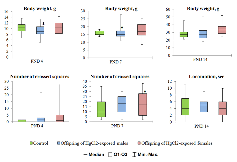
There was statistically significant decreases in the body weight by PND-4 and PND-7 in pups of F1 obtained from HgCl2-exposed males (P=0.005 Vs P=0.038) See Figure 1. The offspring from the females exposed to HgCl2 during all periods of the examination (F1) did not differ from the control group in terms of body weight. In the rat pups of the experimental group (F1), the body weight and motor activity at the age of 14 days were not significantly different from those of the control group (Figure 1). Pups from the F1 offspring (HgCl2-exposed) did not show significant deficits with respect to the righting reflex (PND-4) as compared to that of the controls. Examination of PND-7 indicated an increase in the duration of locomotion in pups of F1 offspring from HgCl2-exposed female rats (P=0.049, Figure 1).

Table 1. Indicators of the behavior of F1 white rats in the "open field"
Indicators Offspring of HgCl2-exposed males Offspring of HgCl2-exposed females
Male Female Male Female
Locomotion, sec 18.5 (12.0-23.5)
22.0 (16.0-26.0)
16.0 (12.0-19.0)
19.0 (12.0-23.0)
17.5 (14.5-23.5)
22.0 (16.0-26.0)
16.5 (12.0-20.0)
19.0 (12-23)
Immobility time, sec 2.0 (1.0-3.5)*
0 (0-1.0)
3.0 (1.5-4.0)*
2.0 (0-3.0)
2.0 (1.0-3.0)*
0 (0-1.0)
1.5 (0-4.0)
2.0 (0-3.0)
Sniffing, sec 25.5 (20.0-33.5)
29.0 (25.0-35.0)
25.5 (20.5-27.5)*
29.0 (26.0-30.0)
23.0 (20.5-25.0)*
29.0 (25.0-35.0)
28.0 (22.0-31.0)
29.0 (26.0-30.0)
Rearing 6.0 (3.0-8.0)
6.0 (5.0-10.9)
6.0 (3.5-8.0)
5.0 (4.0-7.0)
9.0 (5.5-10.0)
6.0 5.0-10.9)
8.0 (5.0-12.0)
5.0 (4.0-7.0)
Grooming 1.0 (0-1.5)
1.0 (0-2.0)
1.0 (0-2.0)
1.0 (0-2.0)
0 (0-1.0)
1.0 (0-2.0)
1.0 (1.0-2.0)
1.0 (0-2.0)
Notes: * = Differences were statistically significant compared to the controls at P<0.05; n = the number of observations; under the line indicators of the corresponding control group.

Figure 1. Body weight and locomotion activity of newborn offspring from females and male rats exposed to HgCl2 during progenesis.  Notes: * = Differences were statistically significant compared to the controls at P<0.05.
PND = Postnatal day. Q1-Q3 = interquartile range. Min = minimum. Max = maximum.


Figure 2. Resident-intruder test results.  Notes: * = Differences were statistically significant compared
 to the controls at P<0.05. Q1-Q3 = interquartile range. Min = minimum. Max = maximum.


Figure 3. The rotarod test results.  Notes: * = Differences were statistically significant
compared to the controls at P<0.05. Q1-Q3 = interquartile range; Min = minimum. Max = maximum.


Figure 4. Dynamics of the development of a conditioned reflex.  Notes: * = Differences
were statistically significant compared to the control at P<0.05.


Figure 5. ENMG parameters of the offspring.  Notes: * = Differences were statistically significant
compared to the control at P<0.05.  ms = milliseconds; mV = millivolt.
Behavioral Changes in Adult Offspring
The inhibition of motor activity in adult F1 offspring of male rats exposed to HgCl2 was evident in open field test.  Significant increases in total duration of immobility in both males and females were documented compared to similar indicators noted in the control group (P=0.037 Vs P=0.037). See Table 1. At the same time, shorter durations of sniffing pattern indicated decreases in the exploratory activity among the female rats’ offspring.
The offspring of HgCl2-exposed females also showed prolonged periods of immobility and reduced exploratory behavior; however, these were only significant in the male offspring (P=0.037 Vs P=0.035). See Table 1. There were no significant differences in the vertical activity and grooming among the study groups. There were decreases in the number of attacks on intruder in the male rats originated from the male rats exposed to HgCl2, as compared to those of the controls (Figure 2). Also, there was no significant difference in social behavior among the female offspring (Figure 2). No significant differences were found in the latency of falling from the rotating rod as compared to those of the offspring of experimental and control rats (Figure 3).
The investigation of the development of the conditioned food-procuring reflex showed that exposure to HgCl2 in females from the parental rats significantly reduced the offspring's reflex. The offspring of both genders from females exposed to HgCl2 did not master the inculcated skills over the allotted time. These animals made significantly fewer correct attempts than those observed in the controls, which may be indicative of memory impairment (Figure 4). The dynamics of learning ability in the male offspring exposed to HgCl2 was not significantly different from that of the control group (Figure 4).
We observed disturbances in peripheral nerves and muscles response of the hind limbs in F1 offspring to electric shock when examined by ENMG. A significant decrease in the amplitude of the M-response and the area of involvement in animals from the offspring of the experimental group was revealed compared to the controls (P=0.05 Vs. P=0.037). See Figure 5. In addition, these rats showed a significant increase in the latency and duration of the M-response, due to longer impulse conduction time along the nerve fibers (P=0.05 Vs P=0.037). See Figure 5.
Discussion
This study was planned to investigate the effect of HgCl2 on the central and peripheral systems of the first-generation offspring of parental rats. The data on impaired fertility and reproductive potential after exposure to HgCl2, and those reviewed from other animal studies [10-18] indicate that organic and inorganic mercury compounds can have adverse effects on human and rodent reproductive systems. To date, numerous studies have been published on the effect of HgCl2 in rodents’ reproductive function, both males and females. The consequences of mercury toxicity in the parental generations of rats vary. The toxic effects can manifest as reproductive losses (still-birth, congenital malformations and/or spontaneous abortions), developmental disorders, and reduced survival rate of the offspring [10]. There are published articles on the reproductive toxicity of methyl mercury chloride (CH3HgCl) when administered to mice, rats or monkeys of the same sex [21, 22]. Currently however, there is insufficient information available on the effects of mercury on the development and health of adult rats’ offspring. In this context, the issue of postnatal development disorders and long-term consequences in adult rats’ offspring exposed to mercury compounds is unclear but of particular relevance.
This study evaluated the long-term adverse effects of HgCl2 in offspring whose parents were exposed to this compound during progenesis. The parent rats were exposed to 0.35 mg/kg of HgCl2 per day, which is similar to the level of long-term human exposure in areas contaminated with mercury [23]. As suggested by Szász, et al. [24], exposure to HgCl2 at 0.8 mg/kg body weight per day in drinking water is tolerated by both pregnant female rats and their pups without showing major signs of toxicity. This includes no motor impairment in adult rats and the average body weight of their newborn offspring is at least 85% of the controls. That study has also demonstrated that the chronic administration of low doses of HgCl2 to female rats throughout pregnancy and lactation enhances 3-AR-induced neocortical epilepsies in their offspring. The incidence of this event increases after the end of breastfeeding and significantly later during puberty [24]. Our study provided evidence that long-term exposure to HgCl2 at low doses in parental rats during progenesis, mainly before fertilization, leads to increased offspring death, failed development, and changes in the CNS functions in the offspring.
Examination of sexually mature male offspring from the first generation of male white rats exposed to HgCl2, revealed low body weight. Also in the experimental group, both male and female offspring showed low motor, orienting and exploratory activities.  In addition, the aggressiveness was reduced in males, which all together suggest the predominance of inhibitory processes in their central nervous system. Also, maternal exposure to HgCl2 before mating leads to impaired cognition in sexually mature offspring, as exemplified by a decline in the ability to learn and act on food-procuring reflex.
This study showed that chronic exposure to HgCl2 at low dosage in the parental rats led to disorders in the peripheral nerves and muscles of adult offspring of both sexes, based on data from stimulation ENMG. In the examined white rats, axonal nerve trunk damages were detected predominantly. These were manifest as a decline in the amplitude of the maximum M-response as compared to those observed in the control animals. Increases in the latency of the studied nerves suggest a slowdown in the speed of impulse conduction along the motor axons of the mixed nerves. Decreases in the duration and amplitude of the M-response, and the area of involvement may also be associated with a decline in the number of functioning motor units in the examined muscles.
The long-term biological effects, as observed in the offspring of the female rats, can be justified by the mercury accumulation, which occurred during the HgCl2 exposure. Physiological stress in mercury-exposed maternal rats leads to its circulation through the bloodstream, penetration through the placental barrier, and exerting adverse effects on the embryo. Also, the duration of HgCl2 in male rats was six weeks, which was comparable to that of their spermatogenesis. Considering that mercury has a mutagenic effect in mammals, we assume that the mechanism of development of CNS disorders in the offspring was mediated mainly by changes in the genetic codes. Concurrently, HgCl2-induced mutations or modifications of the epigenome probably affected the germ cells, which passed steadily through the rats’ generations. It should be noted that the observed effects may be mediated by epigenetic disorders occurring during germ cells formation, leading to distinct changes in individual regions of the rats’ chromosomes. This event can subsequently lead to persistent functional alterations in the gene expression of the offspring. The above mechanism may be mediated by disturbances in the physiology of the offspring of both sublimate-exposed males and exposed females.
Conclusions
The findings of this study provided experimental evidence that long-term exposure of the parental generation to low doses of HgCl2 during progenesis leads to increased mortality and impaired development of the newborn offspring. Also, the exposure alters the animals’ behavior and causes disorders in the peripheral nerves and muscles of the sexually mature offspring. The study results substantiated the need for further investigation into the long-term effects of mercury toxicity in generations of rats, and the mechanism of transmission of such a "chemical load" from generation to generation of the animals.
Conflict of Interests
The authors declare no conflict of interest.
Funding
The study was funded within the framework of a state assignment.
Acknowledgement
The authors wish to acknowledge the support of the professors and staff of Federal State Budgetary Scientific Institution, East Siberian Institute of Medical and Ecological Research in Angarsk, Russia, for their generous support toward the conduction of this study.
Compliance with Ethical Guidelines
All animal experiments were approved by the ethical committee of FSBSI ESIMER (identification code, E37/08; date of approval, 29 May 2008) and carried out in compliance with the rules of humane treatment of animals in accordance with the requirements of the International Recommendations for Biomedical Research Using Animals (WHO, Geneva, 1985), UK Animals (Scientific Procedures) Act (UK, 1986), and National Institutes of Health Guide for the Care and Use of Laboratory Animals (NIH Publications No. 8023, revised 1978).
Authors' Contributions
Conceptualization, L.M.S., and V.A.V.; methodology, L.M.S., E.A.K and V.A.V.; formal analysis, E.A.K., Y.N.L. and V.A.V.; investigations, V.A.V., E.A.K. and Y.N.L.; writing the original draft of the manuscript, L.M.S., V.A.V.; writing, review and editing of the final draft, L.M.S. and V.A.V.; visualization, V.A.V.; project administration, L.M.S. All authors have read and approved the final version of the manuscript prior to submission.
Type of Study: Research | Subject: Special

References
1. Kumar V, Umesh M, Shanmugam MK, Chakraborty P, Duhan L, Gummadi SN, et al. A Retrospection on Mercury Contamination, Bioaccumulation, and Toxicity in Diverse Environments: Current Insights and Future Prospects. Sustainability. 2023;15(18). [DOI:10.3390/su151813292]
2. Arefieva A, Barigina V, Zatsepina O. The present-day ideas about impact of mercuric compounds at cell and system levels. Ekologiia Cheloveka. 2010(8):35.
3. Bezgodov I, Laptev I, Rogaleva V, Efimova N, Andreeva O. Mercury danger assessment at the enterprises of chemical industry in Irkutsk region. Bull VSNC SO RAMN. 2005;40:70-4.
4. Efimova NV, Koval PV, Rukavishnikov VS, Bezgodov IV. Problems associated with mercury pollution of environmental objects. Acta Biomedica Scientifica. 2005;1(39):127-33.
5. Anwar WA, Gabal MS. Cytogenetic study in workers occupationally exposed to mercury fulminate. Mutagenesis. 1991;6(3):189-92. [DOI:10.1093/mutage/6.3.189] [PMID]
6. Lamperti AA, Printz RH. Localization, accumulation, and toxic effects of mercuric chloride on the reproductive axis of the female hamster. Biol Reprod. 1974;11(2):180-6. [DOI:10.1095/biolreprod11.2.180] [PMID]
7. Schurz F, Sabater-Vilar M, Fink-Gremmels J. Mutagenicity of mercury chloride and mechanisms of cellular defence: the role of metal-binding proteins. Mutagenesis. 2000;15(6):525-30. [DOI:10.1093/mutage/15.6.525] [PMID]
8. Bonacker D, Stoiber T, Wang M, Bohm KJ, Prots I, Unger E, et al. Genotoxicity of inorganic mercury salts based on disturbed microtubule function. Arch Toxicol. 2004;78(10):575-83. [DOI:10.1007/s00204-004-0578-8] [PMID]
9. Zasukhina GD, Vasilyeva IM, Sdirkova NI, Krasovsky GN, Vasyukovich L, Kenesariev UI, et al. Mutagenic effect of thallium and mercury salts on rodent cells with different repair activities. Mutat Res. 1983;124(2):163-73. [DOI:10.1016/0165-1218(83)90176-3] [PMID]
10. Schuurs AH. Reproductive toxicity of occupational mercury. A review of the literature. J Dent. 1999;27(4):249-56. [DOI:10.1016/S0300-5712(97)00039-0] [PMID]
11. Lamperti AA, Printz RH. Effects of mercuric chloride on the reproductive cycle of the female hamster. Biol Reprod. 1973;8(3):378-87. [DOI:10.1093/biolreprod/8.3.378] [PMID]
12. Koli S, Prakash A, Choudhury S, Mandil R, Garg SK. Calcium Channels, Rho-Kinase, Protein Kinase-C, and Phospholipase-C Pathways Mediate Mercury Chloride-Induced Myometrial Contractions in Rats. Biol Trace Elem Res. 2019;187(2):418-24. [DOI:10.1007/s12011-018-1379-x] [PMID]
13. Merlo E, Schereider IRG, Simoes MR, Vassallo DV, Graceli JB. Mercury leads to features of polycystic ovary syndrome in rats. Toxicol Lett. 2019;312:45-54. [DOI:10.1016/j.toxlet.2019.05.006] [PMID]
14. Martinez CS, Escobar AG, Torres JG, Brum DS, Santos FW, Alonso MJ, et al. Chronic exposure to low doses of mercury impairs sperm quality and induces oxidative stress in rats. J Toxicol Environ Health A. 2014;77(1-3):143-54. [DOI:10.1080/15287394.2014.867202] [PMID]
15. Adelakun SA, Ukwenya VO, Akingbade GT, Omotoso OD, Aniah JA. Interventions of aqueous extract of Solanum melongena fruits (garden eggs) on mercury chloride induced testicular toxicity in adult male Wistar rats. Biomed J. 2020;43(2):174-82. [DOI:10.1016/j.bj.2019.07.004] [PMID] []
16. Almeer RS, Albasher G, Kassab RB, Ibrahim SR, Alotibi F, Alarifi S, et al. Ziziphus spina-christi leaf extract attenuates mercury chloride-induced testicular dysfunction in rats. Environ Sci Pollut Res Int. 2020;27(3):3401-12. [DOI:10.1007/s11356-019-07237-w] [PMID]
17. Kalender S, Uzun FG, Demir F, Uzunhisarcikli M, Aslanturk A. Mercuric chloride-induced testicular toxicity in rats and the protective role of sodium selenite and vitamin E. Food Chem Toxicol. 2013;55:456-62. [DOI:10.1016/j.fct.2013.01.024] [PMID]
18. Wong EW, Cheng CY. Impacts of environmental toxicants on male reproductive dysfunction. Trends Pharmacol Sci. 2011;32(5):290-9. [DOI:10.1016/j.tips.2011.01.001] [PMID] []
19. Pino-Lopez M, Romero-Ayuso DM. [Parental occupational exposures and autism spectrum disorder in children]. Rev Esp Salud Publica. 2013;87(1):73-85. [DOI:10.4321/S1135-57272013000100008] [PMID]
20. Desrosiers TA, Herring AH, Shapira SK, Hooiveld M, Luben TJ, Herdt-Losavio ML, et al. Paternal occupation and birth defects: findings from the National Birth Defects Prevention Study. Occup Environ Med. 2012;69(8):534-42. [DOI:10.1136/oemed-2011-100372] [PMID] []
21. Khera KS. Reproductive capability of male rats and mice treated with methyl mercury. Toxicol Appl Pharmacol. 1973;24(2):167-77. [DOI:10.1016/0041-008X(73)90136-1] [PMID]
22. Burbacher TM, Mohamed MK, Mottett NK. Methylmercury effects on reproduction and offspring size at birth. Reprod Toxicol. 1987;1(4):267-78. [DOI:10.1016/0890-6238(87)90018-9] [PMID]
23. Huang CF, Liu SH, Hsu CJ, Lin-Shiau SY. Neurotoxicological effects of low-dose methylmercury and mercuric chloride in developing offspring mice. Toxicol Lett. 2011;201(3):196-204. [DOI:10.1016/j.toxlet.2010.12.016] [PMID]
24. Szasz A, Barna B, Gajda Z, Galbacs G, Kirsch-Volders M, Szente M. Effects of continuous low-dose exposure to organic and inorganic mercury during development on epileptogenicity in rats. Neurotoxicology. 2002;23(2):197-206. [DOI:10.1016/S0161-813X(02)00022-0] [PMID]

Add your comments about this article : Your username or Email:
CAPTCHA

Send email to the article author


Rights and permissions
Creative Commons License This work is licensed under a Creative Commons Attribution-NonCommercial 4.0 International License.

© 2024 CC BY-NC 4.0 | Iranian Journal of Toxicology

Designed & Developed by : Yektaweb