Write your message
Volume 17, Issue 4 (October 2023)                   IJT 2023, 17(4): 9-16 | Back to browse issues page


XML Print


Download citation:
BibTeX | RIS | EndNote | Medlars | ProCite | Reference Manager | RefWorks
Send citation to:

Soleymanzadeh Moghadam S, Nobakht M, Mohammadi Z, Rahmani Fard S, Hajian Hossein Abadi F, Mazar Atabaki S et al . Antimicrobial and Cytotoxic Activities of Colloidal Silver and Titanium Dioxide Nanoparticles Against Food-Borne Bacteria: Shigella dysenteriae and Staphylococcus aureus. IJT 2023; 17 (4) :9-16
URL: http://ijt.arakmu.ac.ir/article-1-1232-en.html
1- Antimicrobial Resistance Research Centre, Institute of Immunology and infection diseases, Iran University of Medical Sciences. Tehran, Iran
2- Department of Pharmaceutics, Faculty of Pharmacy, Mazandaran University of Medical Sciences, Sari, Iran
3- Antimicrobial Resistance Research Center, Institute of Immunology and Infectious Diseases, Iran University of Medical Sciences, Tehran, Iran
4- Department of Pharmaceutics, Faculty of Pharmacy, Mazandaran University of Medical Sciences, Sari, Iran , pebrahimnejad@mazums.ac.ir
Full-Text [PDF 1021 kb]   (650 Downloads)     |   Abstract (HTML)  (1335 Views)
Full-Text:   (379 Views)
Introduction
 
The growing emergence of resistant pathogens has created major concerns in the treatment of infectious diseases [1, 2]. The rise in antibiotic resistance may lead to greater bacterial resistance and failed treatments [3]. Drug-resistant bacteria have posed major problems in the design and production of antibiotic drugs [4, 5]. Thus, it is important to control antibiotic resistance which is a major approach to preventing infections [2]. Among novel pharmaceutical approaches, nanomaterials have played major roles in clinical medicine and infectious disease control [6-8]. Some nanoparticles have shown efficacy against a variety of infectious diseases and cancers because of their high surface to volume ratio [8-10]. Colloidal silver nanoparticles (cAg) nanoparticles have shown promising effects as antibacterial drugs and gradually emerged as functional biomaterials to prevent the resultant infections. Silver nanoparticles are effective against both gram-positive and -negative bacteria, molds, fungi and eliminate them [11-13]. They have been widely and successfully used in catheters, implants and wound dressings for therapeutic and prophylactic purposes [14].
Titanium dioxide (TiO2) nanoparticles are effective compounds with a wide range of applications in pharmaceutical industry, such as white pigments for foods, photo catalysts, skin sunscreens, water loss treatment, hydrogen production and antimicrobial activity [13, 15]. The antimicrobial activities of TiO2 and mixed oxide system (MONs) have interesting properties, which are effective against both gram-negative and -positive bacteria [15]. In addition, TiO2 has tunable photocatalytic property, i.e., it can produce reactive oxygen species under ultraviolet light radiation, providing good antimicrobial effect. Also, it kills pathogens by damaging their cell membranes [15]. In addition, TiO2 is extensively used for various purposes, such as strong oxidation, biocompatibility, biochemical and antimicrobial properties [13].
Although there are many reports of antibacterial efficiency of Ag and TiO2 nanoparticles, based on our literature review, no reports was found on the comparison of their efficiency against S. aureus and S. dysenteriae, and their cytotoxicity evaluation on the Hu02 fibroblast cell line. In addition, S. dysenteriae Type-1 is mainly associated with serious life-threatening dysentery in epidemics.
Aim of the Study: This study was planned to search for agents that have antibacterial properties without having toxic effects on the human body cells. We aimed to synthesize Ag and TiO2 nanoparticles, and used a practical method to determine their antibacterial and cytotoxic properties against Staphylococcus aureus and Shigella dysenteriae. Both of these strains are foodborne bacteria [3, 16]. Also, we investigated the gram-negative S. dysenteriae Type-1 because it is one of the main sources of diarrhea in youngsters and elderly people globally, where only a few organisms, i.e., 10-100, are sufficient to cause serious infection [17-19].
Materials and Methods
Materials: The S. dysenteriae Type-1 (ATCC-13313) and S. aureus (ATCC-25923) were obtained from Pasteur Institute, Tehran, Iran. Streptomycin disks (25µg) and tetracycline (100 μg) were obtained from Mast Dignostica GmbH and Mueller Hinton Agar (Reinfeld, Germany). The AgNO3 solution and NaBH4 were purchased from Sigma-Aldrich (Darmstadt, Germany). The MTT assay for cell line (Hu02 fibroblasts) was obtained from the Iranian Biological Resource Center (Tehran, Iran). The cell culture media were high glucose DMEM, fetal bovine serum (Invitrogen), trypsin (Sigma-Aldrich; EUA), and dimethylsulfoxide (DMSO) (Reinfeld, Germany). Isolates of Shigella sonnei resistant to streptomycin and tetracycline, and S. aureus were obtained from Milad Hospital, Tehran, Iran.
Synthesis of cAg Nanoparticles: In this study, 0.2 gram of chitosan was dissolved in 10mL of 1% acetic acid. Then, 6mL of the chitosan solution was poured into a beaker covered with aluminum foil and 50μL of AgNO3 solution (20mM) was added, and the beaker was stirred for 30 minutes. The NaBH4 solution was prepared as follows: 0.383g of NaBH4 was weighed and dissolved in 5mL of deionized water. Thirty minutes later, the AgNO3 and chitosan solutions were well-mixed dropwise. Further, 100μL of NaBH4 was added to the beaker and agitated on a stirrer at 700-800 rpm for 5 minutes. About 90 minutes later, the solution’s color changed from light to dark yellow [20, 21].
Synthesis of cTiO2 Nanoparticles: The synthesis of colloidal TiO2 nanoparticles was done according to the study of S. Mahshid, et al. [22]. Specifically, 5mL of titanium isopropoxide (TTIP) and 15mL isopropanol were mixed. Then, the pH was set to 7.4 by adding HNO3 or NH4OH. After vigorous stirring for peptizing process, the mixture was heated at 60-70°C for 18-20 hours. Next, the precipitate was washed with ethanol and dried at 100°C. Finally, a yellow-white powder was obtained and warmed at 200-800°C for 2 hours [22].
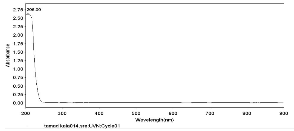
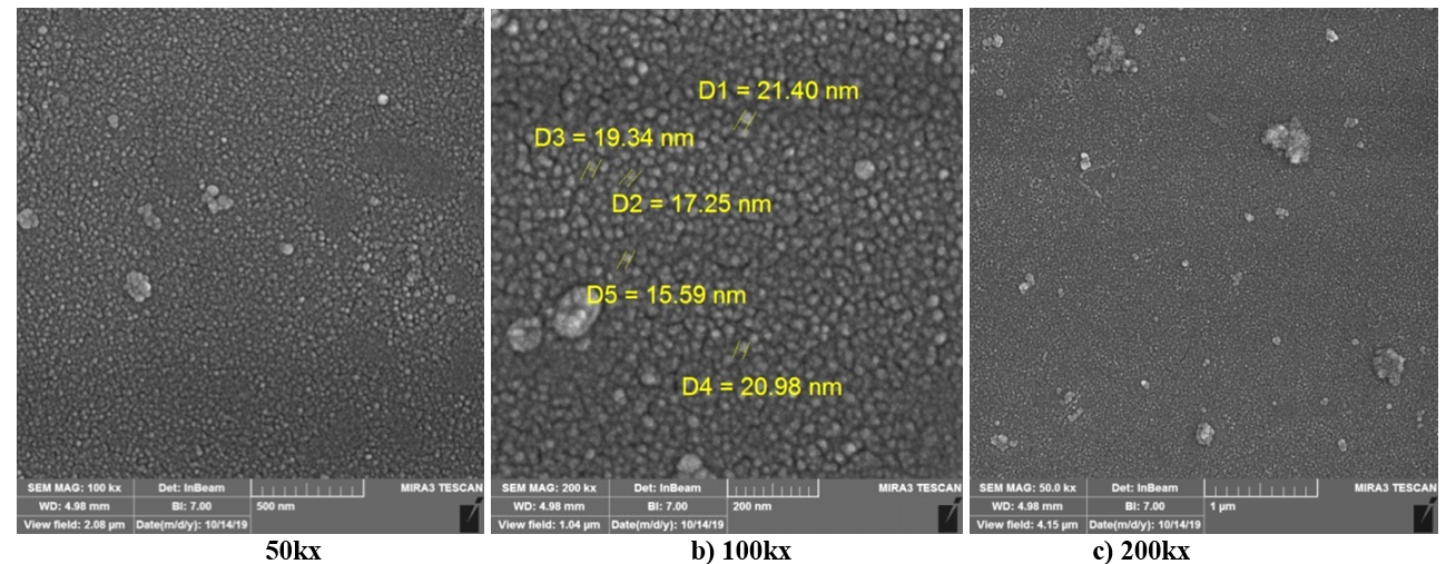
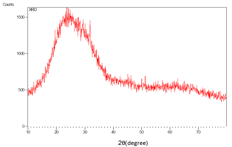
Characterization of Nanoparticles: Several techniques were used to characterize the nanoparticles. The UV–Vis spectra were obtained using a JASCO V-630 spectrophotometer (Jasco C-630, Japan) set in the absorbance range of UV-Vis spectra at 200-800 nm. The UV-visible spectra of the solutions containing Ag nanoparticles had a broad band around 206 nm (Figure 1). Also, the absorption band of the solution with TiO2 nanoparticles was 313 nm (Figure 2). A zetasizer nano ZS (Malvern Instruments Co., London, UK) was used to analyze the hydrodynamic average size in percentage, poly-dispersity index (PDI), and zeta potential of the nanoparticles in the media at 30°C. The measurement was made, using a disposable folded capillary zeta cell at 10mm path length.  Additionally, the morphology of nanoparticles was investigated, using a scanning electron microscope (FE-SEM TESCAN MIRA3) at 50kx, 100kx, and 200kx magnifications. The size of the nanoparticles, the crystal structure of Ag and TiO2 nanoparticles were determined and characterized by X-ray diffraction method (XRD). This method was employed, using a Philips PW1730 diffractometer  to record the X-ray powder diffraction at room temperature, 50 kV, 40 mA and operating CuKα (λ = 1.54060 Å). The data were recorded at 2θ range (10° to 70°). Further, the fineness size related to the Ag and TiO2 nanoparticles was estimated, using Debye–Scherrer equation 1: D = (kλ)/(βcθs) [23]   (1).
In the above formula, D is the diameter of crystallite size, K is a constant, λ is the CuKα wavelength, β is the full width at half maximum (FWHM) of the peak and θ is the Bragg diffraction angle.
Antimicrobial Tests in Colloidal Nanoparticles
(A) Minimum Inhibitory Concentration (MIC): Microdilution was performed according to the CLSI reference method [8, 24]. We used 96-well micro-titer plates. Antimicrobial agents in this experiment included colloidal Ag and TiO2 nanoparticles (100 µg/mL). For each test series, 100μL of Mueller-Hinton broth medium was added to 10 wells in each of the microplates. Then, 100μL antimicrobial agent was added to the first well, and the dilution serial was performed up to the tenth well. Also, wells 11 and 12 were considered as the positive and negative controls. Well-11 contained only the medium while well-12 contained a mixture of the medium and bacterial suspensions. After dilution, the microbial suspension was added to each well and placed in an incubator for 24 hours at 37°C [24]. The first well that prevented the bacterial growth, was considered as the MIC. All tests were done in triplicates and the means were used for the data analyses.
(B) Minimum Bactericidal Concentration (MBC): Based on this method, the well immediately following the first clear well was considered as the MBC, after determining the MIC of the antimicrobial agents. Nevertheless, for more confidence, the MIC well contents and two wells immediately after that were transferred to a dish containing the Muller Hinton Agar medium. Then all plates were incubated for 24 hours and the first dilution that eliminated 99.9% of the bacteria was considered as MBC [25, 26].
Evaluation of Cell Cytotoxicity by MTT Assay: We performed MTT assay based on the depletion of the tetrazolium salt, 3-(4,5-dimethylthiazol-z-yl)-2,5 diphenyl tetrazolium bromide through actively growing cells. This test starts with the breakdown of tetrazolium yellow salt via mitochondrial succinate dehydrogenase enzyme of living cells, and the production of insoluble formazan crystals in purple color. The greater the number of live cells, the more intense would be the color produced. The cytotoxicity of different nanoparticles has been evaluated using Hu02 fibroblast cell line (Biological Resource Center, Tehran, Iran). Briefly, 5×10³ cells in 180μL high glucose DMEM [10% (v/v) FBS] were seeded in 96-well plates. Next, the cells were incubated high glucose at 37°C, 5% CO2 overnight. Then, the cells were treated with Ag and TiO2 nanoparticles (100 µg/mL) for 48 or 72 hours, independently, and each test was done in triplicate. Subsequently, 20μL of MTT solution (5 mg/ml) was added to the wells and incubated at 37°C for another 4 hrs. Next, the media in the wells was washed and the remaining blue formazan crystals were solubilized with 150μL of dimethylsulfoxide (DMSO; Merck, Germany). Indeed, MTT was converted to formazan through metabolically viable cells during the next step. The absorbance of the media was read on an ELISA reader at 570nm. The relative cell viability was derived from three independent experiments and expressed as the means of viable cells compared to the untreated cells (100%) [27].
Results
Characterization of Nanoparticles: The absorption spectra of cAg and cTiO2 nanoparticles are shown in Figures 1 and 2. They demonstrated the highest absorption at 313 and 206 nm, respectively. The average size of Ag and TiO2 nanoparticles were 18.2 ± 0.1 and 20.6 ± 0.2 nm, respectively. The morphologies of nanoparticles are shown in Figures 3 and 4. They had spherical shapes at 15.59 to 21.40nm for Ag (Figure 3) and 20.09 to 30.82nm for TiO2 (Figure 4), all of which with smooth surfaces. Also, to study the crystal structure of nanoparticles, XRD was used as shown in Figures 5 and 6. The XRD pattern of pure colloidal Ag and TiO2 nanoparticles showed a sharp characteristic peak at about 2𝜃 = 24 for Ag and about 2𝜃 = 26∘ for TiO2, respectively, related with their crystal formations.
Evaluation of MIC and MBC: The MIC and MBC (µg/mL) of the cells in cAg and cTiO2 nanoparticles, respectively, were evaluated for the bacteria, S. dysenteriae and S. aureus, the results of which are presented in Table 1. We found that the MIC and MBC of Ag and TiO2 nanoparticlas of S. aureus were similar (12.5 µg/mL). The MICs for Ag and TiO2 nanoparticles against S. dysenteriae, respectively, were 12.5 and 25 µg/mL. Also, the MBC for Ag and TiO2 nanoparticles against S. dysenteriae, respectively, were 25 and 50 µg/ml. See Table 1.
Investigation of MTT Assay: For MTT assay, the cytotoxic effects of the agents (Ag and TiO2 nanoparticles) were evaluated on Hu02 cell line. As shown in Table 2, the amounts among the treatment groups of 48 hours had a significant difference based on one-way ANOVA (df: 3, F:16.91, P=0.001). But the amounts found among the 72hrs test groups were not significantly different from each other (df: 3, F: 2.43, P=0.14). In order to determine the groups that were significantly different, a Tukey’s post-hoc test was performed. The results indicated that the cell viability percentage after 48 hours of treatment with TiO2 nanoparticles was significantly higher than those of the Ag (0.025).
Statistical Analyses
To analyze the data, the means and standard deviations were plotted using 2019 Excel and SPSS, version 28 software. The means’ differences were determined by one-way ANOVA and Tukey's post-hoc test.
 

Table1. Minimum inhibitory concentration (MIC) and minimum bactericidal concentration (MBC) of colloidal Ag and TiO2 nanoparticles.
Bacteria cAg-NPs (µg/mL) cTiO2-NPs (µg/mL)
MIC MBC MIC MBC
S. dysenteriae ATCC 13313 12.5 25 25 50
S. aureus ATCC 25923 12.5 12.5 12.5 12.5
NPs = Nanoparticles
Table 2. Comparison of mean viability percentage of skin fibroblasts after treatment with various agents at 48 or 72 hours as measured by MTT.
Ag Nanoparticles TiO2 Nanoparticles
48 h 72 h 48 h 72 h
65.66±0.57 76±1 87.33±14.1 93.66±3.51
The values were the means ± SDs and the differences in the mean values at P<0.05 was considered significant by
t-test.  Ag NPs (48h) with AgNPs (72h); sig: 0. TiO2-NPs (48h) with TiO2-NPs (72h); sig: 0.49. Ag-NPs with TiO2-NPs (48h); sig: 0.05.  Ag-NPs with TiO2-NPs (72h); sig: 0.001.

Figure 1. UV-visible absorption spectra of synthesized Ag nanoparticles. The absorption peak was obtained in the visible range at 206nm.


Figure 2. UV-visible absorption spectra of the synthesized TiO2 nanoparticles. The absorption peak was obtained in the visible range at 313nm.


Figure 3. Images of colloidal Ag nanoparticles by scanning electron microscopy (SEM).


Figure 4. Images of colloidal TiO2 nanoparticles by scanning electron microscopy (SEM).


Figure 5. X-ray diffraction analysis of colloidal Ag nanoparticles.


Figure 6. X-ray diffraction analysis of colloidal TiO2 nanoparticles.
 
Discussion
Characterization of Nanoparticles: The Ag colloid nanoparticles was confirmed by starting from colorless to darkish brown and UV-Vis was set to 200-900 wavelength, in which the 206 nm peak was obtained for Ag nanoparticles (Figure 1). The corresponding value for cTiO2 nanoparticles was 313 nm (Figure 2). The obtained wavelengths indicated the formation of nanoparticles [28]. The scanning electron microscopy (SEM) showed regular shaped spherical particles. The particles’ surface was smooth and no beads were observed on the surfaces of the samples. These examinations led to the conclusion that the Ag and TiO2 nanoparticles were in an optimal size range commonly used for drug delivery.
Antibacterial Properties: We assessed the antibacterial properties and cytotoxicity of cAg and TiO2 nanoparticles on clinical samples of S. sonei and S. aureus. We found that the MIC and MBC values for Ag and TiO2 nanoparticles were similar (12.5 µg/ml) in the case of S. aureus. In The MICs for Ag and TiO2 nanoparticles against S. sonei were 12.5 and 25 µg/mL, respectively.
Our findings were in agreement with the results of a study conducted by Jayanta Kumar Patra and Kwang-Hyun Baek [29]. They reported that S. aureus showed higher sensitivity to Ag nanoparticles compared to the family of Enterobacteriaceae (MIC=12.5µg/mL) [29]. On the other hand, some studies have shown that the antimicrobial effect of Ag nanoparticles against gram negative bacteria was more potent than that of gram positive bacteria. An earlier study [30] confirmed the effect of TiO2 nanoparticles in eliminating a series of microorganisms, including P. aeruginosa, S. aureus, and E. coli. In this regard, another study [31] reported that MIC and MBC of cAg nanoparticles against S. aureus were 10 µg/mL. The different findings for gram-positive and -negative bacteria is likely due to the greater thickness of the peptidoglycans in the cell walls of gram-positive bacteria [32]. Also, this contradiction may be due to the size of the nanoparticle or the bacterial species.
The nanoparticles’ positive electron charges absorb the negative charges on the plasma membrane of the bacterial cells [33, 34]. Also, the surface adsorption ratio is relatively high because the surface to volume ratio is also high [35]. Further, the nanoparticles can be connected to sulfhydryl, carboxyl, and phosphate groups, affecting the enzymes and proteins of the bacterial respiratory chain [36]. Our study showed that physicochemical and biological properties, and the permeability of Ag nanoparticles were more than that of pure silver. Silver nanoparticles can be anchored to the bacterial cell membranes and cause alterations such as cellular contents drip, cell lysis and bacterial death [32, 37, 38]. In addition, TiO2 nanoparticles cause peroxidation of the cylindrical phospholipids in the bacterial membranes, disturbing the permeability [30].
Cytotoxic Effect: The current study findings showed that TiO2 nanoparticles had less cytotoxic effect (93.66% live cells) than that of Ag on the fibroblasts cell line (87.33% live cells), respectively, following 48 or 72 hr of incubation. Our findings also showed that the cytotoxicity of Ag and TiO2 nanoparticles decreased after 72hr of incubation. The decline in cytotoxicity after 72 hr was significant for Ag nanoparticles compared to 48hr of incubation. In 2018, another study [39] reported a significant rise in the survival of HCT116 cells line (62-68%) induced by TiO2 nanoparticles after 48hr of incubation. In contradiction to the current study, Huang, et al. [40] found that short-term exposure to nanoparticles improved the cell survival in fibroblasts, despite the fact that long-term exposure to TiO2 nanoparticles had no such effects.
In our cytotoxicity assays, the percentage viable fibroblasts was higher after exposure to TiO2 nanoparticles for 48 or 72 hours, compared to a similar treatment with Ag nanoparticles. In this regard, Jin, et al. [41] noted that TiO2 nanoparticles had no significant cytotoxicity on mouse fibroblast cell lines. Another study [42] showed that the increase in the concentration of Ag nanoparticles in normal and tumor human fibroblasts and epithelial cells decreased the cell viability and raised the apoptosis and the generation of reactive oxygen species (ROS).
Nevertheless, other studies have demonstrated that nanoparticles promote oxidative stress, which is responsible for mitochondrial injuries [43]. Further, it has been argued that cells exposed to TiO2 are faced with a dramatic reduction in their respiratory chains followed by inactivation of the cell regulation and signal transduction activities [30]. Earlier studies have also shown that Ag nanoparticles toxicity was significantly different versus various cell species [44]. Overall, these conflicting results suggest that the cytotoxicity of various chemicals largely depends on numerous factors, such as cell types, concentration, size, shape, surface areas, purity, crystal structure, surface charges, agglomeration rates, dosage, and exposure time to nanoparticles [39].
Conclusions
Based on the results of the current study, we discovered the various morphological and physicochemical properties of the two nanoparticles of Ag and TiO2. The results were indicative of the formation and production of small spherical particles at nanoscales. Besides, both Ag and TiO2 nanoparticles demonstrated satisfactory antimicrobial properties. In addition, TiO2 nanoparticles had the lowest toxicity level compared to that of Ag. Future studies are warranted on other microbial strains for systematic investigations of the antibacterial properties and cytotoxicity of these novel nanoparticles.
Conflict of Interests
The authors declare that there was no conflict of interests
Funding
This work was supported by a grant from Iran University of Medical Sciences (grant No.: 26604).
Acknowledgement
The authors wish to thank Ms. Samira Rasouli Koohi, from the Department of Basic and Applied Sciences, School of Engineering, Sapienza University of Rome, Italy for her valuable cooperation with this research.
Compliance with Ethical Guidelines
The concept and protocol of this study were reviewed and approved by the Institutional Review Board of Iran University of Medical Sciences, Tehran, Iran, prior to its conduction (Ethics review & approval No.: IR.IUMS.REC1394.26604).
Authors' Contributions
Writing the original draft: Somayeh Soleymanzadeh Moghadam; Microbiology laboratory tests: Somayeh Soleymanzadeh Moghadam and Farideh Hajian Hossein Abadi; Nano laboratory tests: Samaneh Mazar Atabaki and Soheil Rahmani Fard; Methodology: Maliheh Nobakht and Zahra Mohammadi; Project Administration: Maliheh Nobakht; Supervision: Pedram Ebrahimnejad.  Also, all authors participated in the final evaluation, editing, review and approval of the final draft of the manuscript prior to submission to this journal.
Type of Study: Research | Subject: General

References
1. Sharma J, Chauhan D. Inhibition of Pseudomonas aeruginosa by antibiotics and probiotics combinations-In vitro study. Eur J Exp Biol. 2014;4(6):10-4.
2. Moghadam SS. Comparative effects of granulocyte-colony stimulating factor and colistin-alone or in combination on burn wound healing in Acinetobacter baumannii infected mice. Iran J Microbiol. 2018;10(6):371.
3. Majidpour A, Fathizadeh S, Afshar M, Rahbar M, Boustanshenas M, Heidarzadeh M, et al. Dose-Dependent Effects of Common Antibiotics Used to Treat Staphylococcus aureus on Biofilm Formation. Iranian journal of pathology. 2017;12(4):362-70. [DOI:10.30699/ijp.2017.27993] [PMID] [PMCID]
4. Hentzer M, Riedel K, Rasmussen TB, Heydorn A, Andersen JB, Parsek MR, et al. Inhibition of quorum sensing in Pseudomonas aeruginosa biofilm bacteria by a halogenated furanone compound. Microbiology. 2002;148(Pt 1):87-102. [DOI:10.1099/00221287-148-1-87] [PMID]
5. Safari M. Prevalence of ESBL and MBL encoding genes in Acinetobacter baumannii strains isolated from patients of intensive care units (ICU). Saudi J Biol Sci. 2015;22(4):424-9. [DOI:10.1016/j.sjbs.2015.01.004] [PMID] [PMCID]
6. Chen X, Schluesener HJ. Nanosilver: a nanoproduct in medical application. Toxicology letters. 2008;176(1):1-12. https://doi.org/10.1016/j.toxlet.2003.10.012 [DOI:10.1016/j.toxlet.2007.10.004]
7. Ebrahimnezhad P, Khavarpour M, Khalili S. Survival of Lactobacillus Acidophilus as probiotic bacteria using chitosan nanoparticles. Int J Engineer. 2017;30(4):456-63. [DOI:10.5829/idosi.ije.2017.30.04a.01]
8. Sadeghi-Ghadi Z, Vaezi A, Ahangarkani F, Ilkit M, Ebrahimnejad P, Badali H. Potent in vitro activity of curcumin and quercetin co-encapsulated in nanovesicles without hyaluronan against Aspergillus and Candida isolates. Journal de mycologie medicale. 2020;30(4):101014. [DOI:10.1016/j.mycmed.2020.101014] [PMID]
9. Mir M, Ebrahimnejad P. Preparation and characterization of bifunctional nanoparticles of vitamin E TPGS-emulsified PLGA-PEG-FOL containing deferasirox. Nanosci Nanotechnol Asia. 2014;4(2):80-7. [DOI:10.2174/2210681205666150515000224]
10. Ebrahimnejad P. Development and Validation of an Ion-pair HPLC Chromatography for Simultaneous Determination of Lactone and Carboxylate Forms of SN-38 in Nanoparticles. J Food Drug Anal. 2009;17(4). [DOI:10.38212/2224-6614.2602]
11. Lok CN, Ho CM, Chen R, He QY, Yu WY, Sun H, et al. Silver nanoparticles: partial oxidation and antibacterial activities. Journal of biological inorganic chemistry : JBIC : a publication of the Society of Biological Inorganic Chemistry. 2007;12(4):527-34. [DOI:10.1007/s00775-007-0208-z] [PMID]
12. Klasen HJ. A historical review of the use of silver in the treatment of burns. II. Renewed interest for silver. Burns : journal of the International Society for Burn Injuries. 2000;26(2):131-8. [DOI:10.1016/S0305-4179(99)00116-3] [PMID]
13. Chatterjee T. Antibacterial effect of silver nanoparticles and the modeling of bacterial growth kinetics using a modified Gompertz model. Biochim Biophysic Acta General Subject. 2015;1850(2):299-306. [DOI:10.1016/j.bbagen.2014.10.022] [PMID]
14. Cotton GC, Gee C, Jude A, Duncan WJ, Abdelmoneim D, Coates DE. Efficacy and safety of alpha lipoic acid-capped silver nanoparticles for oral applications. RSC advances. 2019;9(12):6973-85. [DOI:10.1039/C9RA00613C] [PMID] [PMCID]
15. Anaya-Esparza LM, Gonzalez-Silva N, Yahia EM, Gonzalez-Vargas OA, Montalvo-Gonzalez E, Perez-Larios A. Effect of TiO(2)-ZnO-MgO Mixed Oxide on Microbial Growth and Toxicity against Artemia salina. Nanomaterials. 2019;9(7). [DOI:10.3390/nano9070992] [PMID] [PMCID]
16. Brodsky GL, Bowersox JA, Fitzgerald-Miller L, Miller LA, Maclean KN. The prelamin A pre-peptide induces cardiac and skeletal myoblast differentiation. Biochemical and biophysical research communications. 2007;356(4):872-9. [DOI:10.1016/j.bbrc.2007.03.062] [PMID]
17. Boehme C. Comparison of Shigella susceptibility to commonly used antimicrobials in the Temuco Regional Hospital, Chile 1990-2001. Revista Medica de Chile. 2002;130(9):1021-6. [DOI:10.4067/S0034-98872002000900009]
18. Tajbakhsh M, Garcia Migura L, Rahbar M, Svendsen CA, Mohammadzadeh M, Zali MR, et al. Antimicrobial-resistant Shigella infections from Iran: an overlooked problem? The Journal of antimicrobial chemotherapy. 2012;67(5):1128-33. [DOI:10.1093/jac/dks023] [PMID]
19. Gharibi O, Zangene S, Mohammadi N, Mirzaei K, Karimi A, Gharibi A, et al. Increasing antimicrobial resistance among Shigella isolates in the Bushehr, Iran. Pakistan journal of biological sciences : PJBS. 2012;15(3):156-9. [DOI:10.3923/pjbs.2012.156.159] [PMID]
20. Akmaz S. The effect of Ag content of the chitosan-silver nanoparticle composite material on the structure and antibacterial activity. Advances in Materials Science and Engineering. 2013. [DOI:10.1155/2013/690918]
21. Ounkaew A. Green synthesis of nanosilver coating on paper for ripening delay of fruits under visible light. J Environ Chem Engineer. 2021;9(2):105094. [DOI:10.1016/j.jece.2021.105094]
22. Mahshid S, Askari M, Ghamsari MS. Synthesis of TiO2 nanoparticles by hydrolysis and peptization of titanium isopropoxide solution. J Material Process Technol. 2007;189(1-3):296-300. [DOI:10.1016/j.jmatprotec.2007.01.040]
23. Sallehudin TAT, Seman MNA, Chik SMST. Preparation and Characterization Silver Nanoparticle Embedded Polyamide Nanofiltration (NF) Membrane. In MATEC Web of Conferences. 2018;150:02003. [DOI:10.1051/matecconf/201815002003]
24. Lowman W, Aithma N. Antimicrobial susceptibility testing and profiling of Nocardia species and other aerobic actinomycetes from South Africa: comparative evaluation of broth microdilution versus the Etest. Journal of clinical microbiology. 2010;48(12):4534-40. [DOI:10.1128/JCM.01073-10] [PMID] [PMCID]
25. Parvekar P, Palaskar J, Metgud S, Maria R, Dutta S. The minimum inhibitory concentration (MIC) and minimum bactericidal concentration (MBC) of silver nanoparticles against Staphylococcus aureus. Biomaterial investigations in dentistry. 2020;7(1):105-9. [DOI:10.1080/26415275.2020.1796674] [PMID] [PMCID]
26. Farmoudeh A, Shokoohi A, Ebrahimnejad P. Preparation and Evaluation of the Antibacterial Effect of Chitosan Nanoparticles Containing Ginger Extract Tailored by Central Composite Design. Advanced pharmaceutical bulletin. 2021;11(4):643-50. [DOI:10.34172/apb.2021.073] [PMID] [PMCID]
27. Sharifi F, Jahangiri M, Ebrahimnejad P. Synthesis of novel polymeric nanoparticles (methoxy-polyethylene glycol-chitosan/hyaluronic acid) containing 7-ethyl-10-hydroxycamptothecin for colon cancer therapy: in vitro, ex vivo and in vivo investigation. Artificial cells, nanomedicine, and biotechnology. 2021;49(1):367-80. [DOI:10.1080/21691401.2021.1907393] [PMID]
28. Shafiqa A, Aziz AA, Mehrdel B. Nanoparticle optical properties: size dependence of a single gold spherical nanoparticle. J Physic Conference Series. 2018. [DOI:10.1088/1742-6596/1083/1/012040]
29. Patra JK, Baek KH. Antibacterial Activity and Synergistic Antibacterial Potential of Biosynthesized Silver Nanoparticles against Foodborne Pathogenic Bacteria along with its Anticandidal and Antioxidant Effects. Frontiers in microbiology. 2017;8:167. [DOI:10.3389/fmicb.2017.00167] [PMID] [PMCID]
30. Kubacka A, Diez MS, Rojo D, Bargiela R, Ciordia S, Zapico I, et al. Understanding the antimicrobial mechanism of TiO(2)-based nanocomposite films in a pathogenic bacterium. Scientific reports. 2014;4:4134. [DOI:10.1038/srep04134] [PMID] [PMCID]
31. Niakan M. Antibacterial effect of nanosilver colloidal particles and its comparison with dental disinfectant solution against two strains of bacteria. Daneshvar. 2011;19(5):65-72.
32. Qing Y, Cheng L, Li R, Liu G, Zhang Y, Tang X, et al. Potential antibacterial mechanism of silver nanoparticles and the optimization of orthopedic implants by advanced modification technologies. International journal of nanomedicine. 2018;13:3311-27. [DOI:10.2147/IJN.S165125] [PMID] [PMCID]
33. van der Wal A. Determination of the total charge in the cell walls of Gram-positive bacteria. Colloids and surfaces B: Biointerfaces. 1997;9(1-2):81-100. [DOI:10.1016/S0927-7765(96)01340-9]
34. Abbaszadegan A. The effects of different ionic liquid coatings and the length of alkyl chain on antimicrobial and cytotoxic properties of silver nanoparticles. Iran Endodontic J. 2017;12(4):481.
35. Mody VV, Siwale R, Singh A, Mody HR. Introduction to metallic nanoparticles. Journal of pharmacy & bioallied sciences. 2010;2(4):282-9. [DOI:10.4103/0975-7406.72127] [PMID] [PMCID]
36. Mirzajani F, Ghassempour A, Aliahmadi A, Esmaeili MA. Antibacterial effect of silver nanoparticles on Staphylococcus aureus. Research in microbiology. 2011;162(5):542-9. [DOI:10.1016/j.resmic.2011.04.009] [PMID]
37. Seong M, Lee DG. Silver Nanoparticles Against Salmonella enterica Serotype Typhimurium: Role of Inner Membrane Dysfunction. Current microbiology. 2017;74(6):661-70. [DOI:10.1007/s00284-017-1235-9] [PMID]
38. Khalandi B, Asadi N, Milani M, Davaran S, Abadi AJ, Abasi E, et al. A Review on Potential Role of Silver Nanoparticles and Possible Mechanisms of their Actions on Bacteria. Drug research. 2017;67(2):70-6. [DOI:10.1055/s-0042-113383] [PMID]
39. Kukia NR. Bio-effects of TiO2 nanoparticles on human colorectal cancer and umbilical vein endothelial cell lines. Asia Pacific J Cancer Preven. 2018;19(10):2821.
40. Huang S, Chueh PJ, Lin YW, Shih TS, Chuang SM. Disturbed mitotic progression and genome segregation are involved in cell transformation mediated by nano-TiO2 long-term exposure. Toxicology and applied pharmacology. 2009;241(2):182-94. [DOI:10.1016/j.taap.2009.08.013] [PMID]
41. Jin CY, Zhu BS, Wang XF, Lu QH. Cytotoxicity of titanium dioxide nanoparticles in mouse fibroblast cells. Chemical research in toxicology. 2008;21(9):1871-7. [DOI:10.1021/tx800179f] [PMID]
42. Talarska P, Boruczkowski M, Zurawski J. Current Knowledge of Silver and Gold Nanoparticles in Laboratory Research-Application, Toxicity, Cellular Uptake. Nanomaterials. 2021;11(9). [DOI:10.3390/nano11092454] [PMID] [PMCID]
43. Petković J. DNA damage and alterations in expression of DNA damage responsive genes induced by TiO2 nanoparticles in human hepatoma HepG2 cells. Nanotoxicol. 2011;5(3):341-53. [DOI:10.3109/17435390.2010.507316] [PMID]
44. Rohde MM, Snyder CM, Sloop J, Solst SR, Donati GL, Spitz DR, et al. The mechanism of cell death induced by silver nanoparticles is distinct from silver cations. Particle and fibre toxicology. 2021;18(1):37. [DOI:10.1186/s12989-021-00430-1] [PMID] [PMCID]

Add your comments about this article : Your username or Email:
CAPTCHA

Send email to the article author


Rights and permissions
Creative Commons License This work is licensed under a Creative Commons Attribution-NonCommercial 4.0 International License.

© 2025 CC BY-NC 4.0 | Iranian Journal of Toxicology

Designed & Developed by : Yektaweb