Write your message
Volume 17, Issue 4 (October 2023)                   IJT 2023, 17(4): 42-49 | Back to browse issues page


XML Print


Download citation:
BibTeX | RIS | EndNote | Medlars | ProCite | Reference Manager | RefWorks
Send citation to:

M. Abdel Salam H, M.E. Soliman S, H.A. Gadelmawla M, Mahmoud Ashry M A. The Antioxidant and Anti-inflammatory Effects of Lactoferrin Nanoparticles on the Aflatoxin B1-induced Hepatotoxicity in Male Rats. IJT 2023; 17 (4) :42-49
URL: http://ijt.arakmu.ac.ir/article-1-1243-en.html
1- Zoology Department, Faculty of Science, Al-Azhar University, Assuit, Egypt
2- Histology Department, Faculty of Dentistry, Sinai University, Kantara, Egypt
3- Zoology Department, Faculty of Science, Al-Azhar University, Assuit, Egypt , mahmoud_ashry20@yahoo.com
Full-Text [PDF 941 kb]   (417 Downloads)     |   Abstract (HTML)  (1329 Views)
Full-Text:   (304 Views)
Introduction
 
Aflatoxins (AFs) are a category of toxic difuranocoumarin’s secondary metabolites generated by Aspergillus flavus, Aspergillus parasiticus, and Aspergillus nomius that contaminate a variety of foods and feed. AFB1, AFB2, AFG1, AFG2, and their hydroxylated metabolites, particularly Aflatoxin AFM1, are the most common main AFs that contaminate food and feed commodities [1]. Among other major families of AFs found in nature, aflatoxin B1 is the most potent hepatotoxin, mutagen, teratogen, immunosuppressant, and carcinogen [2]. When consumed, it is largely bio-transformed in the liver by CYP3A4 and CYP1A2 isoforms, which are part of the cytochrome P450 (CYP) class of drug metabolizing enzymes. This produces highly reactive metabolites, including AFB1-exo-epoxide [3]. These reactive metabolites attach to macromolecules, resulting in toxicity and mutations. These events cause lipid peroxidation, necrosis, cellular damage and death, deoxyribonucleic acid (DNA) lesions, carcinogenicity, and other genetic illnesses [3].
AFB1 is categorized as a group 1 human carcinogen by the International Agency for Research on Cancer (IARC) [4]. It can lead to bleeding, fibrosis, and cirrhosis in living organisms [5]. It is the primary cause of hepatocarcinoma with negative effects on mutagenicity, teratogenicity, and cancer development [4]. Additionally, a connection between aflatoxin and hepatitis viruses has been suggested [4]. AFB1's negative effects on tissues have been mitigated, using various techniques, including the use of bioactive substances produced from natural resources [4].
All exocrine fluids, including saliva, sweat, tears, and milk, contain lactoferrin (LF), an 80-kDa iron-binding glycoprotein. Milk contains the highest concentration of LF. This compound is involved in promoting lipid metabolism and iron absorption, which was initially identified and refined in 1960. The anti-bacterial, anti-fungal, anti-viral, anti-oxidant, anti-cancer, and anti-inflammatory properties of LF have also been documented among its other physiological effect [6]. Tanaka, et al. observed that bovine LF alleviated colitis induced by dextran sulphate sodium in rats and mice. This effect was attributed to the anti-inflammatory activity of LF, which was linked to the inhibition of pro-inflammatory cytokines, such as TNF-α, IL-1β, and interleukin (IL)-6 [7]. In the rat model of hepatitis induced by carbon tetrachloride, LF inhibited IL-1β [8].
According to another study, in a rat model of systemic lupus erythematosus, LF prevented liver fibrosis and decreased the production of TGF-1β, IL-1β, and TNF-α [9]. Various cancer cell lines, including those of the breast, stomach, head, and neck, have also been shown to respond favorably to LF's anti-tumor properties [10]. According to a randomized placebo-controlled clinical research, consuming 3g of LF daily for 12 months dramatically slowed the formation of colorectal adenomatous polyps without causing any side effects associated with the intervention [11]. However, the effects of LF on hepatotoxicity, fibrosis, and carcinogenesis in NASH have not yet been firmly demonstrated.
Aim of the Study: In this study, we planned to explore how the nanoparticles of LF protected the rat liver from the hepatotoxicity induced by Aflatoxin B1.
Materials and Methods
Chemicals: Aflatoxin B1 (AFTB1) and LF nanoparticles were purchased from Sigma Aldrich (St. Louis, MO, USA).
Animals & Experimental Design: The National Research Center in Egypt provided 40 adult male albino rats (140-200g), which were kept under optimal temperature (25°C) and light conditions (12/12h light/dark cycle) for a week prior to the experiments. This plan allowed for the acclimatization of the animals. The rats received human care in accordance with the institution's standard criteria based on the methods approved by the Ethics Committee of the Faculty of Science, Al-Azhar University, Assuit. The rats were divided randomly into four groups of 10 animals each, after they had been adjusted to the laboratory conditions. For a period of six weeks, each of the following groups of healthy rats received 0.5 mL of water orally.  The four healthy rat groups consisted of the following: Group 1, control rats. Group 2, intraperitoneal rats (ip) administered LF-NPs (50 mg/kg/day).  Group 3, rats intoxicated orally with AFB1 (40 g/kg/week).  Group 4, rats post-injected with LF-NPs for six weeks after being intoxicated with AFB1 at the doses mentioned above.
Blood and Tissue Sampling: At the end of the experimental period, all animals were weighed, fasted for an additional day, and blood samples were taken from the retro-orbital plexus using heparinized, sterile glass capillaries. The blood specimens were then centrifuged at 3000 rpm for 10 minutes to separate the sera. The sera were then divided into aliquots and stored at -80°C pending the completion of biochemical analyses. Following blood collection, the animals were sacrificed via decapitation after being anesthetized with light ether by inhalation. A portion of each animal's liver was removed, washed in normal saline, dried, wrapped in aluminium foil, and stored at -80°C for biochemical analyses and DNA fragmentation. A second portion of the liver samples was immersed in a 10% formalin-saline buffer for histopathological processing and microscopic examinations.
Tissue Homogenization: The liver samples were homogenized in an ice-cold phosphate buffer (50 mM; pH 7.4) to produce a 10% homogenate (w/v). The homogenate was then centrifuged at 5000 rpm for 20 minutes to separate the nuclear and mitochondrial fractions. The supernatant was then divided into aliquots and stored at -80°C until the later biochemical analyses.
Cytokines & Tumor Markers: Tumor necrosis factor alpha (TNF-α), interleukin-1β (IL-1β), CD4, and alpha fetoprotein (AFP) concentrations were measured in the sera, using an ELISA technique (Dynatech Microplate Reader Model MR 5000) and reagent kits (SG-10057, SG-10179, SG-10127, and SG-10128), respectively. These reagents were obtained from SinoGene Clon Biotech Co., Ltd. (Hang Zhou, China).
Biochemical Analyses: The serum aspartate aminotransferase (ASAT), alanine aminotransferase (ALAT), alkaline phosphatase (ALP) and gamma-glutamyl transferase (GGT) activities were determined spectrophotometrically, using reagent kits purchased from Human Gesell Schaft fur Biochemical und Diagnostic GmbH, Germany. The serum albumin, total protein and bilirubin levels were determined, using reagent kits purchased from DiaSys Diagnostic System GmbH, Germany. Further, we assessed the levels of reduced glutathione (GSH), nitric oxide (NO), superoxide dismutase (SOD), and catalase in the liver as well as their respective activity, using reagents obtained from Biodiagnostic (Giza, Egypt). The malondialdehyde (MDA) level, the most prevalent lipid peroxidation end product, was measured biochemically as described by Ruiz-Larnea, et al. [12]. This compound results from the breakdown in biological systems and is an indirect marker for lipid peroxidation.
DNA Fragmentation: The percentage of DNA fragmentation was determined as described earlier [13], by centrifuging the cleaved DNA samples and quantifying the DNA in the supernatant and pellet, using the diphenylamine test to grade the DNA damage. The DNA ratio in the supernatant to the total DNA in both the supernatant and pellet was used to determine the degree of DNA fragmentation. The proportion of fragmented DNA was calculated from reading the absorbance at 578 nm, using the following equation:

DNA fragmentation % = A supernatant A supernatant+A pellet

Histopathological Examinations: The samples for histological examination were stained with hematoxylin and eosin, then embedded in paraffin wax, sliced at 5µ thick sections, and were subsequently examined under light microscopy [14].
Statistical Analyses: Multiple comparisons among the means were carried out, using one way analysis of variance (ANOVA) followed by Tuckey’s post hock test at P0.05 according to Steel and Torrie [15]. The data analyses were performed, using statistical analysis system (SAS) 1998 software; by SAS Institute Inc. (Cary, NC; USA).
Results
The results showed a significant increase in TNF-α, IL1β, AFP, CD4 and hepatic DNA damage levels post AFB1-intoxication, compared to those of the controls. The administration of rats with AFB1 followed by LF treatment led to a marked reduction in the inflammatory cytokines, apoptotic markers (TNF-α, IL1β and CD4) and tumor marker (α-FP) values, which were close to those of normal control group, compared  to the rats intoxicated with AFB1 only (Figure 1).
The data in Table 1 show that the administration of rats with LF-NPs alone did not disturb the activity of serum ASAT, ALAT, ALP, GGT, and bilirubin. However, intoxication with AFB1 led to a significant elevation of these parameters when both groups were compared with the corresponding values in the control group. The injection of LF-NPs after intoxication with AFB1 significantly ameliorated the deteriorations in the parameters.
Similarly, Table 1 shows a significant decrease in the serum total protein and albumin levels, which occurred after treatment with AFB1compared to those of the control group. The administration of rats with LF-NPs after AFB1 injection markedly upregulated the serum total proteins and albumin levels close to those recorded for the normal group, as compared to that of the rats pretreated with AFB1.
Table 2 shows that intoxication of rats with AFB1 led to a significant elevation in the levels of hepatic MDA and NO, which paralleled the marked decline in the GSH, SOD and CAT values, compared to those found for the control group. Treatment of animals with LF-NPs in addition to AFTB1, showed a significant decline in the hepatic MDA and NO levels coupled with a marked restoration in the GSH, SOD and CAT values compared to those of the AFTB1 group.
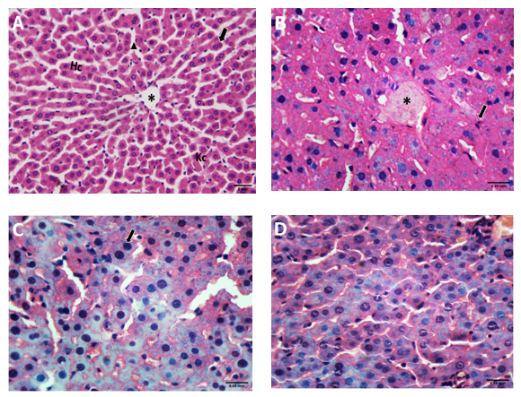
Histopathological Findings: The histopathological examinations of the control group liver tissue samples showed normal structure of hepatic lobules with branching, radiating and anastomosing hepatic cords, and the central veins. The hepatic sinusoids among the hepatic cords were lined with squamous endothelial cells with flat nuclei, and scattered Kupffer cells with dark angular nuclei. The hepatocytes were polygonal in shape with central rounded vesicular nuclei and acidophilic cytoplasm with few binucleated hepatocytes (Figure 2A). The congested central vein with few pyknotic nuclei (black arrow) and reduced hepatic sinusoids were observed in both groups treated with LF (Figure 2B). The AFB1 treated group showed dilated hepatic sinusoids, significant hyperchromatic nuclei and marked amyloid deposits together with increased mitotic pattern and focal hepatocellular necrosis with pycnotic nuclei (Figure 2C). The group that was treated with combined AFB1 and LF exhibited histological improvement in the appearance of the nuclei, with reduced necrosis and pycnosis (Figure 2D).
 

Table 1. The markers of liver function in the controls, AFB1-intoxicated and LF-NPs-treated male rats.
AFB1~ LF-NPs AFB1 LF-NPs Control Liver Function Marker
58.6 ±7.1# 97.4 ±16.3* 22.5 ±2.8 24.8 ±1.2 ALAT (U/L)
68.2 ±7.9# 116.4 ±16.7* 28.7 ±3.7 29.7 ±1.6 ASAT (U/L)
33.6 ±2.0# 50.3 ±4.1* 11.6 ±2.1 11.7 ±1.3 GGT (U/L)
394.4 ±58.4# 516.8 ±95.5* 301.2 ±30.3 302.1 ±25.7 ALP (U/L)
4.2 ±0.34# 3.3 ±0.5* 4.7 ±0.12 4.6 ±0.16 Albumin (g/dl)
7.6 ±0.61# 6.1 ±0.28* 8.36 ±0.28 8.3 ±0.27 Total protein (g/dl)
0.41 ±0.08# 0.79 ±0.06* 0.20 ±0.04 0.28 ±0.03 Bilirubin total (mg/dl)
0.13 ±0.01# 0.22 ±0.001* 0.04 ±0.008 0.05 ±0.007 Bilirubin direct (mg/dl)
Data are presented as means ±SEMs. Data were subjected to one way ANOVA followed by post hoc (Tukey’s) test at P 0.05. * Significantly different from the control group.  # Significantly different from the Aflatoxin B1 group. LF-NPs = lactoferrin nanoparticles.

Table 2. The hepatic levels of malondialdehyde (MDA), nitric oxide (NO), reduced glutathione (GSH), superoxide dismutase (SOD) and catalase (CAT) in controls, AFB1-intoxicated and LF-NPs-treated male albino rats.
AFB1~ LF-NPs AFB1 LF-NPs Control Liver Enzyme
15.2 ±1.5# 26.1 ±2.3* 12.5 ±0.59 12.6 ±0.6 MDA (μmol/g tissue)
65.2 ±2.8# 148 ±5.3* 44.8 ±2.6 45.7 ±4.5 NO (μmol/g tissue)
196 ±12.1# 106 ±4.1* 285 ±6.2 271 ±13.1 GSH (nmol/g tissue )
1817 ±50.4# 903 ±65* 2113 ±71 2030 ±119 SOD (U/g tissue)
9.5 ±1.3# 3.9 ±1.2* 7.9 ±0.4 7.8 ±0.74 CAT (U/g tissue)
Data are presented as means ± SEMs. Data were subjected to one way ANOVA followed by post hoc (Tukey’s) test at P 0.05. * = Significantly different from control group, while # = significantly different from AFB1 (Aflatoxin B1); LF-NPs (lactoferrin nanoparticles).

Figure 1. Serum TNF-α, IL1β, CD4+ and α-FP levels as well as hepatic DNA damage of control, AFTB1-intoxicated and LF NPs-treated male albino rats.
 
*Significantly different from control group. #Significantly different from AFB1-intoxicated group (P≤0.05).


Figure 2.
A-2D: Photomicrographs of liver sections of adult male rats: A: The control group showing normal hepatic lobules with central vein (*), hepatic cord (Hc), binucleated cell (arrow) and hepatic sinusoids (arrow head) with Kupffer cells (Kc). B: Lactoferrin-treated group showing congested central vein (*) and pyknotic nuclei (black arrow). C: AFB1-treated group showing hyperchromatic nuclei (black arrow) and focal necrosis with pycnotic nuclei and severe amyloidosis. D: AFB1+lactoferrin-treated group, showing reduced amyloid cytoplasmic deposits and improved nuclear appearance.

 
Discussion
Animals and humans may be exposed to many xenobiotic toxins, such as aflatoxins, which harm biomolecules and cellular membranes by causing oxidative damage [2]. The liver, a vital organ in humans and animals, plays a major role in the metabolism of xenobiotics, and an established liver toxin is aflatoxin B1. Aflatoxicosis, HCC, necrosis, and cirrhosis are caused by this toxin, inflicting cellular and severe damages on hepatocytes [2]. Through the production of oxidative stress, aflatoxin B1 leads to numerous harmful consequences [2]. Many physiological effects of lactoferrin (LF) have been described, including anti-bacterial, anti-fungal, anti-viral, anti-oxidant, anti-cancer, and anti-inflammatory properties [6]. Therefore, we investigated the protective effects of LF nanoparticles against AFB1-induced hepatotoxicity in rats.
The significant increases in serum levels of ALAT, ASAT, ALP, and GGT found in the current study combined with the reduced albumin and protein levels are clear evidence of the hepatotoxicity caused by AFB1. These results are consistent with recent research showing elevated liver enzymes in the sera of AFB1-intoxicated rats [16]. The biotransformation of AFB1-exo-epoxide and other metabolites into a highly reactive species in the liver by CYP3A4 and CYP1A2 isoforms may be the cause of the raised serum liver enzymes [3]. These compounds are members of the cytochrome P450 (CYP) superfamily of drug metabolizing enzymes. These reactive species bind to macromolecules, result in toxicity and mutations that further lead to lipid peroxidation, necrosis, cellular damage and death, DNA lesions, carcinogenicity, and other genetic illnesses [2]. The LF-NPs play a crucial role in immune responses and protect against various illnesses. Along with its anticancer property, it also demonstrates antibacterial effects against bacterial and parasitic diseases [17]. These nanoparticles have been found to possess a wide range of biological properties, including anti-inflammatory, anticancer, antibacterial, antioxidant, and immune enhancing effects [18]. The use of LF has been helpful in the treatment of such diseases as hepatitis C, viral infections, osteoporosis, and cancers [19].
This study demonstrated that AFB1-induced chronic oxidative stress in the liver of intoxicated rats can directly promote cell necrosis and activate apoptotic pathway [16]. These effects are likely supported by the significant increases in hepatic MDA and NO levels and decline in the anti-oxidative compounds, such as GSH, SOD, and CAT. Thus the presence of AFB1 in the liver is highly likely to be responsible for the elevated hepatic MDA and NO levels. The toxin AFB1 increases the formation and release of excessive levels of ROS, anion superoxide (O2••), hydrogen peroxide (H2O2), hydroxy radicals (OH•), and reactive nitrogen species, e.g., nitric oxide (NO) and peroxynitrite (ONOO-) [20]. These oxidizing compounds also promote lipid peroxidation [20].
The peroxisomal fatty acid, cytochrome P450, and mitochondrial electron transport system are the three primary locations where ROS are produced in living organisms [21]. It is unclear whether AFB1 participates in lipid peroxidation by directly increasing ROS formation or whether it raises the tissue's greater vulnerability to peroxidation and impaired antioxidant defense. Nonetheless, both processes may be involved. Any oxidizing agent with high reactivity to displace hydrogen atoms from methylene groups in polyunsaturated fatty acid may initiate lipid peroxidation [22]. A chemical reaction catalyzed by cyclo-oxygenase can also result in the peroxidation of polyunsaturated fatty acids [16]. Further, the metabolite 8, 9-epoxide promotes and expedites lipid peroxidation, which is followed by a significant loss in membrane stability and inhibition of membrane-bound enzymes [23]. According to Naaz, et al. [24], AFB1 significantly raises the MDA levels in hepatocytes and the liver.  Additionally, the liver and integumentary tissues both have shown an increase in lipid peroxidation triggered by AFB1 [25]. Rastogi, et al. have reported that conjugated diene production increases significantly as a result of disruptions by AFB1 [26].
In the current study, the antioxidant enzymes GSH, SOD, and CAT were shown to significantly decline after treatment with AFB1. We believe this decline is linked to the rough endoplasmic reticulum's substantial reduction in protein synthesis. This might even be a direct and/or indirect reaction to AFB1 buildup in the liver, as found in this study. Toxicologically, ROS have a high capacity to oxidize amino acid side chains, which can lead to the formation of protein cross-links, and oxidation of the protein’s backbone. This causes proteins to disintegrate and develop abnormally modified proteins that build up in cellular organelles [27]. In this study, treatment with LF-NPs reduced the hepatic MDA and NO levels while enhancing GSH, SOD, and CAT. The latter are the two crucial antioxidant and detoxification enzymes linked to intracellular protective mechanisms. Additionally, through activating AFB1, LF-NPs protect the liver [18]. Bovine LF has been shown to have antioxidant properties in a variety of biological and chemical settings [28]. Bovine LF-NPs have been shown to reduce lipid peroxidation by slowing the conversion of H2O2 to OH via sequestering iron by Fenton reaction [28].
In the current study, the serum levels of TNF-α, IL-1β, and α-fetoprotein significantly increased in rats due to the AFB1 administration. These findings support those of another research that was conducted previously [16]. Therefore, we propose that the inflammatory pathway is yet another potential mechanism behind the hepatic damage brought on by AFB1 [29]. The release of inflammatory cytokines and pro-inflammatory gene expression are thought to be involved in the oxidative stress and enhanced ROS production [30].  After exposure to AFB1, previous studies have reported that the mRNA expression of several genes associated to inflammation have been upregulated [31, 32]. The first and most significant mediator of inflammation that contributes to its development is TNF-α [32]. Proinflammatory cytokines, such as IL-1β and nitric oxide synthase (iNOS) are activated by the TNF-α and nuclear factor kappa B (NF- κB) pathways, along with adhesion molecules that promote the migration of leukocytes to the site of inflammation [32].
Lactoferrin is known to exhibit a variety of antioxidant, anti-inflammatory, and hepatoprotective activities [33]. Earlier research has demonstrated that LF protect mice's liver from damage brought on by concanavalin A, acetaminophen, and acetaminophen-induced liver injury [34]. Also, a former study by Tung, et al. has demonstrated the protective effects of LF against the liver fibrosis due to the hepatotoxin dimethylnitrosamine [35]. Tanaka, et al. investigated the manner with which LF reduced pro-inflammatory cytokines, such as TNF-α and IL-1β in a colitis model induced by dextran sulphate sodium in rats and mice [7]. In terms of the liver, another study has shown that LF reduces IL-1β, resulting in the protection of hepatitis in rats induced by carbon tetrachloride [8]. Hepatocellular carcinoma (HCC) is frequently diagnosed, using alpha-fetoprotein (AFP) as a tumor marker. However, recent research has suggested that AFP might be utilized in the diagnosis and evaluation of hepatic fibrosis even in the absence of HCC [36]. As of now, AFB1 has caused considerable increases in the hepatic AFP level, consistent with findings from another study [36], whereas LF-NPs remained at normal level. This outcome demonstrates the hepatoprotective capacity of LF-NPs against hepatotoxicity secondary to AFB1 intoxication. This is corroborated by the study of Aoyama, et al. [6] who demonstrated that the maintenance of AFP level in the liver is an indicator of protection against hepatotoxicity.
Based on the current study’s histopathological findings, AFB1 causes damage to the liver. The evidence in support of this claim are the presence of enlarged hepatic sinusoids, significant hyperchromatic nuclei, noticeable amyloid deposits, increased mitotic pattern, and focal hepatocellular necrosis with pycnotic nuclei. The ability of AFB1 metabolites to damage cell membranes may be the origin of these events. The pathological alterations showed a strong correlation with alterations in the enzymatic activities, confirmed by an earlier investigation conducted by Ahmed, et al. [16]. In this context, our findings suggest that the administration of LF-NPs to rats after AFB1 intoxication offered a significant hepatoprotective effect, confirmed by our biochemical findings and histological analyses. These findings revealed that the hepatoprotective potential is likely through the antioxidant and anti-inflammatory properties, and the subsequent inhibitory effect on NF-κB.
Conclusions
The results of this study indicate that LF-NPs are significantly able to ameliorate the AFTB1-induced liver damage through its anti-inflammatory, and antioxidant mechanisms of the constituents. Therefore, LF-NPs are likely to be effective against various liver diseases and used as a valuable agent in the management of human liver diseases. The elucidation of the efficacies warrants further well-designed and safe studies, conducted in animals and human subjects.
 

List of Abbreviations
Abbreviation Description Abbreviation Description
AFB1 Aflatoxin B1 HCC Hepatocellular carcinoma
AFM1 Aflatoxin M1 IL-1β Interleukin-1beta
AFP Alpha-fetoprotein iNOS Nitric oxide synthase
ALAT Alanine aminotransferase LF NPs Lactoferrin nanoparticles
ALP Alkaline phosphatase MDA Malondialdehyde
ASAT Aspartate aminotransferase NF-κB Nuclear factor kappa B
CAT Catalase NO Nitric oxide
CD4 Cluster of differentiation 4 SOD Superoxide dismutase
GGT Aamma-glutamyl transferase TNF-α Tumor necrosis factor alpha
GSH Reduced glutathione
 
Acknowledgments
The authors wish to appreciate the support and cooperation of the faculty and staff in the Zoology and Histology Departments, Faculties of Science and Dentistry,
Al-Azhar University, Assuit, Egypt.
Ethical Approval & Guidelines
The study was conducted consistent with the guidelines approved by the Ethics Committee of the Faculty of Science, Al-Azhar University, Assuit, Egypt (approval #: AZHAR, 15/2022). All experimental protocols for the care and use of laboratory animals were approved by the University’s Research Ethics Committee. All methods were carried out and reported consistent with relevant university’s guidelines.
Authors’ Contributions
Conceptualization: Mahmoud Ashry, Hussien M. Abdel Salam and Salah M.E. Soliman. Methodology, investigation & writing the original draft: Mohamed H.A. Gadelmawla. Histopathology examinations, supervision, writing, review and editing of the final draft of the manuscript: All authors.
Funding
This study was funded by the authors only.
Availability of Data and Materials
The datasets used and analyzed during the current study are available from the corresponding author on reasonable request.
Conflicts of Interest
The authors declared no potential conflicts of interest with respect to the research, authorship, and publication of this article.
Consent for Publication
Not applicable.
Type of Study: Research | Subject: General

References
1. Raduly Z, Szabo L, Madar A, Pocsi I, Csernoch L. Toxicological and Medical Aspects of Aspergillus-Derived Mycotoxins Entering the Feed and Food Chain. Frontiers in microbiology. 2019;10:2908. [DOI:10.3389/fmicb.2019.02908] [PMID] [PMCID]
2. Zarev Y, Naessens T, Theunis M, Elgorashi E, Apers S, Ionkova I, et al. In vitro antigenotoxic activity, in silico ADME prediction and protective effects against aflatoxin B(1) induced hepatotoxicity in rats of an Erythrina latissima stem bark extract. Food and chemical toxicology : an international journal published for the British Industrial Biological Research Association. 2020;135:110768. [DOI:10.1016/j.fct.2019.110768] [PMID]
3. Rushing BR, Selim MI. Aflatoxin B1: A review on metabolism, toxicity, occurrence in food, occupational exposure, and detoxification methods. Food and chemical toxicology : an international journal published for the British Industrial Biological Research Association. 2019;124:81-100. [DOI:10.1016/j.fct.2018.11.047] [PMID]
4. Loi M, Renaud JB, Rosini E, Pollegioni L, Vignali E, Haidukowski M, et al. Enzymatic transformation of aflatoxin B(1) by Rh_DypB peroxidase and characterization of the reaction products. Chemosphere. 2020;250:126296. [DOI:10.1016/j.chemosphere.2020.126296] [PMID]
5. Coppock RW, Christian RG, Jacobsen BJ. Aflatoxins In R. C. Gupta (Ed.), Veterinary Toxicology: Basic and clinical principles. 3rd ed.: 983-994. Academic Press. 2018. [DOI:10.1016/B978-0-12-811410-0.00069-6]
6. Aoyama Y, Naiki-Ito A, Xiaochen K, Komura M, Kato H, Nagayasu Y, et al. Lactoferrin Prevents Hepatic Injury and Fibrosis via the Inhibition of NF-kappaB Signaling in a Rat Non-Alcoholic Steatohepatitis Model. Nutrients. 2021;14(1). [DOI:10.3390/nu14010042] [PMID] [PMCID]
7. Tanaka H, Gunasekaran S, Saleh DM, Alexander WT, Alexander DB, Ohara H, et al. Effects of oral bovine lactoferrin on a mouse model of inflammation associated colon cancer. Biochemistry and cell biology = Biochimie et biologie cellulaire. 2021;99(1):159-65. [DOI:10.1139/bcb-2020-0087] [PMID]
8. Farid AS, El Shemy MA, Nafie E, Hegazy AM, Abdelhiee EY. Anti-inflammatory, anti-oxidant and hepatoprotective effects of lactoferrin in rats. Drug and chemical toxicology. 2021;44(3):286-93. [DOI:10.1080/01480545.2019.1585868] [PMID]
9. Chen HA, Chiu CC, Huang CY, Chen LJ, Tsai CC, Hsu TC, et al. Lactoferrin Increases Antioxidant Activities and Ameliorates Hepatic Fibrosis in Lupus-Prone Mice Fed with a High-Cholesterol Diet. Journal of medicinal food. 2016;19(7):670-7. [DOI:10.1089/jmf.2015.3634] [PMID]
10. Xiao Y, Monitto CL, Minhas KM, Sidransky D. Lactoferrin down-regulates G1 cyclin-dependent kinases during growth arrest of head and neck cancer cells. Clinical cancer research : an official journal of the American Association for Cancer Research. 2004;10(24):8683-6. [DOI:10.1158/1078-0432.CCR-04-0988] [PMID]
11. Kozu T, Iinuma G, Ohashi Y, Saito Y, Akasu T, Saito D, et al. Effect of orally administered bovine lactoferrin on the growth of adenomatous colorectal polyps in a randomized, placebo-controlled clinical trial. Cancer prevention research. 2009;2(11):975-83. [DOI:10.1158/1940-6207.CAPR-08-0208] [PMID]
12. Ruiz-Larnea MB, Leal AM, Liza M, Lacort M, de Groot H. Antioxidant effects of estradiol and 2 hydroxyestradiol on iron induced lipid peroxidation of rat liver microsome. Steriod. 1994;59:383-8. [DOI:10.1016/0039-128X(94)90006-X] [PMID]
13. Perandones CE, Illera VA, Peckham D, Stunz LL, Ashman RF. Regulation of apoptosis in vitro in mature murine spleen T cells. Journal of immunology. 1993;151(7):3521-9. [DOI:10.4049/jimmunol.151.7.3521]
14. Drury RAB, Wallington EA. Preparation and fixation of tissues. In: Drury RAB, Wallington EA, editors. Carleton's Histological Technique. 5. Oxford: Oxford University Press.1980. 41-54 p.
15. Steel RG, Torrie GH. Principles and Procedures of Statistics: A Biometrical Approach. New York: McGraw Hill.1980. 633 p.
16. Ahmed N, El-Rayes SM, Khalil WF, Abdeen A, Abdelkader A, Youssef M, et al. Arabic Gum Could Alleviate the Aflatoxin B(1)-provoked Hepatic Injury in Rat: The Involvement of Oxidative Stress, Inflammatory, and Apoptotic Pathways. Toxins. 2022;14(9). [DOI:10.3390/toxins14090605] [PMID] [PMCID]
17. Actor JK, Hwang SA, Kruzel ML. Lactoferrin as a natural immune modulator. Current pharmaceutical design. 2009;15(17):1956-73. [DOI:10.2174/138161209788453202] [PMID] [PMCID]
18. Ayob A, Al-Najjar A, Awad A. Amelioration of Bile Duct Ligation Induced Liver Injury by Lactoferrin: Role of Nrf2/HO-1 Pathway. Azhar Int J Pharmaceut Med Sci. 2021;1(3):84-90. [DOI:10.21608/aijpms.2021.78232.1076]
19. Bironzo P, Passiglia F, Novello S. Five-year overall survival of pembrolizumab in advanced non-small cell lung cancer: another step from care to cure? Annals of translational medicine. 2019;7(Suppl 6):S212. [DOI:10.21037/atm.2019.08.91] [PMID] [PMCID]
20. Kodama M, Inoue F, Akao M. Enzymatic and non-enzymatic formation of free radicals from aflatoxin B1. Free radical research communications. 1990;10(3):137-42. [DOI:10.3109/10715769009149882] [PMID]
21. Ray PD, Huang BW, Tsuji Y. Reactive oxygen species (ROS) homeostasis and redox regulation in cellular signaling. Cellular signalling. 2012;24(5):981-90. [DOI:10.1016/j.cellsig.2012.01.008] [PMID] [PMCID]
22. Abdulbaqi NJ, Dheeb BI, Irshad R. Expression of biotransformation and antioxidant genes in the liver of albino mice after exposure to aflatoxin B1 and an antioxidant sourced from turmeric (Curcuma longa). Jordan J Biol Sci. 2018;11(1):93-8.
23. Xu F, Li Y, Cao Z, Zhang J, Huang W. AFB(1)-induced mice liver injury involves mitochondrial dysfunction mediated by mitochondrial biogenesis inhibition. Ecotoxicology and environmental safety. 2021;216:112213. [DOI:10.1016/j.ecoenv.2021.112213] [PMID]
24. Naaz F, Javed S, Abdin MZ. Hepatoprotective effect of ethanolic extract of Phyllanthus amarus Schum. et Thonn. on aflatoxin B1-induced liver damage in mice. Journal of ethnopharmacology. 2007;113(3):503-9. [DOI:10.1016/j.jep.2007.07.017] [PMID]
25. Rastogi S, Dogra RK, Khanna SK, Das M. Skin tumorigenic potential of aflatoxin B1 in mice. Food and chemical toxicology : an international journal published for the British Industrial Biological Research Association. 2006;44(5):670-7. [DOI:10.1016/j.fct.2005.09.008] [PMID]
26. Rastogi S, Shukla Y, Paul BN, Chowdhuri DK, Khanna SK, Das M. Protective effect of Ocimum sanctum on 3-methylcholanthrene, 7,12-dimethylbenz(a)anthracene and aflatoxin B1 induced skin tumorigenesis in mice. Toxicology and applied pharmacology. 2007;224(3):228-40. [DOI:10.1016/j.taap.2007.05.020] [PMID]
27. Sun Y, Huang K, Long M, Yang S, Zhang Y. An update on immunotoxicity and mechanisms of action of six environmental mycotoxins. Food and chemical toxicology : an international journal published for the British Industrial Biological Research Association. 2022;163:112895. [DOI:10.1016/j.fct.2022.112895] [PMID]
28. Kowalczyk P, Kaczynska K, Kleczkowska P, Bukowska-Osko I, Kramkowski K, Sulejczak D. The Lactoferrin Phenomenon-A Miracle Molecule. Molecules. 2022;27(9). [DOI:10.3390/molecules27092941] [PMID] [PMCID]
29. Zhang B, Dai Y, Zhu L, He X, Huang K, Xu W. Single-cell sequencing reveals novel mechanisms of Aflatoxin B1-induced hepatotoxicity in S phase-arrested L02 cells. Cell biology and toxicology. 2020;36(6):603-8. [DOI:10.1007/s10565-020-09547-z] [PMID]
30. Owumi S, Najophe ES, Farombi EO, Oyelere AK. Gallic acid protects against Aflatoxin B(1) -induced oxidative and inflammatory stress damage in rats kidneys and liver. Journal of food biochemistry. 2020;44(8):e13316. [DOI:10.1111/jfbc.13316] [PMID]
31. Li S, Liu R, Xia S, Wei G, Ishfaq M, Zhang Y, et al. Protective role of curcumin on aflatoxin B1-induced TLR4/RIPK pathway mediated-necroptosis and inflammation in chicken liver. Ecotoxicology and environmental safety. 2022;233:113319. [DOI:10.1016/j.ecoenv.2022.113319] [PMID]
32. Jang DI, Lee AH, Shin HY, Song HR, Park JH, Kang TB, et al. The Role of Tumor Necrosis Factor Alpha (TNF-alpha) in Autoimmune Disease and Current TNF-alpha Inhibitors in Therapeutics. International journal of molecular sciences. 2021;22(5). [DOI:10.3390/ijms22052719] [PMID] [PMCID]
33. Sabra S, Agwa MM. Lactoferrin, a unique molecule with diverse therapeutical and nanotechnological applications. International journal of biological macromolecules. 2020;164:1046-60. [DOI:10.1016/j.ijbiomac.2020.07.167] [PMID] [PMCID]
34. Yin H, Cheng L, Holt M, Hail N, Jr., Maclaren R, Ju C. Lactoferrin protects against acetaminophen-induced liver injury in mice. Hepatology. 2010;51(3):1007-16. https://doi.org/10.1096/fasebj.24.1_supplement.759.8 [DOI:10.1002/hep.23476] [PMID]
35. Tung YT, Tang TY, Chen HL, Yang SH, Chong KY, Cheng WT, et al. Lactoferrin protects against chemical-induced rat liver fibrosis by inhibiting stellate cell activation. Journal of dairy science. 2014;97(6):3281-91. [DOI:10.3168/jds.2013-7505] [PMID]
36. Wang X, He Y, Tian J, Muhammad I, Liu M, Wu C, et al. Ferulic acid prevents aflatoxin B1-induced liver injury in rats via inhibiting cytochrome P450 enzyme, activating Nrf2/GST pathway and regulating mitochondrial pathway. Ecotoxicology and environmental safety. 2021;224:112624. [DOI:10.1016/j.ecoenv.2021.112624] [PMID]

Add your comments about this article : Your username or Email:
CAPTCHA

Send email to the article author


Rights and permissions
Creative Commons License This work is licensed under a Creative Commons Attribution-NonCommercial 4.0 International License.

© 2025 CC BY-NC 4.0 | Iranian Journal of Toxicology

Designed & Developed by : Yektaweb