Write your message
Volume 17, Issue 4 (October 2023)                   IJT 2023, 17(4): 1-8 | Back to browse issues page


XML Print


Download citation:
BibTeX | RIS | EndNote | Medlars | ProCite | Reference Manager | RefWorks
Send citation to:

Ilesanmi O, Inala M, Ridwan A. Neuroprotective Effect of Ethylacetate Fraction of Antiaris Africana against Sodium Azide-Induced Neurotoxicity in the Striata of Male Wistar Rats. IJT 2023; 17 (4) :1-8
URL: http://ijt.arakmu.ac.ir/article-1-1244-en.html
1- Department of Biochemistry, Faculty of Science, Federal University Otuoke, Bayelsa State, Nigeria , ilesanmiob@fuotuoke.edu.ng
2- Department of Biology, Faculty of Science, Federal University Otuoke, Bayelsa State, Nigeria
3- Department of Biochemistry, College of Medicine, University of Lagos, Lagos, Nigeria
Full-Text [PDF 751 kb]   (642 Downloads)     |   Abstract (HTML)  (1460 Views)
Full-Text:   (444 Views)
Introduction
Sodium azide (NaN3) is a neurotoxin that is metabolized to nitric oxide, initiating its toxic effects [1, 2]. This compound inhibits the activity of cytochrome C oxidase (complex IV), a mitochondrial enzyme. The findings make some scientists classify NaN3 as a mitochondrial toxin [3]. A major feature of neurodegenerative diseases is a decrease in the activity of respiratory enzymes, such as cytochrome C oxidase. This has enabled neuroscientists to produce neurodegenerative diseases in experimental animals using NaN3 [4]. Biochemical observations during the inhibition of mitochondrial respiratory enzymes, such as depletion of ATP, generation of free radicals, and incomplete oxidation of glucose for energy ultimately lead to a cascade of events, culminating at loss of neurons [5-7].
Exposure of brain tissue to NaN3 is known to activate the release of excitotoxins due to impairments in mitochondrial function. In addition, the disruption of metabolic and cellular disturbances by NaN3 can lead to the destruction of neural synapses [8], memory impairment, and neuronal cell death, as observed in neurodegenerative diseases [9-11]. Sodium azide, a colorless powder, is often used in automobiles and airplanes as part of the safety airbags and ejectors. Concerning the NaN3 toxicity, it readily crosses the blood-brain barrier where it binds to enzymes that contain Fe3+, such as catalase, cytochrome C oxidase, and peroxidase. These enzymes play important roles in neutralizing free radicals and preventing oxidative stress [12]. Exposure to NaN3 causes respiratory irritation, while patients exposed to NaN3 develop hypothermia, hypotension, and bradycardia, which may lead to death if not quickly treated within 12hr [13-16].
Despite the health challenges posed by NaN3, there is no approved drug to neutralize its toxic effects. However, medicinal plants can provide good alternatives in combating the poisonous effects of NaN3. Studies have reported the efficacy of quercetin and other flavonoids in acting as an antidote against NaN3 toxicity [17-19]. Antiaris Africana is one of the underutilized medicinal plants with various pharmacological properties. We have previously investigated the antidotal effect of A. Africana against neurochemicals, such as cyanide and rotenone [20, 21].
Upon the identification of the major flavonoids in the ethylacetate fraction of A. Africana [21], we aimed to investigate the potential effect of the ethylacetate fraction of A. Africana against the NaN3 toxicity to the striata regions in male Wistar rats.
Materials and Methods
Preparation of the Extract: Fresh leaves of A. africana were harvested from the Forest Research Institute of Nigeria (FRIN) at Ibadan. An adequate sample of the plant was identified and authenticated by the Department of Botany, Obafemi Awolowo University, Ile Ife, Nigeria.  The fresh A. Africana leaves were dried in open air and regularly weighed until a constant weight was reached. The dried leaves were then blended and filtered to obtain a fine powder. The powder was dissolved in a mixture of methanol/water (80:20) for 72 hours with constant daily stirring. The sample was then filtered and lyophilized to obtain a dried powder assigned as the crude fraction. This fraction was partitioned, using four different solvents in order of polarity (hexane < dichloroform A. africana.
Experimental Animals: Thirty male Wistar rats were purchased from the animal breeding units of, University of Benin. The animals were allowed to acclimatize in the laboratory environment for two weeks, where they had free access to water and food ad libitum.
Tissue Preparation: After the initial 2-week period, the rats were sacrificed via mild anesthesia and the brains were excised and carefully rinsed in 1.15% potassium chloride (KCl) solution. The striata were carefully sliced out from the rat brains, weighed and homogenized in phosphate buffer (0.1M at pH 7.4). The procedure for the preparation of the striatal mitochondria after fractionation based on the method of Klein-Schwartz, et al. [16].
Experimental Design: The neurotoxicity was induced by incubating the striata samples with 4mM NaN3 according to a modified method described by Gao, et al. [14]. The treatment involved the addition of varying concentrations of the ethyl acetate fraction (5-50μg/ml) as described by an earlier study [15].
Lipid Peroxidation Inhibitory Activity: The lipid peroxidation inhibitory activity was evaluated by measuring the formation of thiobarbituric acid reactive substances (TBARS) according to the method described by Okhawa, et al. [22]. The concentration of malondialdehyde (MDA) in the solution was determined according to the method of Adam-Vizi and Seregi [23]. The reduced glutathione content of the mixture was determined as previously described by Jollow, et al. [24]. The protein carbonyl levels in the samples were quantified according to the method described by Floor and Wetzel [25].
Evaluation of Acetylcholinesterase: The activity of acetylcholinesterase (AChE) was assessed based on the modified method of Ellman, et al. [26]. Briefly, the reaction mixture contained 0.1 ml DTNB and 2.6 ml phosphate buffer (0.1 M, pH 8.0), 0.04 ml of striatal homogenate, and incubated for 5 min. Acetylthio­choline iodide (0.075 M) was added after incubation and the rate of hydrolysis was measured at 420nm continuously for three min. the AChE activity was calculated and expressed in μmol-1 min-1 mg protein-1.
Evaluation of Lactate Dehydrogenase Activity: The effect of the ethylacetate fraction on the activity of lactate dehydrogenase (LDH) in the striata samples was determined according to a previously described valid method [27].
Determination of Mitochondrial Enzymes Integrity: The evaluation of NADH-succinate dehydrogenase (NSD) and NADH-succinate reductase (NSR) activities were measured according to the method of Spinazi, et al. [28], while the activity of NADH-cytochrome C reductase (NCR) was measured according to the method described by Kollareth, et al. [29].
Determination of Monoamine Oxidase Activity: The monoamine oxidase activity (MAO) was measured using the method developed by Holt, et al. [30], and described by Chaudhary and Parvez [31, 32].
Data Analyses: The study data were analyzed using GraphPad Prism (version 6.02). The data from the groups were compared using a one-way analysis of variance (ANOVA). Duncan’s test was utilized as the descriptive statistical test. The data were presented as the means ± standard deviations (SD) of the replicates in each group. The statistical significance level was set at P<0.05.
Results
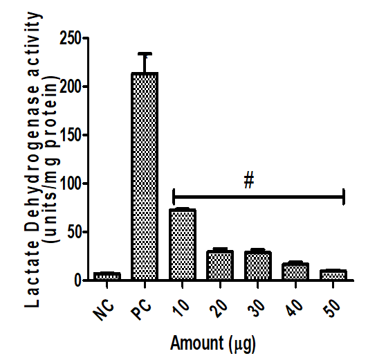
Figure 1 represents the effect of the ethyl acetate fraction of A. Africana on lactate dehydrogenase (LDH) activity on the rat striata intoxicated with sodium azide. The fraction at the used concentrations significantly mitigated the release of LDH from the rat brain tissue as compared to that of the positive control group (P<0.05). The neuroprotective effect was found to be dose-dependent.
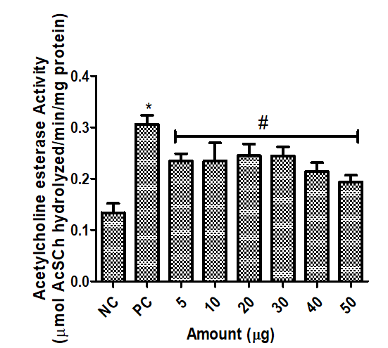
Figure 2 shows the effect of the ethyl acetate fraction of A. Africana and NaN3 on the acetylcholinesterase activity in the rat striatal samples. The data indicated that NaN3 significantly increased the activity of AChE as compared to that of the negative control group (P<0.05). All of the fraction’s concentrations prevented the rise in the AChE activity in response to NaN3 toxicity against the rat striatal samples (P<0.05). The NaN3 fraction at all concentrations significantly inhibited the acetylcholinesterase activity in the striatal samples as compared to that observed in the untreated group (P<0.05).
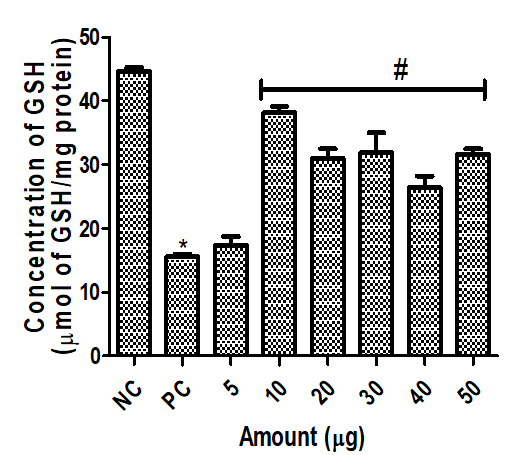
Figure 3 reveals that NaN3 reduced the glutathione (GSH) concentration significantly in the striatal samples as compared to that of the control group (P<0.05). The fraction also increased the GSH (P<0.05). All concentrations of the ethylacetate fraction significantly increased the concentration of GSH as compared to that observed for the untreated positive group (P<0.05). There was no significant difference with respect to the inhibitory effect among the various concentrations of the fraction used.
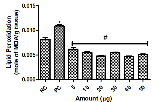
Figure 4 demonstrates that NaN3 significantly increased the activity of lipid peroxidation as measured by the concentration of malondialdehyde (MDA) in the rat striatal samples compared to the controls (P<0.05). The fraction at all concentrations significantly prevented the lipid peroxidation induced by NaN3 in the rat striatal samples (P<0.05).
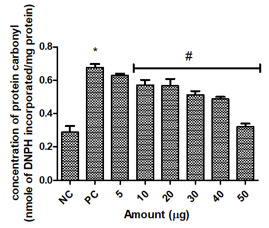
Figure 5 reveals the effect of the fraction on the protein carbonyl levels in the striatal homogenates following exposure to NaN3. This toxin caused a significant rise in the protein carbonyl levels in response to the administered fraction as compared to that observed for the control group. All of the fraction’s concentrations effectively inhibited the generation of protein induced by NaN3 (P<0.05).
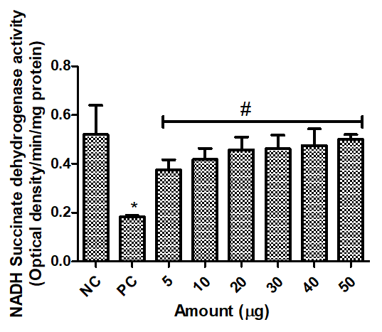
Figure 6 reveals the efficacy of various concentrations of the fraction to reverse the inhibitory effect of NaN3 on the rat brain’s NADH-succinate dehydrogenase (NSD). The data demonstrate that NaN3 at the administered concentration significantly inhibited the NSD activity in the striatal homogenate compared to that of the control group (P<0.05). The treatment with various concentrations of the fraction significantly increased the NSD activity compared to that observed for the rat group that received NaN3 only (P<0.05).
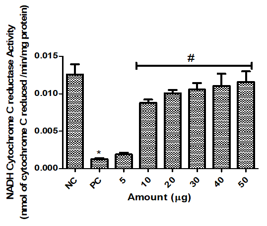
Figure 7 demonstrates the effect of various concentrations of A. Africana fraction on the NADH cytochrome C oxidase activity in the striatal homogenate in response to NaN3. The data indicated that NaN3 at the given concentrations caused a significant inhibition of NADH cytochrome C oxidase activity compared to that of the controls (P<0.05).
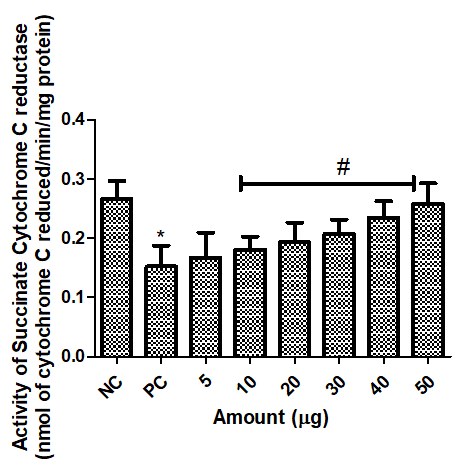
Figure 8 presents the role of various concentrations of the A. africana fraction in reversing the inhibitory effect of NaN3 on the succinate cytochrome C reductase (SCR) levels in the rat striatal homogenate samples. The data indicated that NaN3 caused a significant inhibition of SCR as compared to that of the control group (P<0.05). The findings also showed that the fraction at all concentrations except for 5µg, was able to significantly prevent the inhibition of SCR in a dose-dependent manner (P<0.05).
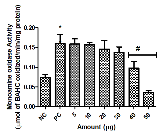
Figure 9 demonstrates the effect of the A. Africana fraction in reversing the inhibitory effect of NaN3 on monoamine oxidase (MAO) in the striatal homogenate. The NaN3 caused a significant increase in the MAO activity. The results indicated that the fraction at 40 and 50µg only was effective at inhibiting the rise in the MAO activity induced by NaN3 (P<0.05).

Figure 1. Protective effect of the A. africana fraction on lactate dehydrogenase (LDH) activity of the rats’ striata samples exposed to sodium azide (NaN3).  The data are presented as the means ± standard deviations per goup (n=5). *P<0.05: NaN3 vs Control, #P<0.05: NaN3 vs EFA. NC = negative control; PC = positive control.

Figure 2. Anticholinesterase activities of Ethyl acetate fraction of Antiaris africana in the striatum region of rats exposed to NaN3 neurotoxicity. The data are presented as means ± standard deviations per group (n=5). *p<0.05: NaN3 vs Control, #p<0.05: NaN3 vs EFA. NC = negative control; PC = positive control.

Figure 3. Protective effect of Ethyl acetate fraction of A. africana on reduced GSH concentration in the striata of rats exposed to NaN3 neurotoxicity. The data are presented as means ± standard deviations per group (n=5). *P<0.05: NaN3 vs Control, #P<0.05: NaN3 vs EFA. NC = negative control; PC = positive control.

Figure 4. Protective effect of Ethyl acetate fraction of A. africana on lipid peroxidation in the striata region of rats exposed to NaN3 neurotoxicity. Data are presented as means ± standard deviations per group (n=5). *P<0.05: NaN3 vs Control, #P<0.05: NaN3 vs EFA. NC = negative control; PC = positive control.

Figure 5. The protective Effect of the fraction on protein carbonyl in the positive control group on the rats striata exposed to NaN3. The data are presented as means ± standard deviations per group (n=5). *p<0.05: NaN3 vs Control, #p<0.05: NaN3 vs EFA. NC = negative control; PC = positive control.


Figure 6. The effect of the A. africana fraction on the rat brain’s NADH-succinate dehydrogenase exposed to NaN3. The data are presented as means ± standard deviations per group (n=5). *P<0.05: NaN3 vs Control, #P<0.05: NaN3 vs EFA. NC = negative control; PC = positive control.

Figure 7. The effect of the A. africana fraction on NADH-cytochrome C reductase activity on the rat striata exposed to NaN3. The data are presented as means ± standard deviations per group (n=5). *P<0.05: NaN3 vs Control, #P<0.05: NaN3 vs EFA. NC = negative control; PC = positive control.

Figure 8. The effect of the A. africana fraction on Succinate-cytochrome C reductase on rat striata exposed to NaN3. The data are presented as means ± standard deviations per group (n=5). *P<0.05: NaN3 vs Control, #P<0.05: NaN3 vs EFA. NC = negative control; PC = positive control.

Figure 9. The effect of the A. africana on the monoamine oxidase (MAO) activity in rat striata exposed to NaN3.  The data are presented as means ± standard deviations per group (n=5). *P<0.05: NaN3 vs Control, #P<0.05: NaN3 vs EFA. NC= negative control; PC=positive control.

Discussion
Studies have widely reported the neurotoxicity of NaN3 especially in Alzheimer’s disease secondary to the toxicity in animal models [31-34]. As an inhibitor of mitochondrial cytochrome C oxidase, which leads to ATP depletion, it is one of the models that help discover new drugs to treat mitochondria-related disorders [35]. This study investigated the neuroprotective effect of the ethylacetate fraction of A. Africana against typical neurotoxins, such as NaN3.  This was attempted because currently there is no report on the potential effect of A. Africana in protecting against sodium azide neurotoxicity. Specifically, we evaluated the activities of nine different enzymatic or non-enzymatic processes in the rat brain striatal samples in response to NaN3 toxicity in the presence or absence of the A. Africana fraction.
Lactate dehydrogenase (LDH) is a glycolytic enzyme involved in energy production, and is compartmentalized in the striata under normal conditions [36]. However, under neuronal damaging condition, LDH is released from the tissue [37]. Patients with Alzheimer disease (AD) have high levels LDH in their blood circulation due to neuronal cytotoxicity, hence its role as a diagnostic marker for AD [38]. Our results indicated that NaN3 caused a significant increase in the striatal LDH level compared to that of the control group (Figure 1). This finding suggests that NaN3 induced LDH leakage from the rats’ striata. This finding is consistent with those reported by two earlier studies [39, 40]. The former studies had suggested that NaN3 might have a significant role in causing mitochondrial damage likely due to the generation of ROS and alterations in the bioenergetic processes of neurons.
Investigation of acetylcholinesterase (AChE) activity is one of the hallmarks of diagnosing AD, since the high activity of this enzyme has consistently been reported in the brain tissues of patients with AD [41]. This enzyme is involved in the deactivation of acetylcholine, a major neurotransmitter involved in brain functions. Patients challenged with AD have low Ach, causing memory impairment. Therefore, chemicals and toxins that increase AChE activity are good condidates for generating AD in animal models [42]. From the experiment, NaN3 caused a significant increase in the AChE level in the rat striatal samples at the given doses (Figure 2). This finding is consistent with that of other investigations, where the use of NaN3 increased the choline concentration in the brain. Choline is the product of the catalytic breakdown of acetylcholine, which suppresses the reconstitution of the neurotransmitter, acetylcholine.
Reduced glutathione is the major non-enzymatic antioxidant in the brain, the deficiency of which has been linked to a number of neurodegenerative diseases [43]. It serves as an electron donor to free radicals, neutralizing them, and thereby preventing them from damaging biomolecules [44]. However, under oxidative stress, the GSH concentration is not enough to neutralize the generated ROS or RNS. The oxidative effect of NaN3 was reflected in the current study, as it significantly reduced the GSH concentration (Figure 3). Once the GSH level is low, the generated ROS readily interacts with biomolecules to gain stability, which is reflected in the high concentrations of MDA and PC. The importance of GSH in preventing and maintaining healthy cells cannot be overemphasized. It is likely that GSH is included in food supplements or added to drug regimens toward the treatment and slowing down the neurodegenerative disease progression [45, 46].
Consistent with previous reports [47, 48], the current study demonstrated that exposure of the striatal regions to NaN3 at the given dosage triggered the generation of various reactive species. Specifically, we documented high levels of malondialdehyde and protein carbonyl, which are products of lipid and protein oxidation [49]; see Figures 4 and 5. A major mechanism of NaN3 toxicity is the generation of reactive species secondary to mitochondrial damages. These unstable reactive species are attracted to biomolecules, such as lipids and proteins, which are rich in electrons to form stable molecules. During the process, they damage functional molecules that can be detrimental to the well-being of the organisms or humans. Pathological investigations of the brain in patients, suffering from neurodegenerative diseases have shown increased levels of MDA and PC, indicating that reactive oxygen species play important roles in the etiology of certain neurological diseases.
Further, NaN3 has been proven to be toxic to mitochondria, since the inhibition of cytochrome C oxidase (CCO) disrupts the cellular respiratory chain, leading to mitochondrial damages. In addition, it results in ATP depletion and other biochemical processes that ultimately cause cell injury and death [50]. As a mitochondrial toxin that inhibits CCO, we planned to investigate the effect of NaN3 on the rats’ brain tissue. We discovered that NaN3 caused a significant decline in the activities of NADH succinate dehydrogenase (NSD), NADH cytochrome C reductase (NCR), and succinate cytochrome C reductase (SCR) (Figures 6-8). The findings strongly suggest that NaN3 induces a significant decline in the oxidative phosphorylation process in the mitochondria of the rat’s brain striatal region. Based on our findings, the mechanism of NaN3 action starts from the inhibition of CCO. This prevents electron transfer between cytochrome and other molecules, such as NADH and succinate, and then leads to the generation of free radicals, such as superoxide dismutase and hydroxyl groups.
Monoamine oxidase (MAO) is an important enzyme, and its activity levels have been implicated in the etiology of neurodegenerative diseases [51, 52]. Its activity has been reported to deplete the neuronal cells of hormones, such as dopamine and serotonin by oxidizing them [53]. The results indicate that NaN3 at the administered dose significantly increase the MAO activity.
Deficient levels of antioxidants system will enable the radicals to attack other respiratory enzymes, causing energy depletion and mitochondrial injury. While there is no approved antidote against NaN3, the search for a suitable neutralizer of NaN3 toxicity is still underway. Using the ethylacetate fraction of A. africana, which is rich in polyphenols [21] as a NaN3 antagonist, demonstrated that it was effective in inhibiting the toxicity against the rats’ striata.
The fraction improved the antioxidant status in the striatal regions as observed by the higher GSH concentrations compared to that of the untreated group. Similarly, it resulted in low MDA and PC concentrations, compared to that of the untreated group. The findings suggest that the A. Africana fraction can inhibit lipid peroxidation by acting as an electron donor and preventing the reactive species from oxidizing proteins. This prevents oxidative-related striatal injury in the rat brains. The antioxidant effects of the A. africana fraction has been reported previously [16, 54], hence supporting its ability to reduce oxidative stress. These properties are highly likely to be responsible for the pharmacological activity of A. africana in the treatment or prevention of cancer, microbial infections, and some brain-related disorders.
In addition, the fraction prevented the leakage of LDH from the striatal regions after exposure to NaN3. In this context, natural plant antioxidants have been effective in enhancing the bioenergetic processes in animal models [55, 56]. Thus our findings provide novel evidence that the A. africana fraction contains important phytochemicals able to prevent losses of bioenergetic enzymes. The inhibitory effect of EFA on MAO activity is a reflection of the potential of A. africana to improve brain function and another scientific confirmation in support of the ethnomedicinal applications of the plant in treating neurological disorders [53].
Lastly, earlier investigations have shown that A. africana fractions act as antidote in preventing the neurotoxicity of chemicals, such as rotenone and cyanide that are linked to mitochondrial damage [7, 21]. Consistently, our current findings support the fact that the A. africana fraction was able to protect the mitochondrial enzymes involved in the respiratory chain. This fact was also reflected in the improved activities of NSR, NCR, and SCR as compared to those of the untreated group. As reported by another investigation on the mechanism of protection [53], the fraction used in the current study is likely to improve the associated enzymatic activities by preventing NaN3 from inhibiting important enzymes. Alternatively, the fraction may block the NaN3 metabolism into such active compounds as nitric oxide (NO) or scavenge the NO generated from NaN3, thereby blocking NO from binding to mitochondrial respiratory enzymes.
Conclusions
The findings of the current study support the fact that A. africana fraction was able to protect the mitochondrial enzymes involved in the respiratory chain. This fact was also reflected in the improved activities of NSR, NCR, and SCR as compared to those found for the untreated group. As reported by other investigations on the mechanism of protection, the fraction used in this study is likely to improve the associated enzymatic activities by preventing NaN3 from inhibiting important enzymes. Alternatively, the fraction may block the NaN3 metabolism into such active compounds as nitric oxide (NO) or scavenge the NO generated from NaN3, thereby blocking NO from binding to mitochondrial respiratory enzymes. Elucidation of further biochemical and toxicological mechanisms involved in the neuroprotective effect of the ethylacetate fraction of Antiaris africana awaits future research.
Conflicts of Interest
Authors declare no conflicts of interest in conducting this study.
Funding
This research was supported by Tetfund under the institutional based research support, at Federal University Otuoke, Nigeria; 2021.
Acknowledgements
The authors wish to appreciate the laboratory staff of Dr. Asogwa, where parts of this study’s experiments were conducted.
Ethical Considerations
Compliance with ethical guidelines: All of the rats used in this study were healthy and treated according to the guidelines of the Helsinki Declaration of 1975 for the care and use of laboratory animals. The experimental design was approved by the Ethics Committee on Animal Research and Treatment (ART) of the Federal University Otuoke, Nigeria (Approval code: ART2022019).
Authors' Contributions
OBI designed, conduct the experiment, data analysis and preparation of manuscript, MI conduct the experiment, and manuscript preparation, RAL prepared the manuscript and data analysis.
Type of Study: Research | Subject: General

References
1. Smith CJ, Hopmans P, Cook FJ. Accumulation of Cr, Pb, Cu, Ni, Zn and Cd in soil following irrigation with treated urban effluent in Australia. Environmental pollution. 1996;94(3):317-23. [DOI:10.1016/S0269-7491(96)00089-9] [PMID]
2. Su P, Zhang J, Wang S, Aschner M, Cao Z, Zhao F, et al. Genistein alleviates lead-induced neurotoxicity in vitro and in vivo: Involvement of multiple signaling pathways. Neurotoxicology. 2016;53:153-64. [DOI:10.1016/j.neuro.2015.12.019] [PMID]
3. Gao C, Chang P, Yang L, Wang Y, Zhu S, Shan H. Neuroprotective effects of hydrogen sulfide on sodium azide-induced oxidative stress in PC12 cells. Int J Molecular Med. 2018;41(1):242-50. [DOI:10.3892/ijmm.2017.3227]
4. Le Blanc-Louvry I, Laburthe-Tolra P, Massol V, Papin F, Goulle JP, Lachatre G, et al. Suicidal sodium azide intoxication: An analytical challenge based on a rare case. Forensic science international. 2012;221(1-3):e17-20. [DOI:10.1016/j.forsciint.2012.04.006] [PMID]
5. Ahmad MH, Fatima M, Ali M, Rizvi MA, Mondal AC. Naringenin alleviates paraquat-induced dopaminergic neuronal loss in SH-SY5Y cells and a rat model of Parkinson's disease. Neuropharmacology. 2021;201:108831. [DOI:10.1016/j.neuropharm.2021.108831] [PMID]
6. Tefera TW, Steyn FJ, Ngo ST, Borges K. CNS glucose metabolism in Amyotrophic Lateral Sclerosis: a therapeutic target? Cell & bioscience. 2021;11(1):14. [DOI:10.1186/s13578-020-00511-2] [PMID] [PMCID]
7. Ilesanmi OB, Akinmoladun AC, Josiah SS, Olaleye MT, Akindahunsi AA. Modulation of key enzymes linked to Parkinsonism and neurologic disorders by Antiaris africana in rotenone-toxified rats. Journal of basic and clinical physiology and pharmacology. 2019;31(3). [DOI:10.1515/jbcpp-2019-0014] [PMID]
8. Liu F, Zou Y, Liu S, Liu J, Wang T. Electro-acupuncture treatment improves neurological function associated with downregulation of PDGF and inhibition of astrogliosis in rats with spinal cord transection. Journal of molecular neuroscience : MN. 2013;51(2):629-35. [DOI:10.1007/s12031-013-0035-3] [PMID]
9. Luque-Contreras D, Carvajal K, Toral-Rios D, Franco-Bocanegra D, Campos-Pena V. Oxidative stress and metabolic syndrome: cause or consequence of Alzheimer's disease? Oxidative medicine and cellular longevity. 2014;2014:497802. [DOI:10.1155/2014/497802] [PMID] [PMCID]
10. Marino A, Battaglini M, Moles N, Ciofani G. Natural Antioxidant Compounds as Potential Pharmaceutical Tools against Neurodegenerative Diseases. ACS omega. 2022;7(30):25974-90. [DOI:10.1021/acsomega.2c03291] [PMID] [PMCID]
11. Ahmed MAE, Fahmy HF. Histological study on the effect of sodium azide on the corpus striatum of albino rats and the possible protective role of L-carnitine. Egypt J Histol. 2013;36(1):39-49. [DOI:10.1097/01.EHX.0000424089.76006.d7]
12. Smith RP, Loius CA, Kruszyna R, Kruszyna H. Acute neurotoxicity of sodium azide and nitric oxide. Fundament Appl Toxicol. 1991;17:120-7. [DOI:10.1016/0272-0590(91)90244-X] [PMID]
13. Graham JDP. Actions of sodium azide. British J Pharmacol Chemotherap. 1949;4(1):1. https://doi.org/10.1111/j.1476-5381.1956.tb01018.x https://doi.org/10.1111/j.1476-5381.1966.tb01868.x [DOI:10.1111/j.1476-5381.1949.tb00508.x] [PMID] [PMCID]
14. Haas JM, Marsh WM, Jr. Sodium azide: a potential hazard when used to eliminate interferences in the iodometric determination of sulfur. American Industrial Hygiene Association journal. 1970;31(3):318-21. [DOI:10.1080/0002889708506248] [PMID]
15. Trout D, Esswein EJ, Hales T, Brown K, Solomon G, Miller M. Exposures and health effects: An evaluation of workers at a sodium azide production plant. America J Indust Med. 1996;30(3):343-50. https://doi.org/10.1002/(SICI)1097-0274(199609)30:3<343::AID-AJIM13>3.0.CO;2-W [DOI:10.1002/(SICI)1097-0274(199609)30:33.0.CO;2-W]
16. Klein-Schwartz W, Gorman RL, Oderda GM, Massaro BP, Kurt TL, Garriott JC. Three fatal sodium azide poisonings. Medical toxicology and adverse drug experience. 1989;4(3):219-27. [DOI:10.1007/BF03259998] [PMID]
17. Gao C, Chang P, Yang L, Wang Y, Zhu S, Shana H. Neuroprotective effects of hydrogen sulfide on sodium azide-induced oxidative stress in PC12 cells. Intl J Molecular Med. 2018;41:242-50. [DOI:10.3892/ijmm.2017.3227]
18. Motafeghi F, Mortazavi P, Shahsavari R. Evaluation of the protective role of hydroalcoholic extract of ginger and n-acetylcysteine on genetic disorder caused by sodium azide on human blood lymphocytes by micronucleus method. Stud Med Sci. 2023;34(1):46-57. [DOI:10.52547/umj.34.1.46]
19. Olajide OJ, Enaibe BU, Bankole OO, Akinola OB, Laoye BJ, Ogundele OM. Kolaviron was protective against sodium azide (NaN3) induced oxidative stress in the prefrontal cortex. Metabolic brain disease. 2016;31(1):25-35. [DOI:10.1007/s11011-015-9674-0] [PMID]
20. Ilesanmi OB, Akinmoladun AC, Elusiyan CA, Ogungbe IV, Olugbade TA, Olaleye MT. Neuroprotective flavonoids of the leaf of Antiaris africana Englea against cyanide toxicity. Journal of ethnopharmacology. 2022;282:114592. [DOI:10.1016/j.jep.2021.114592] [PMID]
21. Ilesanmi OB, Adewunmi R, Alawode TT, Komolafe KC, Odewale TT, Akinmoladun AC. Alteration of NADH Succinate Dehydrogenase Activity and Redox Status by Different Solvent Fractions of Antiaris Africana in the Brain of Rats Exposed to Rotenone. Biomed J Sci Technic Res. 2019;13(2):1-7. [DOI:10.26717/BJSTR.2019.13.002371]
22. Ohkawa H, Ohishi N, Yagi K. Assay for lipid peroxides in animal tissues by thiobarbituric acid reaction. Analytical biochemistry. 1979;95(2):351-8. [DOI:10.1016/0003-2697(79)90738-3] [PMID]
23. Adam-Vizi V, Seregi A. Receptor independent stimulatory effect of noradrenaline on Na,K-ATPase in rat brain homogenate. Role of lipid peroxidation. Biochemical pharmacology. 1982;31(13):2231-6. [DOI:10.1016/0006-2952(82)90106-X] [PMID]
24. Jollow DJ, Mitchell JR, Zampaglione N, Gillette JR. Bromobenzene-induced liver necrosis. Protective role of glutathione and evidence for 3,4-bromobenzene oxide as the hepatotoxic metabolite. Pharmacology. 1974;11(3):151-69. [DOI:10.1159/000136485] [PMID]
25. Floor E, Wetzel MG. Increased protein oxidation in human substantia nigra pars compacta in comparison with basal ganglia and prefrontal cortex measured with an improved dinitrophenylhydrazine assay. Journal of neurochemistry. 1998;70(1):268-75. [DOI:10.1046/j.1471-4159.1998.70010268.x] [PMID]
26. Ellman GL, Courtney KD, Andres V, Jr., Feather-Stone RM. A new and rapid colorimetric determination of acetylcholinesterase activity. Biochemical pharmacology. 1961;7:88-95. [DOI:10.1016/0006-2952(61)90145-9] [PMID]
27. Allen M, Millett P, Dawes E, Rushton N. Lactate dehydrogenase activity as a rapid and sensitive test for the quantification of cell numbers in vitro. Clinical materials. 1994;16(4):189-94. [DOI:10.1016/0267-6605(94)90116-3] [PMID]
28. Spinazzi M, Casarin A, Pertegato V, Salviati L, Angelini C. Assessment of mitochondrial respiratory chain enzymatic activities on tissues and cultured cells. Nature protocols. 2012;7(6):1235-46. [DOI:10.1038/nprot.2012.058] [PMID]
29. Kollareth DMJ, Muralidhara M. Neuroprotective efficacy of a combination of Fish oil and Ferulic acid against 3-nitropropionic acid-induced oxidative stress and neurotoxicity in rats: behavioral and biochemical evidence. APNM. 2003;39(4):1-10.
30. Holt A, Sharman DF, Baker GB, Palcic MM. A continuous spectrophotometric assay for monoamine oxidase and related enzymes in tissue homogenates. Analytical biochemistry. 1997;244(2):384-92. [DOI:10.1006/abio.1996.9911] [PMID]
31. Chaudhary S, Parvez S. An in vitro approach to assess the neurotoxicity of valproic acid-induced oxidative stress in cerebellum and cerebral cortex of young rats. Neuroscience. 2012;225:258-68. [DOI:10.1016/j.neuroscience.2012.08.060] [PMID]
32. Zhang RY, Zhang X, Zhang L, Wu YC, Sun XJ, Li L. Tetrahydroxystilbene glucoside protects against sodium azide-induced mitochondrial dysfunction in human neuroblastoma cells. Chinese herbal medicines. 2021;13(2):255-60. [DOI:10.1016/j.chmed.2020.11.007] [PMID] [PMCID]
33. Zhang Y, Huang N, Lu H, Huang J, Jin H, Shi J, et al. Icariin protects against sodium azide-induced neurotoxicity by activating the PI3K/Akt/GSK-3beta signaling pathway. PeerJ. 2020;8:e8955. [DOI:10.7717/peerj.8955] [PMID] [PMCID]
34. Mahdi O, Baharuldin MTH, Nor NHM, Chiroma SM, Jagadeesan S, Moklas MAM. Chemicals used for the induction of Alzheimer's disease-like cognitive dysfunctions in rodents. Biomed Res Ther. 2019;6(11):3460-84. [DOI:10.15419/bmrat.v6i11.575]
35. Suzuki Y, Taguchi K, Hanyu S, Kure T, Enoki Y, Otagiri M. Oxidized liposomal artificial red blood cells rescue azide-poisoned mice from lethal toxidrome by recovering cytochrome c oxidase activity. J Drug Delivery Sci Technol. 2022;71:103282. [DOI:10.1016/j.jddst.2022.103282]
36. Liu X, Wei Q, Yang X, Wang X, Zhang J, Xu R, et al. Lipidomics Reveals Dysregulated Glycerophospholipid Metabolism in the Corpus Striatum of Mice Treated with Cefepime. ACS chemical neuroscience. 2021;12(23):4449-64. [DOI:10.1021/acschemneuro.1c00608] [PMID]
37. Alamro AA, Alsulami EA, Almutlaq M, Alghamedi A, Alokail M, Haq SH. Therapeutic Potential of Vitamin D and Curcumin in an In Vitro Model of Alzheimer Disease. Journal of central nervous system disease. 2020;12:1179573520924311. [DOI:10.1177/1179573520924311] [PMID] [PMCID]
38. Olajide OJ, Asogwa NT, Moses BO, Oyegbola CB. Multidirectional inhibition of cortico-hippocampal neurodegeneration by kolaviron treatment in rats. Metabolic brain disease. 2017;32(4):1147-61. [DOI:10.1007/s11011-017-0012-6] [PMID]
39. Jayasena T, Poljak A, Braidy N, Smythe G, Raftery M, Hill M, et al. Upregulation of glycolytic enzymes, mitochondrial dysfunction and increased cytotoxicity in glial cells treated with Alzheimer's disease plasma. PloS one. 2015;10(3):e0116092. [DOI:10.1371/journal.pone.0116092] [PMID] [PMCID]
40. Walczak-Nowicka LJ, Herbet M. Acetylcholinesterase Inhibitors in the Treatment of Neurodegenerative Diseases and the Role of Acetylcholinesterase in their Pathogenesis. International journal of molecular sciences. 2021;22(17). [DOI:10.3390/ijms22179290] [PMID] [PMCID]
41. Utkin YN. Aging Affects Nicotinic Acetylcholine Receptors in Brain. Central nervous system agents in medicinal chemistry. 2019;19(2):119-24. [DOI:10.2174/1871524919666190320102834] [PMID]
42. Akinrinde AS, Fapuro J, Soetan KO. Zinc and ascorbic acid treatment alleviates systemic inflammation and gastrointestinal and renal oxidative stress induced by sodium azide in rats. Beni Suef Univ J Basic Appl Sci. 2021;10(1):1-11. [DOI:10.1186/s43088-021-00108-9]
43. Moustapha A. Neurodegenerative diseases: potential effect of glutathione. In Glutathione System and Oxidative Stress in Health and Disease. Intech Open. 2020. [DOI:10.5772/intechopen.92240]
44. Christopher Kwon YI, Xie W, Zhu H, Xie J, Shinn K, Juckel N, et al. gamma-Glutamyl-Transpeptidase-Resistant Glutathione Analog Attenuates Progression of Alzheimer's Disease-like Pathology and Neurodegeneration in a Mouse Model. Antioxidants. 2021;10(11). [DOI:10.3390/antiox10111796] [PMID] [PMCID]
45. Lizzo G, Migliavacca E, Lamers D, Frezal A, Corthesy J, Vinyes-Pares G, et al. A Randomized Controlled Clinical Trial in Healthy Older Adults to Determine Efficacy of Glycine and N-Acetylcysteine Supplementation on Glutathione Redox Status and Oxidative Damage. Frontiers in aging. 2022;3:852569. [DOI:10.3389/fragi.2022.852569] [PMID] [PMCID]
46. Guloyan V, Oganesian B, Baghdasaryan N, Yeh C, Singh M, Guilford F, et al. Glutathione Supplementation as an Adjunctive Therapy in COVID-19. Antioxidants. 2020;9(10). [DOI:10.3390/antiox9100914] [PMID] [PMCID]
47. Lekchand Dasriya V, Samtiya M, Dhewa T, Puniya M, Kumar S, Ranveer S, et al. Etiology and management of Alzheimer's disease: Potential role of gut microbiota modulation with probiotics supplementation. Journal of food biochemistry. 2022;46(1):e14043. [DOI:10.1111/jfbc.14043] [PMID]
48. Rodriguez-Garcia A, Garcia-Vicente R, Morales ML, Ortiz-Ruiz A, Martinez-Lopez J, Linares M. Protein Carbonylation and Lipid Peroxidation in Hematological Malignancies. Antioxidants. 2020;9(12). [DOI:10.3390/antiox9121212] [PMID] [PMCID]
49. Kolar D, Kleteckova L, Skalova K, Brozka H, Kalous M, Vales K. Glycolytic and Krebs cycle enzymes activity in rat prefrontal cortex, hippocampus, and striatum after single and repeated NMDA inhibition by MK-801. Neurotoxicology. 2022;90:35-47. [DOI:10.1016/j.neuro.2022.02.005] [PMID]
50. Cho SG, Du Q, Huang S, Dong Z. Drp1 dephosphorylation in ATP depletion-induced mitochondrial injury and tubular cell apoptosis. American journal of physiology Renal physiology. 2010;299(1):F199-206. [DOI:10.1152/ajprenal.00716.2009] [PMID] [PMCID]
51. Chen LW, Wang YQ, Wei LC, Shi M, Chan YS. Chinese herbs and herbal extracts for neuroprotection of dopaminergic neurons and potential therapeutic treatment of Parkinson's disease. CNS & neurological disorders drug targets. 2007;6(4):273-81. [DOI:10.2174/187152707781387288] [PMID]
52. Mathang DC, Namasivayam A. Effect of chronic sublethal cyanide administration on brain neurotransmitters and behaviour in rats. J Occup Health. 2000;42:88-90. [DOI:10.1539/joh.42.88]
53. Adewunmi R, Ilesanmi OB, Crown OO, Komolafe KC, Akinmoladun AC, Olaleye TM. Attenuation of KCN-induced Neurotoxicity by Solvent Fractions of Antiarisafricana Leaf. Europe J Med Plant. 2018;23(2):1-11. [DOI:10.9734/EJMP/2018/41054]
54. Kuete V, Vouffo B, Mbaveng AT, Vouffo EY, Siagat RM, Dongo E. Evaluation of Antiaris africana methanol extract and compounds for antioxidant and antitumor activities. Pharmaceut Biol. 2009;47(11):1042-9. [DOI:10.3109/13880200902988595]
55. Filippov MA, Tatarnikova OG, Pozdnyakova NV, Vorobyov VV. Inflammation/bioenergetics-associated neurodegenerative pathologies and concomitant diseases: a role of mitochondria targeted catalase and xanthophylls. Neural regeneration research. 2021;16(2):223-33. [DOI:10.4103/1673-5374.290878] [PMID] [PMCID]
56. Min HY, Pei H, Hyun SY, Boo HJ, Jang HJ, Cho J, et al. Potent Anticancer Effect of the Natural Steroidal Saponin Gracillin Is Produced by Inhibiting Glycolysis and Oxidative Phosphorylation-Mediated Bioenergetics. Cancers. 2020;12(4). [DOI:10.3390/cancers12040913] [PMID] [PMCID]

Add your comments about this article : Your username or Email:
CAPTCHA

Send email to the article author


Rights and permissions
Creative Commons License This work is licensed under a Creative Commons Attribution-NonCommercial 4.0 International License.

© 2025 CC BY-NC 4.0 | Iranian Journal of Toxicology

Designed & Developed by : Yektaweb