Write your message
Volume 17, Issue 4 (October 2023)                   IJT 2023, 17(4): 35-41 | Back to browse issues page


XML Print


Download citation:
BibTeX | RIS | EndNote | Medlars | ProCite | Reference Manager | RefWorks
Send citation to:

Vali A, Soleimani Badie M, Dastan D, Moradkhani S. Cytotoxicity of the Extracts of Lamiaceae Plant Species against a Breast Cancer Cell Line. IJT 2023; 17 (4) :35-41
URL: http://ijt.arakmu.ac.ir/article-1-1251-en.html
1- Department f Pharmacognosy, School of Pharmacy, Medicinal Plants and Natural Product Research Center, Hamadan University of Medical Sciences, Hamadan, Iran
2- Department f Pharmaceutical Biotechnology, School of Pharmacy, Hamadan University of Medical Sciences, Hamadan, Iran
3- Department f Pharmacognosy, School of Pharmacy, Medicinal Plants and Natural Product Research Center, Hamadan University of Medical Sciences, Hamadan, Iran , shirin.moradkhani@yahoo.com
Full-Text [PDF 534 kb]   (614 Downloads)     |   Abstract (HTML)  (1329 Views)
Full-Text:   (388 Views)
Introduction
 
Breast cancer is the primary cause of death from cancer in women globally, and the high medical costs have a major impact on the healthcare systems of many countries. Considering the possible side effects of the available treatments and drug resistance in tumor cell line, the search for developing new therapeutic methods and more effective chemotherapy agents is rising worldwide [1].
Many medicinal plants have shown antibreast cancer activity in different types of in vivo and in vitro studies. In this context, several investigations have been conducted into the anti-proliferative effects of medicinal plants that belong to the Lamiaceae family [2].  These plants include:  Salvia spinosa (S. spinosa) [3], Salvia multicaulis (S.multicaulis) [4], Stachys lavandulifolia (S. lavandulifolia) [5], Stachys inflata (S.inflata) [6], Thymus daenensis (T.daenensis) [7], Origanum vulgare (O.vulgare) [8], Melissa officinalis (M.officinalis) [9], Phlomisolivieri (Ph.olivieri) [10], Phlomis bruguieri  (Ph.brugurieri) [10], and Nepeta crispa  (N.crispa) [11]. Hamadan province, located in the Zagros Mountains in western Iran, is a natural habitat for 6000 herbaceous species, among which, 315 medicinal plants from 71 families and 209 genera have been identified. Amongst these medicinal plants, 59 species have been utilized in traditional medicine for various therapeutic purposes [12]. The production of phyto-constituents in plants is influenced by many environmental factors, such as growing seasons, climate, locations, nutrients and soil types. Besides plant maturity, the method used for storage and processing are other influential factors [13].
Aim of the Study: Considering the high prevalence of breast cancer, the importance of Lamiaceae family for having cytotoxicity against the growth of cancer cells,  the present study was conducted to investigate the cytotoxic effects of the hydro-alcoholic extracts of ten plant species on the MCF-7 breast cancer cell line through MTT assay. The tested plant species were S. spinosa, S. multicaulis, T. daenensis, N. crispa, S. inflata, S. lavandulifolia, Ph. olivieri, Ph. brugurieri, O. vulgare, and M. officinalis that grow in Hamadan province.  In addition, we studied the cytotoxic activity of different fractions and essential oil of the most effective plant species, on cancer (MCF-7) and normal (HEK293) cell lines.
Materials and Methods
Preparation of Plant Species: The flowering aerial parts of S. multicaulis, S. spinosa, T. daenensis, N. crispa, S. inflata, S. lavandulifolia, Ph. olivieri, Ph. brugurieri, O. vulgare, and M. officinalis were collected from their natural habitats in Hamadan province in western Iran in June 2017. The plant samples were identified and subsequently given a voucher number by the Department of Pharmacognosy, School of Pharmacy, Hamadan University of Medical Sciences, Hamadan, Iran.
Extraction and Fractionation: The aerial parts of the plants were dried at room temperature in the shade, and were then powdered by a mechanical grinder. The extraction was done by maceration method, using 80% hydro-ethanol. All solvent-free extracts were stored in the dark at 4°C until the subsequent cytotoxic analyses [14]. Finally, based on the results of our preliminary screening, five grams of the solvent-free extract of N.crispa was dissolved in 100-mL of distilled water and fractionated with 100-mL of each solvent with three replications to prepare n-hexane fraction (HFR), chloroform fraction (ChFR), and ethyl acetate fraction (EaFR). The remaining part in water was considered as a hydro-alcoholic fraction (HAFR). The solvent free fractions were then stored in a refrigerator until further analyses [15].
Preparation of Essential Oil: We used hydro-distillation method for the isolation of the essential oil of N.crispa. Anhydrous sodium sulfate was used for drying the obtained oil [16].
Cell Culture: The MCF-7 human breast cancer cell line and HEK-293 normal cell line were purchased from the Pasteur Institute of Iran (Tehran, Iran). The cell lines were cultured in RPMI-1640 medium supplemented with 10% fetal bovine serum (FBS) and 1% Pen-Strep in sterile flasks. The cultures were maintained in an incubator at 37°C under humidified atmosphere of 95% and 5% CO2 [17].
Cytotoxicity Assay: We conducted MTT assay to evaluate the cytotoxic activity of the hydro-ethanolic extracts of the ten plant species against MCF-7 cell lines (10-800µg/mL). For the essential oil and fractions of N.crispa, the test was done on both HEK293 and MCF-7 cell lines (10-400µg/mL). Briefly, the cultivated cells (7×10³ cells/well) that were dissolved in 1% dimethyl sulfoxide (DMSO) in 96-well culture plates were treated with the extracts, the fractions and essential oil, separately for 48hr. Then, 20-µL of the 5mg/mL MTT solution was added to each well, and the plates were incubated for four hours at 37°C. After removing the MTT solution, 150-µL of DMSO was added to each part to dissolve the formazan crystals. The plates were gently shaken for 10-min at room temperature, and the absorbance was read at 570nm on an ELISA reader. All tests were carried out in triplicates. The results were expressed as the percent of viability compared to the control conditions (100% viability) and finally reported as IC50 values for each sample. The IC50 value was measured at the concentration of each sample that inhibited 50% of the cells’ viability. The viability of tumor cells was calculated using the following formula:
% Viability = (Optical density of treated cells/Optical density of control cells) × 100 [18].
Also, by dividing the IC50 value of normal cells by that of the cancer cells, the selectivity index (SI) of the samples was calculated. The higher amount of SI value, demonstrates the higher selective effect on the cancer cells [5].
Phytochemical Screening: Considering the results of cytotoxicity assay, the n-hexane fraction of the aerial parts of N.crispa was subjected to silica gel column chromatography and thin-layer chromatography (Sigel) plates based on the standard procedures observed in previous studies to identify different classes of the secondary metabolites [19].
Statistical Analyses: The data were obtained from three separate experiments were presented as the means ±SDs. Student t-test was used for statistical comparisons in the experiments on SPSS software for Windows (version 21; SPSS, Inc., Chicago, IL, USA). The statistical significance of the differences among the data was set at P<0.05.
Results
Extract Yield and Cytotoxicity Effects: The in vitro cytotoxic effects of the hydro-alcoholic extracts were obtained for the aerial parts of the ten selected plant species were determined on the breast cancer cell line (MCF-7), using the MTT assay. Table 1 shows the extraction yields of the aerial parts of the plants, ranging from 16.95% to 25.22%. Table 1 also illustrates the voucher number of the plants and the IC50 values of the related extracts. Based on the results, the hydro-alcoholic extract of N.crispa displayed the highest cytotoxicity against the MCF-7 cells with the least IC50 value being 59 ±3.4 μg/mL.
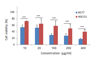
Cytotoxic Activity, Selective Indices and Extraction Yields of N.crispa: After the initial screening, the hydro-alcoholic extract of N.crispa was fractionated based on the increasing polarity of the solvents. Besides, the essential oil of N.crispa was prepared by hydro-distillation method. Then, the cytotoxic effects of the essential oil and different fractions of N.crispa were evaluated against HEK293 and MCF-7 cells, using the MTT assay. The cell viability of MCF-7 cell line and HEK293 normal cells were compared to determine the selective index (SI) of the essential oil and different fractions of N.crispa. The results are shown in Table 2 and Figures1-5.
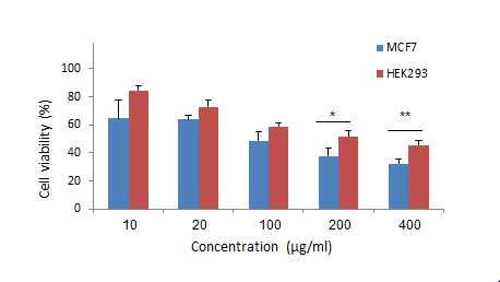
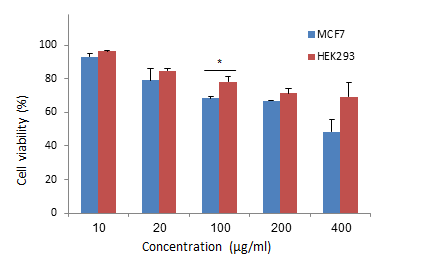
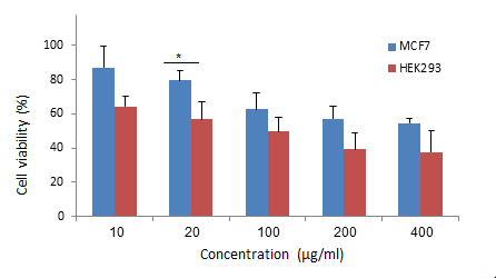
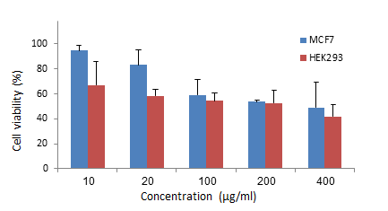
As shown in Figure 1, a significant difference in the cell viability was observed at concentrations of 200 and 400 μg/mL of the n-hexane fraction compared to the control group. As shown in Table 2, the n-hexane fraction of N.crispa had the highest cytotoxicity effect against MCF-7 cells with the IC50 value being 65.47 ±4.3μg/mL and the SI value of 3.64 among the fractions. The chloroform fraction demonstrated weak cytotoxicity against both normal and cancer cell lines (Figure 2). As shown in Figure 3, a significant difference in the cell viability was only observed at 20μg/mL of the ethyl acetate fraction compared to those of the control group. Although the hydro-alcoholic fraction at 100, 200, and 400 μg/mL reduced the cell viability of the MCF-7 cells in a dose-dependent manner, the differences were not statistically significant (Figure 4). The MTT test results also revealed that the essential oil of the aerial parts of N. crispa induced a noteworthy inhibitory effect on MCF-7 cells with the IC50 value of 18.15 ±2.7 μg/mL and the SI value of 9.69, compared to the HEK293 normal cell line (P<0.001) (Figure 5). Phytochemical Screening:  The preliminary phyto-chemical screening was performed on the n-hexane fraction (HFR) of the aerial parts of N.crispa using thin-layer and silica gel column chromatography plates. Based on the obtained data, the n-hexane fraction of N.crispa contained terpenoids, particularly the triterpene components.
Discussion
As a threat to the health of communities, breast cancer disrupts the lives of many women and their families worldwide [20]. Medicinal plants have been directly or indirectly used as the source of many current anticancer drugs [2]. In western Iran, Hamadan province is rich in valuable plant species, especially those belonging to the Lamiaceae family [5, 12]. The current study is the first screening attempt on the anti-cancer effects of ten plant species from the Lamiaceae family that grow in Hamadan province, the results of which are shown in Table 1. Various studies have investigated the cytotoxicity of some of these plants in recent years (see Table 1). The findings of the current study are consistent with those reported in previous investigations.  There are similarities to some extent between the results of the present study and those obtained in previous studies. However, the differences in some cases can be justified by the regions where the plants grow, the methods and solvents of the extraction, the types of cell line and the incubation periods used in cytotoxicity assays. Another endemic plant that was tested in the present study was N.crispa.  The hydro-alcoholic extract of N.crispa showed the lowest IC50 value (59 ±3.4 µg/mL) among the ten examined species (Table 1). The cytotoxic activity of the N.crispa extract against the MCF-7 cell line has not been reported in earlier studies. The different fractions of the hydro-alcoholic extract of N.crispa were investigated for their possible cytotoxicity against the MCF-7 cell line.  Among the examined fractions, the n-hexane fraction showed the highest cytotoxic activity, with the IC50 value being 65.47 ± 4.3µg/mL (Table 2).
The phyto-chemical screening methods based on silica gel column chromatography and TLC fingerprint of n-hexane fraction revealed the presence of terpenoids, especially triterpenes. This result suggests the cytotoxicity of terpenoids and lipophilic compounds in the Nepeta genus. Several in vitro and in vivo studies have demonstrated that terpenoids inhibited human cancers' growth and cell proliferation, using multiple molecular pathways [21]. Badrhaddad, et al. have reported the cytotoxic effect of hexane, chloroform, ethyl acetate and aqueous fractions of N.crispa extract on K562 cell line (Chronic Myelogenous Leukemia) with the IC50 values of 72, 49, 40 and 160µg/mL [11].
The difference between the effective fraction in that study and the current study can be due to the difference in the cell line and also the difference between the phyto-constituents of the wild plant versus the grown plant. In the present work, the essential oil of N.crispa showed the highest cytotoxicity against the MCF-7 cell line with the IC50 value of 18.15 ±2.7µg/mL and selectivity index of 9.69 compared to the hydro-alcoholic extract and the fractions. Previously, we examined the chemical constituents of the essential oil and reported the presence of 1,8-cineol (44.25%) and 4aα,7α,7aα-Nepetalactone (24.72%) [22]. Also, the essential oil of some Nepeta genus has been investigated for its cytotoxicity on cancer cell lines in earlier studies.
In 2017, Sharifi-rad, et al. reported an IC50 value of 32.56 ±3.23 µg/mL for the essential oil of N.schiraziana Boiss. They also reported that the main constituents of the essential oil were 1,8-cineole (33.67%), germacrene D (11.45%), β-caryophyllene (9.88%), caryophyllene oxide (7.34%) [23]. Based on the study conductred by Shakeri, et al., the essential oil of N.sintenisii Bornm (EONSi) Collected from Khorasan Razavi province, Iran, contained the following constituents: 4aα,7α,7aβ-Nepetalactone (51.74%), β-Farnesene (12.26%), 4aα,7α,7aα-Nepetalactone (8.01%), Germacrene-D (5.01%), and 4aα,7β,7aα-Nepetalactone (3.71%); which constituted 95.81% of the extracted oil.  The in-vitro cytotoxic activity was investigated on MCF-7 cell lines (IC50 value = 43.75 µg/mL) [24]. The essential oil of N.ucrainica L. spp. Kopetdaghensis (EONU) has been studied by shakeri, et al. They reported that this plant contained germacrene D (53.0%),  bicyclogermacrene  (6.4%), -bourbonene (4.3%), -elemene (3.3%), spathulenol (3.2%), cubenol (2.8%), trans-caryophyllene  (2.8%), germacrene A (2.1%), and -cadinene (2.0%). Besides, the EONU was cytotoxic against MCF-7 with the IC50 value of 50 µg/mL (18). Kahkeshani, et al. studied the essential oil of N.menthoides Boiss & Buhse (EONM) from Ardabil, Iran. The major components of EONM were 4a-α,7β,7a-α-nepetalactone (18.39%), 4a-α,7α,7a-α-nepetalactone (17.57%), 1,8-cineol (16.66%), and geranyl acetate (7.0%). They also reported EONM was cytotoxic against T47D with the IC50 value of 19.37 ±4.92 µg/mL [25]. In another study on EONM conducted by Kahkeshani, et al., the major component of EONM was 1,8-cineol (70.06%). The cytotoxic activities of EONM and 1,8-cineol were examined against three breast cancer cell lines (MCF-7, MDA-MB-231 and T47D) with the (IC50 values being 0.424, 1.243, 1.934μg/mL for the essential oil and 1.231, 2.130, 2.727μg/mL for 1,8-cineole, respectively) [26]. They also concluded that the essential oil was more effective than 1,8-cineole and etoposide, which implies the probable additive effects of the essential oil components that led to the inhibition of cancer cells. Kladniew, et al. in 2014 reported that 1,8-cineole inhibited the proliferation of human liver cancer cell line (HePG2) and adenocarcinoma of human alveolar basal epithelial cells (A549) [27]. Nepetalactone was the major component of the flower, leaf, and stem oils of several species in the Nepeta genus. Some studies were performed on the antibacterial activities of nepetalactone, but its cytotoity has not been studied yet [28]. Besides the IC50, selective index (SI) is one of the essential elements in the comparison of anticancer agents (plants). Selective index defines the selectivity of the anticancer materials on cancer cells. The higher amount of SI value demonstrates the higher selective effect on cancer cells. In the present study, EONC showed the SI of 9.69 for MCF-7 cell line, which means that EONC is 10 times more selective against cancerous cells. Although there are no reports on selectivity for Nepeta genus. 
Besides Motaghed, et al. in 2022 examined the antioxidant properties of the extract and essential oil of N.crispa using various methods (FRAP, Beta carotene-linoleic acid and DPPH). In all of the assays conducted, the essential oil had less activity than the extract [29]. It seems that the essential oil of N.crispa which shows a better cytotoxic effect on MCF-7 cell line but less antioxidant activity in-vitro. This finding is not consistent with those of previous reports. For example, Khalighi, et al. investigated the antioxidant and cytotoxicity of plant species belonging to Fabaceae family and concluded that the agents were capable of scavenging free radicals and preventing cancer development [29]. In another study conducted by Ranjbaran, et al. in 2022 on Fe2+ chelating activity of different fractions of N.crispa, the hexane fraction of methanolic extract showed the highest activity compared to other fractions [30]. The present study also demonstrated that the hexane fraction is the most cytotoxic one of the hydro-alcoholic extract from N.crispa. This finding is consistent with the study conducted by Khalighi, et al. [29]. The variations on the findings suggest the need for well-designed investigations on each plant.
 

Table 1. The percentage yields and IC50 values of the hydro alcoholic extracts of the ten plant species from the Lamiaceae family against the MCF-7 cell line.
Plants Voucher Number Extraction Yields (%) IC50 Values (μg/ml) IC50 Values (μg/ml) (Ref. No.)
Salvia spinosa 184 19.52 % 420  ± 9.8 154.07±61.88(3)
Salvia multicaulis 5 18.574 % 596 ± 10.8 51.1±2.4(4)
Thymus daenensis 6 25.22 % 260  ±7.5 84.1(7)
Nepeta crispa 72 21.54 % 59 ±3.4 Not reported
Stachys inflata 174 19.952 % 396  ± 6.9 >300(6)
Stachyslavandulifolia 178 16.948 % >800 >100(5)
Origanum vulgare 193 20.586 % 450  ± 13.2 231.46±1.15(8)
Phlomisolivieri 73 20.738 % 591  ± 12.1 >1000(10)
Phlomisbrugurieri 81 19.01 % 547  ± 9.6 726.3±12.7(10)
Melissa officinalis 346 19.392 % 419  ± 7.8 35.52±1.462(9)

Table 2.  IC50 values for essential oil and four different fractions of Nepetacrispa in MCF-7 cell line
Fraction Extraction Yields (%) IC50 Values (μg/ml) Selectivity Index (SI) HEK293/MCF-7
n-Hexane 18.1% 65.47 ± 4.3 3.64
Chloroform 13.06% >400 4.46
Ethyl acetate 31.8% >400 0.15
Hydroalcoholic 37.32% 278  ± 4.9 0.60
Essential oil 0.5% 18.15 ± 2.7 9.69


Figure 1. The cytotoxic effect of different concentrations of the n-hexane fraction on MCF-7 and HEK-293 cell lines using MTT assay. The results were reported as the percent viability compared to the control cells and as the means ± SDs (n=3). P<0.05: * and P<0.01: ** indicates a significant difference in cell viability between cancer and normal cell lines.


Figure 2. The cytotoxic effect of different concentrations of the chloroform fraction on MCF-7 and HEK-293 cell lines using MTT assay. The results were reported as the percentage viability compared to the control cells and as mean ± SD (n=3).  P < 0.05: * indicates a significant difference in cell viability between cancer and normal cell lines.


Figure 3. The cytotoxic effect of different concentrations of the ethyl acetate on MCF-7 and HEK-293 cell lines using MTT assay. The results were reported as the percentage viability compared to the control cells and as mean ± SD (n=3).  P < 0.05: * indicates a significant difference in cell viability between cancer and normal cell lines.


Figure 4. The cytotoxic effect of different concentrations of the hydro-alcoholic fraction on MCF-7 and HEK-293 cell line using MTT assay. The results were reported as the percentage viability compared to the control cells and as mean ± SD (n=3(


Figure 5. The cytotoxic effect of different concentrations of the essential oil on MCF-7 and HEK-293 cell lines using MTT assay. The results were reported as the percentage viability compared to the control cells and as mean ± SD (n=3).  P < 0.05: *, P < 0.01: **, and P < 0.001: *** indicate a significant difference in cell viability between cancer and normal cell lines.
Conclusions
The screening of the ten plant species belonging to Lamiaceae family revealed that Nepeta crispa had the most cytotoxicity against MCF-7 breast cancer cell line. This preliminary study implied the high in vitro potency of the essential oil and n-hexane fraction of N.crispa against the proliferation of MCF-7 breast cancer cell line, which have not been reported for this plant to date. Preliminary phytochemical screening methods showed the presence of triterpenoids in the n-hexane fraction of the hydro-alcoholic extract of N.crispa. According to our previous study, the essential oil of N.crispa contained 70% 1,8-cineol. Besides the essential oil had the most cytotoxicity effect and the least antioxidant activity.  The bioactive constituents can be used as potential candidates to develop new anti-breast cancer formulations. Future research should consider the isolation and identification of the active components derived from N.crispa.
Ethical Considerations
This study was approved by the Ethics Committee, Hamadan University of Medical Sciences, Hamadan, Iran (Code: IR. UMSHA.REC.1397.334)

Funding
This study was funded by School of Pharmacy, Hamadan University of Medical Sciences, Hamadan, Iran (Grant No. 9705162784).
Authors' Contributions
Study design, supervision and data analysis: Shirin Moradkhani, Meysam Soleimani Badie, Dara Dastan; Data collection: Azadeh Vali.
Conflicts of Interest
The authors declared no conflicts of interest with any internal of external entities.
Acknowledgments
This article was extracted from the thesis materials of Azadeh Vali toward the fulfillment of Pharm. D. Degree. The authors are grateful to the Vice-chancellor for Research and Technology, Hamadan University of Medical Sciences, for the financial support of this work (Grant No. 9705162784).
Type of Study: Research | Subject: Special

References
1. Nolan J, Dunne SS, Mustafa W, Sivananthan L, Kiely PA, Dunne CP. Proposed hypothesis and rationale for association between mastitis and breast cancer. Medical hypotheses. 2020;144:110057. [DOI:10.1016/j.mehy.2020.110057] [PMID]
2. Calderon-Montano JM, Martinez-Sanchez SM, Jimenez-Gonzalez V, Burgos-Moron E, Guillen-Mancina E, Jimenez-Alonso JJ, et al. Screening for Selective Anticancer Activity of 65 Extracts of Plants Collected in Western Andalusia, Spain. Plants. 2021;10(10). [DOI:10.3390/plants10102193] [PMID] [PMCID]
3. Abu-Dahab R, Afifi F, Kasabri V, Majdalawi L, Naffa R. Comparison of the antiproliferative activity of crude ethanol extracts of nine salvia species grown in Jordan against breast cancer cell line models. Pharmacognosy magazine. 2012;8(32):319-24. [DOI:10.4103/0973-1296.103664] [PMID] [PMCID]
4. Pirbalouti AG, Hashemi M, Ghahfarokhi FT. Essential oil and chemical compositions of wild and cultivated Thymus daenensis Celak and Thymus vulgaris L. Indust Crops Product. 2013;48:43-8. [DOI:10.1016/j.indcrop.2013.04.004]
5. Behzad S, Pirani A, Mosaddegh M. Cytotoxic activity of some medicinal plants from Hamedan district of Iran. Iran J Pharmaceut Res. 2014;13(Suppl):199.
6. Delnavazi MR, Saiyarsarai P, Jafari-Nodooshan S, Khanavi M, Tavakoli S, Hadavinia H. Cytotoxic flavonoids from the aerial parts of Stachys lavandulifolia Vahl. Pharmaceut Sci. 2018;24(4):332-9. [DOI:10.15171/PS.2018.47]
7. Esmaeili S, Irani M, Moazzeni Zehan H, Keramatian B, Tavakoli Harandi Z, Hamzeloo-Moghadam M. Cytotoxic activity of some ethnic medicinal plants from southwest of Iran. J Pharmacog Res. 2016;3:43-7.
8. Grbovic F, Stankovic MS, Curcic M, Dordevic N, Seklic D, Topuzovic M, et al. In Vitro Cytotoxic Activity of Origanum vulgare L. on HCT-116 and MDA-MB-231 Cell Lines. Plants. 2013;2(3):371-8. [DOI:10.3390/plants2030371] [PMID] [PMCID]
9. Khallouki F, Breuer A, Akdad M, Laassri FE, Attaleb M, Elmoualij B. Cytotoxic activity of Moroccan Melissa officinalis leaf extracts and HPLC-ESI-MS analysis of its phytoconstituents. Future J Pharmaceut Sci. 2020;6(1):1-12. [DOI:10.1186/s43094-020-00037-x]
10. Sarkhail P, Sahranavard S, Nikan M, Gafari S, Eslami-Tehrani B. Evaluation of the cytotoxic activity of extracts from six species of Phlomis genus. J Appl Pharmaceut Sci. 2017;7(2):180-4.
11. Badrhadad A, Piri K, Mansouri K. Anti-proliferative effects of some fractions of Elaeagnus angustifolia L. flower and aerial part of Nepeta crispa L. on K562 leukemic cells. Iran J Med Aromatic Plant Res. 2015;31(5):881-90.
12. Mosaddegh M, Esmaeili S, Hassanpour A, Malekmohammadi M, Naghibi F. Ethnobotanical study in the highland of Alvand and Tuyserkan, Iran. Res J Pharmacog. 2016;3(1):7-17.
13. Li Y, Kong D, Fu Y, Sussman MR, Wu H. The effect of developmental and environmental factors on secondary metabolites in medicinal plants. Plant physiology and biochemistry : PPB. 2020;148:80-9. [DOI:10.1016/j.plaphy.2020.01.006] [PMID]
14. Kerdar T, Moradkhani S, Dastan D. Phytochemical and biological studies of scrophularia striata from Ilam. Jundishapur J Natur Pharmaceut Product. 2018;13(3):e62705. [DOI:10.5812/jjnpp.62705]
15. Moradkhani S, Kobarfard F, Ayatollahi SA. Phytochemical Investigations on Chemical Constituents of Achillea tenuifolia Lam. Iranian journal of pharmaceutical research : IJPR. 2014;13(3):1049-54.
16. Kiani M, Firozian F, Moradkhani S. Formulation and physicochemical evaluation of toothpaste formulated with Thymus vulgaris essential oil. J Herbmed Pharmacol. 2017;6(3):130-5.
17. Hamedeyazdan S, Sharifi S, Nazemiyeh H, Fathiazad F. Evaluating antiproliferative and antioxidant activity of Marrubium crassidens. Advanc Pharmaceut Bullet. 2014;4(Suppl 1):459.
18. Shakeri A, Khakdan F, Soheili V, Sahebkar A, Rassam G, Asili J. Chemical composition, antibacterial activity, and cytotoxicity of essential oil from Nepeta ucrainica L. spp. kopetdaghensis. Indust Crop Product. 2014;58:315-21. [DOI:10.1016/j.indcrop.2014.04.009]
19. Ayeni EA, Abubakar A, Ibrahim G, Atinga V, Muhammad Z. Phytochemical, nutraceutical and antioxidant studies of the aerial parts odoi: 10.15171/jhp.2018.12f Daucus carota L.(Apiaceae). J Herbmed Pharmacol. 2018;7(2):68-73. [DOI:10.15171/jhp.2018.12]
20. Ferlay J, Colombet M, Soerjomataram I, Mathers C, Parkin DM, Pineros M, et al. Estimating the global cancer incidence and mortality in 2018: GLOBOCAN sources and methods. International journal of cancer. 2019;144(8):1941-53. [DOI:10.1002/ijc.31937] [PMID]
21. Yang H, Dou QP. Targeting apoptosis pathway with natural terpenoids: implications for treatment of breast and prostate cancer. Current drug targets. 2010;11(6):733-44. [DOI:10.2174/138945010791170842] [PMID] [PMCID]
22. Abdoli P, Moradkhani S, Dastan D. Comparative analysis of Nepeta crispa essential oil composition in flowering and vegetative stages. Am J Phytomed Clin Ther. 2016;4:106-12.
23. Sharifi-Rad J, Ayatollahi SA, Varoni EM, Salehi B, Kobarfard F, Sharifi-Rad M. Chemical composition and functional properties of essential oils from Nepeta schiraziana Boiss. Farmacia. 2017;65(5):802-12.
24. Shakeri A, Khakdan F, Soheili V, Sahebkar A, Shaddel R, Asili J. Volatile composition, antimicrobial, cytotoxic and antioxidant evaluation of the essential oil from Nepeta sintenisii Bornm. Indust Crop Product. 2016;84:224-9. [DOI:10.1016/j.indcrop.2015.12.030]
25. Kahkeshani N, Hadjiakhoondi A, Navidpour L, Akbarzadeh T, Safavi M, Karimpour-Razkenari E, et al. Chemodiversity of Nepeta menthoides Boiss. & Bohse. essential oil from Iran and antimicrobial, acetylcholinesterase inhibitory and cytotoxic properties of 1,8-cineole chemotype. Natural product research. 2018;32(22):2745-8. [DOI:10.1080/14786419.2017.1378202] [PMID]
26. Kahkeshani N, Razzaghirad Y, Ostad SN, Hadjiakhoondi A, Shams Ardekani MR, Hajimehdipoor H. Cytotoxic, acetylcholinesterase inhibitor and antioxidant activity of Nepeta menthoides Boiss & Buhse essential oil. J Essen Oil Bear Plant. 2014;17(4):544-52. [DOI:10.1080/0972060X.2014.929040]
27. Rodenak Kladniew B, Polo M, Montero Villegas S, Galle M, Crespo R, Garcia de Bravo M. Synergistic antiproliferative and anticholesterogenic effects of linalool, 1,8-cineole, and simvastatin on human cell lines. Chemico-biological interactions. 2014;214:57-68. [DOI:10.1016/j.cbi.2014.02.013] [PMID]
28. Shafaghat A, Oji K. Nepetalactone content and antibacterial activity of the essential oils from different parts of Nepeta persica. Natur Product Communicat. 2010;5(4):625-8. [DOI:10.1177/1934578X1000500427]
29. Khalighi-Sigaroodi F, Ahvazi M, Hadjiakhoondi A, Taghizadeh M, Yazdani D, Khalighi-Sigaroodi S. Cytotoxicity and antioxidant activity of 23 plant species of Leguminosae family. Iran J Pharmaceut Res. 2012;11(1):295.
30. Ranjbaran P, Moradkhani S. Iron Chelating Activity of Nepeta Crispa Willd., an Endemic Plant in the West of Iran. Avicenna J Med Biochem. 2022;10(1):65-70. [DOI:10.34172/ajmb.2022.09]

Add your comments about this article : Your username or Email:
CAPTCHA

Send email to the article author


Rights and permissions
Creative Commons License This work is licensed under a Creative Commons Attribution-NonCommercial 4.0 International License.

© 2025 CC BY-NC 4.0 | Iranian Journal of Toxicology

Designed & Developed by : Yektaweb