Write your message
Volume 18, Issue 1 (January 2024)                   IJT 2024, 18(1): 45-51 | Back to browse issues page


XML Print


Download citation:
BibTeX | RIS | EndNote | Medlars | ProCite | Reference Manager | RefWorks
Send citation to:

Jimat R T, Ethica S N, Rahmani N, Yulianti S E, Rachmayati R, Nuryati N, et al . Alginate Lyase from Streptomyces olivaceus is a Safe and Effective Antibiofilm in Male Wistar Rats (Rattus norvegicus). IJT 2024; 18 (1) :45-51
URL: http://ijt.arakmu.ac.ir/article-1-1274-en.html
1- Magister Study Program of Clinical Laboratory Science, Postgraduate Program, Universitas Muhammadiyah Semarang
2- Research Center for applied Microbiology, Organization Research of Life Sciences and Environment, National Research and Innovation Agency
3- Research Center for Biosystematics and Evolution, Organization Research of Life Sciences and Environment, National Research and Innovation Agency
4- Deputy for Regional Research and Innovation, National Research and Innovation Agency
5- Research Center for Deep Sea, Earth Sciences and Maritime Research Organization, National Research and Innovation Agency
6- Department of Biomedical Sciences, Faculty of Medicine, Universitas Muhammadiyah Semarang , mayadianr@unimus.ac.id
Full-Text [PDF 743 kb]   (622 Downloads)     |   Abstract (HTML)  (1449 Views)
Full-Text:   (422 Views)
Introduction
Infection with biofilm-producing microorganisms is a health problem throughout the world [1]. The United States’ National Institutes of Health (NIH) has reported that 80% of cases of chronic infection with pathogenic microorganisms are associated with biofilm formation. Therefore, degradative agents are needed to fight against biofilms produced by pathogenic bacteria [2]. The formation of biofilm in cell-protecting polymer matrices can make bacteria resistant to antibiotics. Biofilm matrices need to be destroyed to make antibiotics function optimally.
Alginate lyase is a compound that can be developed into an antibiofilm agent. It is an enzyme that is able specifically to degrade the extracellular polymer matrices of biofilms [3, 4]. Alginate is one of the components of the Pseudomonas aeruginosa biofilm polymer matrix, which can be a target for degradation by alginate lyase. Degradation of alginate structure has the potential to increase the phagocytic ability of immune cells and reduce the virulence of pathogenic bacteria [5], as well as enhancing the therapeutic effect of anti-pseudomonal agents [6]. Alginate lyase also has the potential to degrade biofilms in the lungs caused by Mycobacterium smegmatis. Also, alginate lyase has the potential to develop as anti-biofilm agent to eradicate tuberculosis (TB) disease [7].
All medicinal ingredients, including anti-biofilm from enzymes, undergo a metabolic process to produce metabolites which may be toxic to hepatocytes [8]. Liver is an organ that functions to detoxify and inactivate drugs into substances that are not harmful to the body. Liver damages due to drugs can occur secondary to the loss of the liver's ability to regenerate cells, which undergoes permanent and necrotic damages [9]. Therefore, all medicinal substances including anti-biofilm agents need to be tested for their toxicity in vivo or clinically to rule out their toxic effects on the liver.
Cellular damages to the liver can be detected by biochemical analyses of the tissue and serum. The enzymatic examinations include transaminases, such as Aspartate Transaminase (AST) or Serum Glutamic Oxaloacetic Transaminase (SGOT) and Alanine Transaminase (ALT) and Serum Glutamic Pyruvic Transaminase (SGPT). These enzymes are produced by hepatocytes, and can be used to monitor damages to liver cells [10]. Liver damage can occur due to spontaneous toxicity or accumulation of drugs that are used in the long term [11]. This causes immunological processes in the body which ultimately lead to injuries to the liver tissue [12]. Apart from drugs, disorders of the liver immunological processes can also be caused by viral or bacterial infections, or by alcohol consumption. Functional disorders of the immunological processes can cause liver damage, leading to degeneration, intracellular accumulation, necrosis, inflammation, regeneration and fibrosis [13, 14]. These damages can be observed microscopically in the liver tissue.
Recently, there has been much interest in using marine bacteria that produce enzymes with significant biological activities. One of the bacteria from seaweed, Streptomyces luridiscabiei can produce a strong enzyme with the specific activity of 108.6 U/mg, molecular weight of 28.6 kDa, consisting of 260 amino acids. The enzyme, characterized as AlyDS44, is capable of hydrolyzing sodium alginate to produce alginate disaccharide and trisaccharide as the final products [15]. The initial screening results revealed the potential of alginate lyase from the Streptomyces olivaceus found in marine sediments on Serena Kecil Island, Bitung City, North Sulawesi, Indonesia. The alginate lyase can degrade M. smegmatis biofilms, the IC50 and IC90 levels of which have already been determined [7]. However, the toxicity tests for the alginate lyase from S. olivaceus have not been reported previously.
The current study conducted an in vivo test using experimental animals to determine the toxic effects of the alginate lyase from S. olivaceus, especially for its development as an oral anti-biofilm agent. The Wistar rats were chosen in this study because they are the most recommended animals for various in vivo experimental purposes.
Materials and Methods
Research Setting
This study was conducted at the Experimental Animal Laboratory, Universitas Muhammadiyah Semarang in Indonesia. It was approved by the Health Research Bioethics Commission, Faculty of Medicine, Universitas Sultan Agung Semarang, and given the registration #: 213/VI/2023; by the Bioethics Commission. The crude extract of alginate lyase, being the research object, was obtained from the microorganism known as Streptomyces olivaceus (S. olivaceus).
Preparation of Alginate Lyase Crude Extract
The S. olivaceus isolates were rejuvenated on ISP2 agar media (4 g yeast extract, 10 g wheat extract, and 4 g-glucose using 30 g artificial sea water in 1L distilled H2O) and incubated at 28℃ for 4 days. As much as 10 µL of S. olivaceus colonies were taken and pre-cultured using liquid ISP2 media for three days at 28℃ and centrifuged at 190 rpm. A total of 5% and 7.5% of the preculture results were inoculated into the alginate lyase production medium (ISP2 medium without glucose) and added sodium alginate as the substrate. The alginate lyase activity was determined from the culture, using 5% inoculum S. olivaceus at 5,558 U/mL, which was originally derived from 7.5% inoculum at 5,348 U/mL. The culture process was carried out under the same conditions as the pre-culture but extended for seven days. The enzyme harvesting was performed by centrifuging the S. olivaceus culture product at 4℃ and 12,000 rpm for 20 minutes [16]. The enzyme concentrate was stored at the Genomics Laboratory of the National Research and Innovation Agency before transporting it to the research laboratory.
Animal Treatments
In this study, a total of 20 male Wistar rats were used, which were divided into four groups five rats each. The rats with an initial weight of 180-300 g were first acclimatized under laboratory conditions for seven days, given pellet food and drinking water ad libitum. After the acclimatization period, the animals were randomly divided into:
a) technical control group (TC), given food and drinking water only; b) negative control group (NC), given alginate lyase solvent, i.e., phosphate buffer saline (PBS) 1x; c) treatment group 1 (E1), given alginate lyase concentrate at 0.63 g/kg BW; and d) treatment group 2 (E2), given alginate lyase concentrate at 20.85 g/kg BW. The dosage of alginate lyase used in this study was predetermined as being effective. The dosage of 0.63 g/kg BW was the IC50 and that of 20.85 g/kg BW was the IC90 to degrade the M. smegmatis biofilm [7].
Animal Body Weight and Behavior
Changes in the rats’ body weight (gain or loss) were determined systematically during the 7-day treatment period. Also, the behavioral changes observed in the test rats were regularly documented. The behavioral changes included tremor, pain gesture, eyes rolling, earlobe reflex, salivation, hyperactivity, and mortality [17].
Measurement of AST and ALT Levels
The blood AST and ALT levels were measured before treatment and on the 7th day after the treatments were complete. The blood sampling was carried from the retro-orbital vasculature because relatively large amounts of blood samples could be obtained. A total of 1-2 mL of blood was collected in microtubes and incubated for 30 minutes at room temperature. The blood samples were then centrifuged for 10 minutes at 3000 rpm to obtain the sera. The AST and ALT levels were determined, using a kinetic method adapted from the International Federation of Clinical Chemistry (IFCC) [18].
Histopathological Examinations
On the 8th day, rats were sacrificed under ether anesthesia, then the liver was dissected. The macroscopic observations of the liver were carried out before washing the tissue with physiological NaCl solution.  After washing, the livers were cut into the right and left lobes, and then fixed in 10% neutral buffer formalin (NBF). The tissue samples were processed for microscopic histopathological examinations through stages of fixation, dehydration, cleaning, impregnation, embedding, and cutting into sections until the tissue samples were ready for staining with hematoxylin-eosin. After sectioning, the liver slides were examined under light microscopy at 400x magnification, by observing sets of 100 optic fields for each animal group. The histopathological appearances of the rat liver samples were assessed by documenting the level of hepatocytes damage in the area near the central vein. The level of cellular damages were determined based on the degree of changes in the histopathological structures of liver cells, using the Manja Roenigk scoring system (score: 1/2/3/4) [19, 20].
Data Analyses
The data representing the body weight, AST, ALT levels, and Manja Roenigk scores were analyzed for normality, using the Kolmogorov-Smirnov and homogeneity, using Levene tests [21]. The analyses of changes in the body weight, AST, and ALT levels before and after treatment were also performed, using paired t-test. The differences in Manja Roenigk scores among the treatment groups were determined, using One-way ANOVA. Changes in the animal behaviors during the treatments and the histopathological alterations in the liver cells were analyzed descriptively.
Results
Body Weight and Behavioral Changes
Changes in the rats’ body weight were documented in each group. During the treatments, animals in the technical control group demonstrated a significant gain in their body weight. Meanwhile, the administration of alginate lyase reduced the body weights insignificantly (Table 1). The control and treated rats were observed for changes in their behaviors due to toxicity. During the alginate lyase treatment, none of the rats showed signs of poisoning, nor did the enzyme cause death in the rats (Table 2).
Examinations of AST and ALT Levels
The administration of alginate lyase significantly reduced the AST and ALT levels in the rats’ sera. However, the PBS solvent significantly reduced the AST levels. The AST/ALT ratio in the control group that were given alginate lyase and PBS was above 2.0, compared to that of the E1 and E2 groups being below that level (Table 3).

Table 1. Changes in body weight of Wistar rats during treatment
Before Treatment After Treatment Paired Samples t-test
TC 267.0 ± 40.18 282.6 ± 35,52 0.003*
NC 222.8 ± 24.26 246.8 ± 16,05 0.210
E1 197.6 ± 32.73 188.0 ± 29.33 0.228
E2 201.8 ± 30.34 197.0 ± 32.97 0.352
Data in table are presented as Mean ± SD
Note: TC=Technical Control (No Treatment); NC=Negative Control (PBS 1x solvent); E1=Treatment of alginate lyase 0.63 g/kg BW; E2=Treatment of alginate lyase Dose 20.85 g/kg BW; *Significant different.

Table 2. Changes in Wistar rat behavior during treatment
Observation Day 1 Day 2 Day 3 Day 4 Day 5 Day 6 Day 7
Tremors - - - - - - -
Painful - - - - - - -
Eye N N N N N N N
Ear reflex N N N N N N N
Salivation - - - - - - -
Hyperactivity - - - - - - -
Mortality - - - - - - -
Note: Observations were made on rats in all treatment groups; (-) = No symptoms were found; (N) = Normal

Table 3. Changes in the AST and ALT levels of the rats during treatment
Mean ± SD (Before vs After) AST/ALT Ratio
AST ALT
NC 129.0 ± 49.65 vs 83.4 ± 19.83 67.8 ± 16.39 vs 36.8 ± 19.32* 2.3
E1 156.0 ± 44.37 vs 68.8 ± 12.03* 64.8 ± 19.63 vs 34.8 ± 6.46* 2.0
E2 129.6 ± 26.05 vs 55.2 ± 15.27* 64.2 ± 15.06 vs 29.8 ± 9.83* 1.8
Notes: NC = Negative Control (PBS 1x solvent); E1 = Alginate lyase treatment 0.63 g/kg BW; E2 = Alginate lyase treatment 20.85 g/kg BW; *Significant different using paired samples t-test.

Table 4. Results of liver rats histopathological scoring using the Manja Roenigk score
Group Mean ± SD Overall Conclusion One Way ANOVA
TC 3.90 ± 0.03 Normal 0,000a
NC 2.94 ± 0.22b Parenchymal degeneration
E1 2.83 ± 0.15b Parenchymal degeneration
E2 2.81 ± 0.23b Parenchymal degeneration
Notes: TC = Technical control (no treatment); NC = Negative control (PBS 1x solvent); E1 = Alginate lyase treatment 0.63 g/kg BW; E2 = Alginate lyase treatment 20.85 g/kg BW; a significant value between groups;
b post-hoc analysis, significant with TC.


Figure 1. Rat liver samples after treatment.
Notes: TC = Technical control (no treatment); NC = Negative control (PBS 1x solvent); E1 = Alginate lyase treatment. Lyase 0.63 g/kg BW; E2 = Alginate lyase treatment 20.85 g/kg BW.


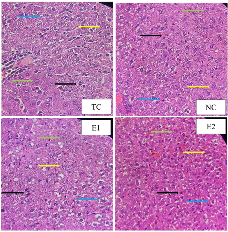
Figure 2. Microscopic histopathological features of the rat liver samples stained with H&E and examined under light microscopy at 400x magnification.
Note: TC = Technical Control (No Treatment); NC = Negative Control (PBS 1x solvent); E1 = Alginate lyase treatment 0.63 g/kg BW; E2 = Alginate lyase treatment 20.85 g/kg BW; Green arrows = Normal cells; Blue arrows = Hydropic degeneration; Yellow arrows = Parenchymal degeneration; Black arrows = Necrotic cells.
Histopathological Examinations
Macroscopic examinations of the rat liver samples were carried out by observing changes in color, surface appearance, and consistency of the tissue. The macroscopic results of the liver tissue samples did not show any changes or abnormalities after 7 days of treatment. The samples were not hardened, and appeared brownish-red in color with smooth surfaces (Figure 1). Based on the microscopic histopathological findings from the liver cells, all treatment groups exhibited cellular abnormalities, such as hydropic and parenchymal degenerations, and cell necrosis (Figure 2). However, after calculating the scores, it was noted that there were differences in the number of abnormal liver cells among all treatment groups. The administration of alginate lyase at both doses caused parenchymal degeneration in the liver cells. This damage was significantly different as compared to that of the technical control group, which had generally retained the normal appearance of the liver cells (Table 4).
Discussion
This study evaluated the safety of the alginate lyase by conducting toxicity tests on the liver tissue samples from male Wistar rats aged 2-4 months. Our reasons for choosing rats were the ease of acquisition and handling, price, and their relatively large size. Minimizing the biological variations and other factors that could influence the research was also carried out by equalizing race, age, body weight, environment, and rat food. White Wistar rats (Rattus norvegicus) offer the advantage of showing fast response plus being easy to adapt. Apart from that, rats are omnivores, have body organs, and almost the same nutritional needs as humans [22].
Toxicity can also be observed from changes in the body weight of test rats; hence, the insignificant weight loss in the animals’ body weight after seven days of Alginate lyase treatment (P≥0.05) (Table 1), which is normal. A substance is believed to have toxic effect if it causes a significant weight loss of 10% or greater than the initial body weight [23]. In this regard, the administration of alginate lyase did not affect the rats’ body weight. Apart from body weight, changes in the rats’ behavior or clinical signs may also be considered as the evidence of toxicity. Clinical symptoms include half-closed eyes, piloerection, and low motor activity was most strongly associated with pathological findings [24]. In this study, no behavioral changes were found such as tremors, pain expression, eyes abnormalities and earlobe reflexes, salivation, hyperactivity, or death (Table 2). The rats’ behavior after the alginate lyase administration was normal, again suggestive of the enzyme lacking toxicity, which is reported for the first time by this study.
To observe liver damage after the administration of alginate lyase, we investigated changes in AST and ALT levels in the rats’ sera. We found that the AST values decreased significantly after administering the alginate lyase at 0.63 g/kg BW and 20.85 g/kg BW. The ALT levels also showed a significant reduction after administering either alginate lyase or 1x PBS solvent. However, the decline in the ALT and AST levels was still within the normal reference range (Table 3) [18]. Changes in ALT and AST levels can be caused by physiological or extra-hepatic conditions. These include persistent tachycardia; intense physical exercise; prolonged fasting; vitamin B6 deficiency; hyperthyroidism; hyperthermia; obesity; and excessive protein intake. Low ALT and AST values are known to be good, although it may also suggest renal dysfunction [25]. Apart from pathophysiological factors, ALT and AST levels can also be influenced by providing food intake to the animals ad libitum, and changes in the temperature and humidity of the rats’ environment [26].
A high ALT level in the sera is directly related to the extent of cellular damage. This parameter usually increases within the first 12 hours after the damage has occurred and remains high for the next five days. Rises in AST and/or ALT can be attributed to damages in the liver cell walls, and can be interpreted as a marker of impaired liver cells integrity. However, changes in AST and ALT levels up to 300 U/L are not specific to liver disorders alone. Therefore, the DeRitis AST/ALT ratio value can be seen as the severity of liver cell damage [27, 28]. The normal value of DeRitis ratio is below 1.0. If the ratio is greater than 1.0, there may be an indication of liver damage, and if it is greater than 2.0, it could be a sign of alcoholic liver disease [29]. Our DeRitis ratio calculation showed a value of 2.0 in rats that were administered alginate lyase at 0.63 g/kg BW; and 1.8 in rats given the same enzyme at 20.85 g/kg BW. However, because the AST and ALT values remained at normal values, it may be concluded that no liver cell damage occurred in the rats due to the alginate lyase.
Macroscopically, there were no signs of liver damage found in the current study. The rats’ liver samples both in the control and treatment groups had normal appearances (Figure 1). Liver cell damage can be interpreted from the Manja Roenigk scores. In the rats’ liver samples that were only given food and drink, some histopathological features of cell abnormalities, such as parenchymal and hydropic degenerations were visible. However, the Manja Roenigk score showed overall normality of the liver. In the rat liver cells, given 1x PBS solvent, alginate lyase at 0.63 and 20.85 g/kg BW caused more cellular abnormalities than in the normal control (Figure 2). This is supported by a Manja Roenigk score of around 3, concluding that PBS 1x and alginate lyase cause parenchymal degeneration in liver cells (Table 4).
Parenchymal degeneration is the mildest cellular changes characterized by swelling and turbidity of the cytoplasm. Parenchymal degeneration is reversible because it occurs only in mitochondria and endoplasmic reticula due to oxidative stress [30]. It is often caused by such factors, as stress due to less-than-ideal cage conditions, low appetite and body weight, fatigue, and other internal factors, such as low endurance in the animals [31]. Parenchymal degeneration can also occur as the adaptive response in the rats treated with drugs. Finally, because it is reversible, the symptoms usually last a short period in response to exposure to substances [32]. In the current study, the alginate lyase was in its crude form. Therefore, further research is warranted on the effects of this enzyme when a highly pure form of alginate lyase is available. Also, additional investigation shall be conducted to assess the long-term toxicity of this enzyme.
Overall, the findings of the current study demonstrated the safety of alginate lyase at 0.63 g/kg BW and 20.85 g/kg BW in rats (Rattus norvegicus). Based on the results, alginate lyase was shown to be safe because it did not cause changes in the body weight and behavior of rats, and did not cause liver damages. This enzyme has the potential of being developed into an effective oral anti-biofilm agent in the future.
Conflict of Interests
The authors had no conflict of interests to disclose in conducting this study.
Compliance with Ethical Guideline
This study was approved by the Health Research Bioethics Commission, Faculty of Medicine, Universitas Sultan Agung Semarang. The registered ethical approval code assigned to this study was: 213/VI/2023/Bioethics Commission.
Funding
This research was financially supported by Rumah Program, Organization of Research in Life Sciences and Environment, National Research and Innovation Agency during the fiscal year 2022-2023 with the grant number: 9/III/HK/2022.
Authors' Contributions
RTJ (data collection and analysis, drafting the manuscript); SNE and NR (conception and design, data analysis, funding responsibility); SEY, RR, NN, AA, SR, PL, YY, and DSZ (conception and data collection); MDR (conception and design, data analysis, drafting the manuscript). All authors read and approved the final draft of the manuscript prior to submission to this journal.
Acknowledgments
This research was financially supported by Rumah Program, Organization of Research in Life Sciences and Environment, National Research and Innovation Agency fiscal year 2022-2023 with the registered number being: 9/III/HK/2022.
Type of Study: Research | Subject: General

References
1. Founou RC, Founou LL, Essack SY. Clinical and economic impact of antibiotic resistance in developing countries: A systematic review and meta-analysis. PLoS One. 2017;12(12):e0189621. [DOI:10.1371/journal.pone.0189621] [PMID] []
2. Topka-Bielecka G, Dydecka A, Necel A, Bloch S, Nejman-Falenczyk B, Wegrzyn G, et al. Bacteriophage-Derived Depolymerases against Bacterial Biofilm. Antibiotics (Basel). 2021;10(2). [DOI:10.3390/antibiotics10020175] [PMID] []
3. Ethica SN. Bioprospection of alginate lyase from bacteria associated with brown algae Hydroclathrus sp. as antibiofilm agent: a review. ACL Bioflux. 2021;14(4):1974-89.
4. Ethica SN, Zilda DS, Oedjijono O, Muhtadi M, Patantis G, Darmawati S, et al. Biotechnologically potential genes in a polysaccharide-degrading epibiont of the Indonesian brown algae Hydroclathrus sp. J Genet Eng Biotechnol. 2023;21(1):18. [DOI:10.1186/s43141-023-00461-5] [PMID] []
5. Roy R, Tiwari M, Donelli G, Tiwari V. Strategies for combating bacterial biofilms: A focus on anti-biofilm agents and their mechanisms of action. Virulence. 2018;9(1):522-54. [DOI:10.1080/21505594.2017.1313372] [PMID]
6. M. Haroon A, M. Daboor S. Nutritional status, antimicrobial and anti-biofilm activity of Potamogeton nodosus Poir. Egyptian Journal of Aquatic Biology and Fisheries. 2019;23(2):81-93. [DOI:10.21608/ejabf.2019.29301]
7. Sa'adah AL. Aktivitas antibiofilm alginat liase Streptomyces olivaceus pada jaringan tikus wistar (Rattus norvegicus) terinfeksi Mycobacterium smegmatis: Magister Study Program of Clinical Laboratory Science Universitas Muhammadiyah Semarang; 2023.
8. Mandasari AA, Wahyuningsih SPA, Darmanto W. Uji Toksisitas akut polisakarida krestin dari ekstrak Coriolus versicolor dengan parameter kerusakan hepatosit, enzim SGPT dan SGOT pada mencit. Jurnal Sain Veteriner. 2015;33(1):69-74.
9. Purba M, Surjanto S, Batubara R. Keamanan teh gaharu (Aquilaria malaccencis Lamk) dari pohon induksi terhadap toksik oral. Wahana Forestra: Jurnal Kehutanan. 2018;13(1):1-11. [DOI:10.31849/forestra.v13i1.1399]
10. Firdausi KM. Uji toksisitas subkronik ekstrak air daun katuk (Sauropus androgynus (L) Merr) terhadap kadar enzim transaminase (AST dan ALT) hepar tikus (Rattus norvegicus) betina: Universitas Islam Negeri Maulana Malik Ibrahim; 2015.
11. Murti FK, Amarwati S, Wijayahadi N. Pengaruh ekstrak daun kersen (Muntingia calabura) terhadap gambaran mikroskopis hepar tikus wistar jantan yang diinduksi etanol dan soft drink. Diponegoro Medical Journal (Jurnal Kedokteran Diponegoro). 2016;5(4):871-83.
12. Murray RK, Granner DK, Victor W. Biokimia Harper: Harper's illustrated biochemistry. In: Soeharsono R, editor. Harper's illustrated biochemistry. Jakarta: BP FKUI; 2014.
13. Shariffah-Muzaimah SA, Idris AS, Madihah AZ, Dzolkhifli O, Kamaruzzaman S, Maizatul-Suriza M. Characterization of Streptomyces spp. isolated from the rhizosphere of oil palm and evaluation of their ability to suppress basal stem rot disease in oil palm seedlings when applied as powder formulations in a glasshouse trial. World J Microbiol Biotechnol. 2017;34(1):15. [DOI:10.1007/s11274-017-2396-1]
14. Puspita Sari D, Hadisusanto S, Istriyati I. Struktur Histologis Hepar, Intestinum, dan Ren Burung Cerek Jawa (Charadrius javanicus Chasen 1938) Dengan Kontaminasi DDT di Delta Sungai Progo Yogyakarta. Biogenesis: Jurnal Ilmiah Biologi. 2014;2(2):126-31. [DOI:10.24252/bio.v2i2.479]
15. Nguyen TNT, Chataway T, Araujo R, Puri M, Franco CMM. Purification and Characterization of a Novel Alginate Lyase from a Marine Streptomyces Species Isolated from Seaweed. Mar Drugs. 2021;19(11). [DOI:10.3390/md19110590] [PMID] []
16. Zilda DS, Yulianti Y, Sholihah RF, Subaryono S, Fawzya YN, Irianto HE. A novel Bacillus sp. isolated from rotten seaweed: Identification and characterization alginate lyase its produced. Biodiversitas Journal of Biological Diversity. 2019;20(4):1166-72. [DOI:10.13057/biodiv/d200432]
17. Sabbani V, Ramesh A, Shobharani S. Acute Oral Toxicity Studies of Ethanol Leaf Extracts of Derris Scandes & Pulicaria Wightiana in Albino Rats. International Journal Of Pharmacological Research: India. 2015;5(1):1-5.
18. Hasan KMM, Tamanna N, Haque MA. Biochemical and histopathological profiling of Wistar rat treated with Brassica napus as a supplementary feed. Food Science and Human Wellness. 2018;7(1):77-82. [DOI:10.1016/j.fshw.2017.12.002]
19. Fitri NMA, Haeni L, Mardliyah E. Pengaruh Pemberian Ekstrak Etanol Kulit Batang Kelor (Moringa oleifera Lam.) sebagai Hepatoprotektor. JIMKI: Jurnal Ilmiah Mahasiswa Kedokteran Indonesia. 2018;6(2):55-62.
20. López Panqueva RdP. Aspectos morfológicos de la enfermedad hepática inducida por drogas. Revista Colombiana de Gastroenterología. 2014;29(4):449-60. [DOI:10.22516/25007440.445]
21. Rinaldi SF, Mujianto B. Metodologi Penelitian dan Statistik. Bahan Ajar TLM. Jakarta: KEMENKES RI Pusat Pendidikan SDM Kesehatan; 2017.
22. Wolfensohn S, Lloyd M. Handbook of Laboratory Animal Management and Welfare. 3rd ed. USA: Blackwell Publishing; 2013.
23. Abdulwahid SJ, Goh MY, Ebrahimi M, Mohtarrudin N, Hashim ZB. Sub-Acute Oral Toxicity Profiling of the Methanolic Leaf Extract of Clinacanthus Nutans in Male and Female Icr Mice. International Journal of Pharmacy and Pharmaceutical Sciences. 2018;10(12). [DOI:10.22159/ijpps.2018v10i12.28075]
24. Silva AV, Norinder U, Liiv E, Platzack B, Oberg M, Tornqvist E. Associations between clinical signs and pathological findings in toxicity testing. ALTEX. 2021;38(2):198-214. [DOI:10.14573/altex.2003311] [PMID]
25. Kuntz E, Kuntz H-D. Hepatology Textbook and Atlas. Germany: Spinger Medizin Verlag Heidelberg; 2008. [DOI:10.1007/978-3-540-76839-5]
26. Sari HK, Budirahardjo R, Sulistyani E. Kadar Serum Glutamat Piruvat Transaminase (SGPT) pada Tikus Wistar (Rattus norvegicus) Jantan yang Dipapar Stresor Rasa Sakit berupa Electrical Foot Shock selama 28 Hari (The Level of Serum Glutamic Pyrufic Transaminase [SGPT] on a Wistar [Rattus norvegicus]). Pustaka Kesehatan. 2015;3(2):205-11.
27. Rosida A. Pemeriksaan Laboratorium Penyakit Hati. Berkala Kedokteran. 2016;12(1). [DOI:10.20527/jbk.v12i1.364]
28. Parmar K, Singh G, Gupta G, Pathak T, Nayak S. Evaluation of De Ritis ratio in liver-associated diseases. International Journal of Medical Science and Public Health. 2016;5(9). [DOI:10.5455/ijmsph.2016.24122015322]
29. Ndrepepa G. De Ritis ratio and cardiovascular disease: evidence and underlying mechanisms. Journal of Laboratory and Precision Medicine. 2023;8:6-. [DOI:10.21037/jlpm-22-68]
30. Maulina M. ZAT zat yang mempengaruhi histopatologi hepar. Sulawesi: Unimal Press; 2018.
31. Nugrahanti MP, Armalina D, Partiningrum DL, Fulyani F. The effect of ranitidine administration in graded dosage to the degree of liver damage: A study on Wistar rats with acute methanol intoxication. Hum Exp Toxicol. 2021;40(3):497-503. [DOI:10.1177/0960327120954529] [PMID]
32. Stanek M, Rotkiewicz T, Sobotka W, Bogusz J, Otrocka-Domagała I, Rotkiewicz A. The effect of alkaloids present in blue lupine (Lupinus angustifolius) seeds on the growth rate, selected biochemical blood indicators and histopathological changes in the liver of rats. Acta Veterinaria Brno. 2015;84(1):55-62. [DOI:10.2754/avb201585010055]

Add your comments about this article : Your username or Email:
CAPTCHA

Send email to the article author


Rights and permissions
Creative Commons License This work is licensed under a Creative Commons Attribution-NonCommercial 4.0 International License.

© 2025 CC BY-NC 4.0 | Iranian Journal of Toxicology

Designed & Developed by : Yektaweb