Write your message
Volume 18, Issue 2 (May 2024)                   IJT 2024, 18(2): 78-83 | Back to browse issues page

Ethics code: IR.QUMS.REC.1399.546


XML Print


Download citation:
BibTeX | RIS | EndNote | Medlars | ProCite | Reference Manager | RefWorks
Send citation to:

Asoudeh-Fard A, Najafipour R, Hosseini A H, Razi F, Salahshourifar I, Zareian Jahromi M, et al . Novel Effects of Phycocyanin Extract on the Regulation of Apoptosis Genes in Human Colorectal Cancer Cells. IJT 2024; 18 (2) :78-83
URL: http://ijt.arakmu.ac.ir/article-1-1294-en.html
1- Laboratory for Vascular Translation Science, Cardiovascular Bioengineering, University Sorbonne, Paris North, Paris, France, and the Research Center for Pharmaceutical Nanotechnology, Tabriz University of Medical Sciences, Tabriz, Iran
2- Genetics Research Center, University of Social Welfare and Rehabilitation Science, Tehran, Iran
3- Student Research Committee, School of Medicine, Qazvin University of Medical Sciences, Qazvin, Iran
4- Faculty of Convergent Sciences and Technologies, Islamic Azad University of Science and Research, Tehran, Iran
5- Assistant Professor, Department of Biology, Science and Research Branch, Islamic Azad University, Tehran, Iran
6- Assistant Professor, Department of Biology, Jahrom Branch, Islamic Azad University, Jahrom, Iran.
7- Rayan Novin Pajoohan Pars, Biotechnology Company Rep. and Shiraz University of Medical Sciences, Shiraz, Iran
8- Associate Professor, Department of Biochemistry and Genetics, School of Medicine, Qazvin University of Medical Sciences, Qazvin, Iran , hosseinpiry@gmail.com
Full-Text [PDF 440 kb]   (250 Downloads)     |   Abstract (HTML)  (985 Views)
Full-Text:   (298 Views)

Introduction

Colorectal cancer (CRC) is among the most common cancers worldwide and causes more than half a million deaths annually [1]. It is responsible for a large number of cancer-related deaths, ranking second after lung cancer in the Western world [2, 3]. The incidence of CRC varies in different regions of the world. Epidemiological data indicates that men are more affected by CRC than women [4, 5]. Studies suggest that the incidence of CRC in Iran ranks third for men and fourth for women [4]. Thus, while genetics play a significant role in the development of colon cancer, environmental factors possibly have the greatest impact on cancer prevalence [6, 7].
Some studies suggest that up to 90% of the mentioned malignancies are associated with dietary factors. Two dietary hypotheses have gained more attention: first, high consumption of animal foods, and second, low dietary fiber intake may contribute to CRC [5], Additionally, antioxidants, folate, calcium, and vitamin D are believed to have protective effects. Finally, excessive food consumption and a sedentary lifestyle have been hypothesized to increase the risk of developing CRC [8].
The treatment for CRC depends on the location of the tumor in the colon or rectum and the stage of the disease. Common treatments for CRC include surgery, chemotherapy, biological therapy, or radiation therapy. Some patients may receive a combination of these treatments [5, 9]. Inhibition of apoptosis is among the most important mechanisms affecting cancer cell proliferation. Several factors regulate apoptosis, such as cytochrome-C, caspase-3, and Bax [5, 10]. Although the mentioned methods in treating CRC are very helpful in preventing the progression of the disease, they are associated with certain side effects and high costs and may need to be more effective. Therefore, it is crucial to find a more appropriate, less expensive, and complication-free method for treating this disease [11].
Phycocyanin is a major pigment compound found in Spirulina, a type of microalgae used in many countries as a dietary supplement with well-established nutritional and therapeutic values [12]. Phycocyanin consists of two main components: protein subunits and chromophores. The protein subunits are arranged in a hexameric structure, forming a ring composed of six subunits. Each subunit consists of a long alpha-helix and several beta-sheets which fold together to create a compact structure. The chromophores are the pigments responsible for the blue-green color of phycocyanin, which are connected to the protein subunits via covalent bonds. The chromophores are made up of two parts: a chromophore core and a phycobilin moiety. The chromophore core is a ring structure that absorbs light in the blue-green region of the spectrum. The phycobilin moiety is a long, flexible chain that extends from the chromophore core and interacts with adjacent subunits to form the hexameric structure [13].
Phycocyanin has several unique properties that make it useful for various applications. It exhibits potent antioxidant and anti-inflammatory characteristics that benefit human health. Some studies have suggested its potential as a natural food colorant, as well as in the development of new drugs and cosmetics [14]. Additionally, phycocyanin demonstrates high stability and resistance to heat, light, and pH changes, making it suitable for use in various industrial processes [15]. Numerous studies have reported that phycocyanin can directly inhibit inflammation in different diseases, such as inflammatory bowel disease, atherosclerosis [16], lung disease [17], and Alzheimer’s disease [18].
It is well-established that reactive oxygen species (ROS) play an important role in various  pathological processes, including inflammatory and neurodegenerative diseases, atherosclerosis, cancer, and reperfusion injury [19]. Additionally, B-cell lymphoma-2 family proteins (Bcl-2), which were first identified as proto-oncogenes in B cells from follicular lymphoma, play a key role in controlling cancer cell death (apoptosis) [20]. Furthermore, the PI3K/AKT signaling pathway and the expression of AKT can specifically affect various cellular processes that inhibit apoptosis, promote cell growth, and increase cell proliferation. These signaling pathways are regulated negatively by a tumor suppressor gene called phosphatase and tensin homolog (PTEN) [21]. The increased activation of AKT signaling and the decreased PTEN are linked to 60-70% of colon cancer cases [22, 23].
Aim of the Study: This study aims to investigate how phycocyanin affects various pathological conditions regarding its antioxidant and anti-inflammatory properties and evaluate its potential as a therapeutic agent for CRC. By investigating the impact of phycocyanin on AKT/PTEN and Bcl-2/Bax signaling pathways, this study may also provide insights into the molecular mechanisms responsible for its anti-cancer properties.

Materials and Methods

Chemicals: Phycocyanin extract was purchased from Bio-Green )Isfahan, Iran). Initially, phycocyanin powder (112 KDa) solution was prepared in PBS and immediately put in the refrigerator for the next step to evaluate its deterrence effects on cancer cells [24]
Cell Culture: The HT-29 cancer cell line (ATCC Number: HTB-38) and normal HUVEC cells (ATCC Number: CRL-1730) were used in this study. The cells were cultivated in DMEM medium containing 10% of the inactive fetal bovine serum (FBS) and (penstrep 1%) at 37°C, 5% CO2, and 96% humidity for 24 h.
Treatment with Phycocyanin: The initial concentration of phycocyanin was prepared according to the following formula. Duplicated serialized 25-75-150-250-300 (µg)/ml was then added to cancer cells and exposed to the different concentrations for 24 h [25].
Cellular Toxicity of the Extracts Against Normal Cells: The cytotoxic effects of phycocyanin extract on normal HUVEC and HT-29 cancer cells were evaluated using the MTT assay. The cells were transferred to a 96-cell cultivation plate and treated with various phycocyanin extract dilutions for 24 h. Each concentration was evaluated three times. At first, the supernatant was removed from the wells, and 200 μL of the MTT solution were added and incubated for three hours. Subsequently, the solution was discarded, and 200 μL of isopropanol were added to each well and incubated for 15 min. The absorbance was then measured at 590 nm using an Elisa plate reader [5].
RNA Extraction and Real-time PCR: In this study section, the HT-29 cells were cultivated in a six-well plate at a specific concentration of 1.0×10⁶ cells per ml. The cells were then exposed to tetraselmis concentrations. RNA extraction was performed using TRI reagent (Sigma Aldrich). After determining the RNA concentration with a spectrophotometer, cDNA was synthesized using the cDNA Synthesis Kit (AMPIGENE®). Real-time PCR (RT-PCR) was then performed on the Bcl-2, Bax, AKT, PTEN, and β-actin genes to assess their expression levels (Table 1). The PCR products obtained from the amplification process were separated on a 2% agarose gel via electrophoresis to separate DNA fragments based on size and electrical charge. Ethidium bromide staining was used to visualize the DNA bands under ultraviolet rays. After 40 cycles, the mRNA expression level was calculated using the (-ΔΔct)^2 method. [∆ct=ct(target)-ct (control)]. It should also be noted that the RT-qPCR main mixture tube contained 1 µl of each primer, 10 µl Cyber Green, 5 µl cDNA, and 3 µl DNase without H2O.

Table 1. Forward and reverse primers designed for different genes
GAAGGACGGGAAGCAGGCGGC AKT F
CCTCCTCCAGGCAGCCCTT AKT R
AAGGCACAAGAGGCCCTAGATTTCT PTEN F
ACTGAGATTGCAAGTTCCGCCA PTEN R
CCTGTGCACCAAGGTGCCGGAACT BAX F
CCACCCTGGTCTTGGATCCAGCCC BAX R
GATGTGATGCCTCTGCGAAG Bcl-2 F
CATGCTGATGTCTCTGGAATCT Bcl-2 R

Flow Cytometry Analyses: To detect apoptosis and necrosis in phycocyanin-treated HT-29 cells, the samples were first prepared according to the manufacturer’s protocol. Cells were then treated with phycocyanin for 48 h, harvested, and centrifuged for five minutes. Subsequently, the cells were washed with PBS and resuspended in Annexin 5 binding buffer. After that, the cells were stained with Annexin V-FITC for 15 min and then incubated at room temperature in the dark. A PI binding buffer was used to resuspend the cells, and the cells were prepared for apoptosis analysis.

Results

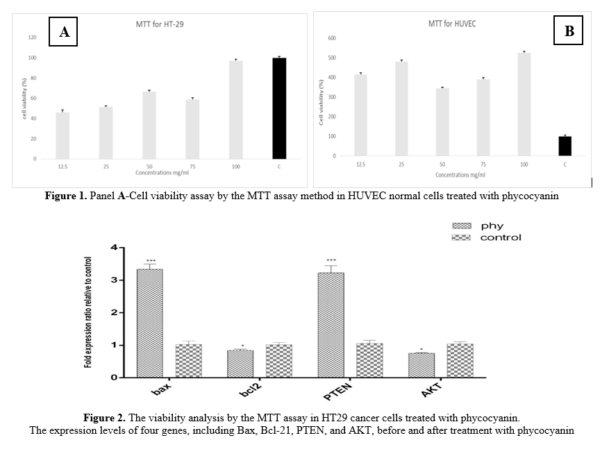
Cytotoxicity Measurement: The MTT assay revealed the potential cytotoxic effects of the phycocyanin extract on human cancer (HT-29) and normal (HUVEC) cell lines. These findings were based on treating HT-29 and HUVEC cell lines at varying doses of the phycocyanin extract (12.5, 25, 50, 75, and 100 mg/ml). The IC50 value of the extract for the HT-29 cell line was 12.5 mg/ml. The results demonstrated a significant decrease in the number of living cells after treating the human cancer cell line with the phycocyanin extract, while no toxic effect was observed in the normal cell line (HUVEC). Therefore, these results confirmed the effective inhibition of growth in HT-29 with the phycocyanin extract without posing a toxic effect on the normal human cell line. You can refer to Figures 1A and 1B for further details.
Effect of Extract on Gene Expression: The study confirmed the effect of phycocyanin extract on apoptosis signaling pathways, involving AKT/PTEN and Bax/Bcl-2 genes in the HT-29 cancer cell line. The RT-PCR results demonstrated that the phycocyanin extract significantly increased the expression of PTEN and Bax by up to 2.8-fold in the HT-29 cell line (P<0.01). Additionally, the phycocyanin extract at 12.5 mg/ml did not affect the expression of Bcl-2 or AKT genes, which induces apoptosis (P<0.05). This suggests that the phycocyanin extract positively regulated the AKT/PTEN genes without negatively affecting the expression of Bax/Bcl-2 genes in the human colon cell lines (See Figure 2).


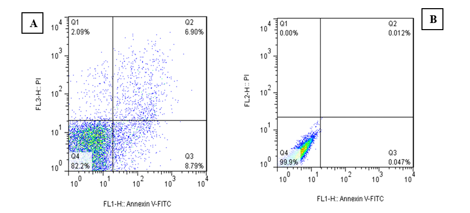
Flow Cytometry Assessment: The induction of apoptosis in the HT-29 cell line after treatment with the phycocyanin extract at IC50 concentrations is presented in Figure 3. The quantitative analysis of the flow cytometry data from the HT-29 cell line revealed that the phycocyanin extract caused apoptosis in 8.79% of the cells. In addition, 2.9% of treated HT-29 cells underwent necrosis compared to 0%, and no necrosis was detected in the control cells.

Figure 3. Flow cytometry was performed to explore the rate of apoptosis by phycocyanin on HT29 cell lines for 24 h. Panels A & B: The phycocyanin could trigger apoptosis (both early and late) in HT29 cells after 24 h of treatment.

Discussion

This study aimed to investigate the potential of phycocyanin as a natural anticancer agent and apoptosis activator for CRC treatment. In recent years, natural compounds have gained attention as potential anticancer agents due to their safety profile and potential effectiveness [26].
The results of this study demonstrated that phycocyanin treatment up-regulated PTEN and Bax genes while down-regulating AKT and Bcl-2 genes. This suggests that phycocyanin can effectively induce apoptosis in colon cancer cells. In light of these findings, phycocyanin is recommended as a herbal treatment and a helpful supplement for CRC. Natural compounds such as phycocyanin could offer new avenues for cancer therapy, particularly due to their ability to target specific genes involved in apoptosis [27].
The results of the MTT assay indicated that phycocyanin may have a cytotoxic effect on HT-29 and HUVEC cells. This is a significant finding, as it suggests that phycocyanin could be a potential treatment choice for CRC. The fact that phycocyanin did not show any toxic effect on normal cells is also promising, as it indicates that it may have a selective cytotoxic effect on cancer cells (Figures 1A and 1B).
The RT-PCR results also provided further insights into the mechanism of action of phycocyanin. The negative regulation of AKT by PTEN, commonly mutated in most cancers, suggests that phycocyanin may inhibit the AKT signaling pathway in colon cancer cells. This is significant because the AKT signaling pathway is known to promote cell survival and proliferation, and its dysregulation has been implicated in the development and progression of CRC.
Additionally, the observed changes in the ratio of Bax/Bcl-2 indicated that phycocyanin may trigger apoptosis in colon cancer cells via the intrinsic pathway. Bcl-2, an anti-apoptotic protein, is significantly overexpressed in many colon cancers. Consequently, targeting this pathway by inhibiting Bcl-2 and activating Bax could present a promising therapeutic approach for management of colon cancer [28].
The expression of AKT and PTEN is observed in most CRC cases, making the signaling pathway a potential target for treatment. The activity of AKT was negatively regulated by the tumor suppressor protein PTEN, which is frequently mutated in most cancers [29]. In addition to the positive regulation of PTEN and the negative regulation of AKT, it was associated with significant changes in the ratio of Bax/Bcl-2. It should be noted that Bcl-2 is an anti-apoptotic protein, while Bax, as a pro-apoptotic protein, can be related to the intrinsic pathway of apoptosis [28].
Bcl-2 has been observed in about 30-94% of colon cancers [30]. Therefore, targeting this pathway by inactivating Bcl-2 and activating Bax seems to be a promising treatment for colon cancer. The expression of AKT and PTEN has been observed in 60-70% of CRCs [31]. The inhibition of this signaling pathway is suggested as a potential target in CRC.
Although the findings of this study show promise, further research is necessary to completely understand the mechanisms behind phycocyanin’s anticancer effects and its potential as a therapeutic agent. Clinical trials should be carried out to evaluate its safety and efficacy in humans. Nevertheless, this study offers valuable insights into the potential benefits of phycocyanin for colon cancer treatment.
Conclusions
In conclusion, phycocyanin extract, considering its effects on the AKT/PTEN, Bax/BCL-2, and inducing apoptotic genes (Bax and PTEN) holds promise for enhancing the clinical efficacy and outcomes, while phycocyanin has minimal or no side effects. Therefore, it may be an effective therapeutic agent to treat human colon cancer in the future. Continued research may lead to the identification of new treatments that can boost the immune system’s response to cancer and enhance the prognosis for patients with colon cancer.
Conflict of Interests
The authors declare they had no conflicts of interest.
Funding
This research project was financially supported by the Deputy for Research and Technology, and the Student Research Committee of Qazvin University of Medical Sciences, Qazvin, Iran.
Acknowledgement
The authors wish to acknowledge the Deputy for Research and Technology, Qazvin University of Medical Sciences, for financially supporting this study.
Compliance with Ethical Guidelines
This study was confirmed by the Ethics Committee of Qazvin University of Medical Science, Iran (Code: IR.QUMS.REC.1399.546). This study did not conduct any experiments on humans or animals.
Authors' Contributions

HP and AP designed the study; SAHH, AAF, and FR performed the laboratory tests and data collection; HP and IS carried out the data analysis; FR, SAHH, RN, and MZJ drafted the initial manuscript. All authors reviewed and approved the final version of the manuscript prior to submission for publication.

Type of Study: Research | Subject: Special

References
1. Curado M-P, Edwards B, Shin HR, Storm H, Ferlay J, Heanue M, et al. Cancer incidence in five continents, Volume IX: IARC Press, International Agency for Research on Cancer. 2007. [Link]
2. Alexiusdottir KK, Möller PH, Snaebjornsson P, Jonasson L, Olafsdottir EJ, Björnsson ES, et al. Association of symptoms of colon cancer patients with tumor location and TNM tumor stage. Scandinavian Journal of Gastroenterology. 2012;47(7):795-801. [doi:10.3109/00365521.2012.672589]
3. Jemal A, Bray F, Center MM, Ferlay J, Ward E, Forman D. Global Cancer Statistics. CA: A Cancer Journal for Clinicians. 2011;61(2):69-90. [doi:10.3322/caac.20107]
4. Hamdan NT, Jwad BA, Jasim SA. Synergistic anticancer effects of phycocyanin and Citrullus colocynthis extract against WiDr, HCT-15 and HCT-116 colon cancer cell lines. Gene Reports. 2021;22:100972. [doi:10.1016/j.genrep.2020.100972]
5. Hashiguchi Y, Muro K, Saito Y, Ito Y, Ajioka Y, Hamaguchi T, et al. Japanese Society for Cancer of the Colon and Rectum (JSCCR) guidelines 2019 for the treatment of colorectal cancer. Int J Clin Oncol. 2020;25:1-42. [doi:10.1007/s10147-019-01485-z]
6. Cannon-Albright LA, Skolnick MH, Bishop DT, Lee RG, Burt RW. Common inheritance of susceptibility to colonic adenomatous polyps and associated colorectal cancers. N Engl J Med. 1988;319(9):533-7. [doi:10.1056/NEJM198809013190902]
7. Doll R, Peto R. The causes of cancer: quantitative estimates of avoidable risks of cancer in the United States today. JNCI: Journal of the National Cancer Institute. 1981;66(6):1192-308. [doi:10.1093/jnci/66.6.1192]
8. Giovannucci E, Willett WC. Dietary factors and risk of colon cancer. Annals of Medicine. 1994;26(6):443-52. [doi:10.3109/07853899409148367]
9. Keum N, Giovannucci E. Global burden of colorectal cancer: emerging trends, risk factors and prevention strategies. Nat Rev Gastroenterol Hepatol. 2019;16(12):713-32. [doi:10.1038/s41575-019-0189-8] [pmid: 31455888]
10. Zhang X, Hu P, Ding SY, Sun T, Liu L, Han S, et al. Induction of autophagy-dependent apoptosis in cancer cells through activation of ER stress: an uncovered anti-cancer mechanism by anti-alcoholism drug disulfiram. Am J Cancer Res. 2019;9(6):1266. [pmid: 31285958]
11. Arbabi M, Hasanpoor H, Arabzadeh S, Maghami P. The Synergic effect of Abujahl watermelon extract (Citrullus colocynthis) and Phycocyanin on apoptosis inducton in human colon cancer cell line (HT-29). Cellular and Molecular Research (Iranian Journal of Biology). 2020;33(2):126-135. [Link]
12. De Rivera CG, Miranda-Zamora R, Diaz-Zagoya J, Juárez-Oropeza M. Preventive effect of Spirulina maxima on the fatty liver induced by a fructose-rich diet in the rat, a preliminary report.Life Sciences. 1993;53(1):57-61. [doi:10.1016/0024-3205(93)90611-6]
13. Wu XJ, Yang H, Chen YT, Li PP. Biosynthesis of fluorescent β subunits of c-phycocyanin from spirulina subsalsa in escherichia coli, and their antioxidant properties. Molecules. 2018;23(6):1369. [doi:10.3390/molecules23061369]
14. Fernández-Rojas B, Hernández-Juárez J, Pedraza-Chaverri J. Nutraceutical properties of phycocyanin. Journal of Functional Foods. 2014;11:375-92. [doi:10.1016/j.jff.2014.10.011]
15. Chaiklahan R, Chirasuwan N, Bunnag B. Stability of phycocyanin extracted from Spirulina sp.: Influence of temperature, pH and preservatives. Process Biochemistry. 2012;47(4):659-64. [doi:10.1016/j.procbio.2012.01.010]
16. Strasky Z, Zemankova L, Nemeckova I, Rathouska J, Wong RJ, Muchova L, et al. Spirulina platensis and phycocyanobilin activate atheroprotective heme oxygenase-1: a possible implication for atherogenesis. Food & Function. 2013;4(11):1586-94. [doi:10.1039/c3fo60230c]
17. Zhang DL, Wu JH, Xie LM, Liu SQ. Molecular mechanism of C-phycocyanin in alleviation of acute lung injury in septic rats. Basic & Clinical Medicine. 2014;34(7):891. [Link]
18. Liu Y, Jovcevski B, Pukala TL. C-Phycocyanin from spirulina inhibits α-synuclein and amyloid-β fibril formation but not amorphous aggregation. Journal of Natural Products. 2019;82(1):66-73. [Link]
19. Kehrer JP. Free radicals as mediators of tissue injury and disease. Critical Reviews in Toxicology. 1993;23(1):21-48. [doi:10.3109/10408449309104073]
20. Romay C, Gonzalez R, Ledon N, Remirez D, Rimbau V. C-phycocyanin: a biliprotein with antioxidant, anti-inflammatory and neuroprotective effects. Current Protein and Peptide Science. 2003;4(3):207-16. [doi:10.2174/1389203033487216]
21. Danielsen SA, Eide PW, Nesbakken A, Guren T, Leithe E, Lothe RA. Portrait of the PI3K/AKT pathway in colorectal cancer. Biochimica et Biophysica Acta (BBA) - Reviews on Cancer. 2015;1855(1):104-21. [doi:10.1016/j.bbcan.2014.09.008]
22. Alizadeh S, Esmaeili A, Omidi Y. Anti-cancer properties of Escherichia coli Nissle 1917 against HT-29 colon cancer cells through regulation of Bax/Bcl-xL and AKT/PTEN signaling pathways. Iranian Journal of Basic Medical Sciences. 2020;23(7):886-93. [doi: 10.22038/ijbms.2020.43016.10115] [pmid: 32774810]
23. Pandurangan AK. Potential targets for prevention of colorectal cancer: a focus on PI3K/Akt/mTOR and Wnt pathways. Asian Pacific Journal of Cancer Prevention. 2013;14(4):2201-05. [doi:10.7314/APJCP.2013.14.4.2201]
24. Patel A, Mishra S, Pawar R, Ghosh P. Purification and characterization of C-Phycocyanin from cyanobacterial species of marine and freshwater habitat. Protein Expression and Purification. 2005;40(2):248-55. [doi:10.1016/j.pep.2004.10.028]
25. Tantirapan P, Suwanwong Y. Anti-proliferative effects of C-phycocyanin on a human leukemic cell line and induction of apoptosis via the PI3K/AKT pathway. Journal of Chemical and Pharmaceutical Research. 2014;6(5):1295-301. [Link]
26. Wang J, Jiang YF. Natural compounds as anticancer agents: Experimental evidence. World J Exp Med. 2012;2(3):45. [doi:10.5493/wjem.v2.i3.45] [pmid: 24520533]
27. Jiang L, Wang Y, Yin Q, Liu G, Liu H, Huang Y, et al. Phycocyanin: a potential drug for cancer treatment. J Cancer. 2017;8(17):3416. [doi:10.7150/jca.21058] [pmid: 29151925]
28. Qian S, Wei Z, Yang W, Huang J, Yang Y, Wang J. The role of BCL-2 family proteins in regulating apoptosis and cancer therapy. Front Oncol. 2022;12:985363. [doi:10.3389/fonc.2022.985363]
29. Gao F, Huang W, Zhang Y, Tang S, Zheng L, Ma F, et al. Hes1 promotes cell proliferation and migration by activating Bmi-1 and PTEN/Akt/GSK3β pathway in human colon cancer. Oncotarget. 2015;6(36):38667. [doi:10.18632/oncotarget.5484] [pmid: 26452029]
30. Ismail NI, Othman I, Abas F, H. Lajis N, Naidu R. Mechanism of apoptosis induced by curcumin in colorectal cancer. Int J Mol Sci. 2019;20(10):2454. [doi:10.3390/ijms20102454]
31. Cheng H, Jiang X, Zhang Q, Ma J, Cheng R, Yong H, et al. Naringin inhibits colorectal cancer cell growth by repressing the PI3K/AKT/mTOR signaling pathway. Experimental and Therapeutic Medicine. 2020;19(6):3798-804. [doi:10.3892/etm.2020.8649]

Add your comments about this article : Your username or Email:
CAPTCHA

Send email to the article author


Rights and permissions
Creative Commons License This work is licensed under a Creative Commons Attribution-NonCommercial 4.0 International License.

© 2025 CC BY-NC 4.0 | Iranian Journal of Toxicology

Designed & Developed by : Yektaweb